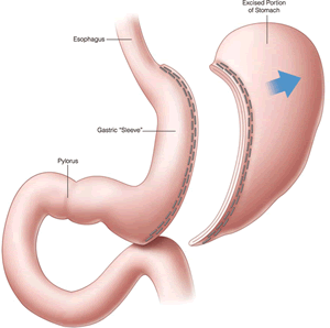
Global Treatment Services Pvt. Ltd.

Global Treatment Services

Leg amputation

Leg amputation is the surgical removal of legs either above or below the knee. This surgery is carried out as a result of poor blood circulation because of damage or narrowing of the arteries. As a result the blood cells in these region get deprived of oxygen and nutrients which in turn lead to infection and necrosis of the tissues.

IMG-20190417-WA0001

Amputation is also carried out due to injuries from accidents and burns, infections which are incurable with antibiotics,blood clots, cancerous tumor at the region, diabetes and nerve thickening and to prevent the disease from spreading farther into the body. The purpose of the surgery is to restore one’s ability to function and improving total health.

The procedure for amputation is done under general anesthesia  or with spinal anesthesia.

 TYPES

  1. Above the knee amputation: lt is also called a trans femoral amputation. Here the amputation occurs at the middle part of the thigh bone. After this type of amputation knee joint will not be present. Here walking with a prosthesis post surgery will be difficult.
  2. Below knee amputation: It also known as a trans tibial amputation and is the most common type. Here the knee joint will be present and walking with a prosthesis is more comfortable.
  3. Hemipelvic amputation: Here the hip-joint and part of the pelvis is amputated. It also known as a trans pelvic amputation. It is mostly done in case of a malignant tumor at the region. Here walking with a prosthesisis difficult
  4. Toe amputation: Here the toe is amputated due to poor supply of blood to the toe.
  5. Partial foot amputation: Here the toes and part of the long bones of the foot are amputated. This is also known as a trans metatarsal amputation.
  6. Disarticulation: Here amputation occurs through a joint. It helps to preserve the growth plate in the bone in a child.

Blood pressure

Blood pressure is the measurement of the pressure of the blood in the artery. A high or low blood pressure is a symptom of an underlying medical condition. Blood pressure has a normal range which is 120/80 mm Hg. The highest value of normal range is reached when the  heart muscle contracts and pumps blood, a cycle called systole.The lowest value of normal range is reached when the  the heart relaxes and refills with blood, a cycle called diastole. Based on the circumstances the blood pressure of a person may vary. Blood pressure above 140/90 mm Hg is considered as high.  A high blood pressure can put your arteries , muscles and heart at risk. It can also affect eyes kidneys and legs .It can also end up in stroke and heart attacks. The blood pressure of a person do not remain constant throughout the day. It fluctuates depending on the work done, the amount of fluid in the body , medicines in the blood stream and time of the day. It also varies with emotions sleep and exercise.

IMG-20190401-WA0000

The blood pressure is measure using a sphygmomanometer. It uses the height of a column of mercury to reflect the circulating pressure and is measured in millimetres of mercury. Today full automated digital blood pressure monitors are also available which makes it easier to check the BP at home without the help of a doctor or medical practitioner.

A high blood preassure is also called hypertension. There are many factors that contribute to hypertension. They are being overweight, reduced or lack of  physical activity, consumption of alcohol, smoking old age and stress. Family history of high blood pressure is also generally a factor for hypertension. An undiagnosed and untreated hypertension can lead to  coronary heart disease (CHD). Hypertension can be prevented or reduced by following a healthy lifestyle. Choices like regular exercise, balanced diet with reduced salt and fat, eating  plenty of vegetables and fruits, quitting smoking and alcohol consumption and maintaining a healthy weight  etc can help cntrol high blood pressure.


Tuberculosis (TB)

Tuberculosis (TB) is a disease caused by bacteria called Mycobacterium tuberculosis. The bacteria usually attack the lungs, but they can also damage other parts of the body.

file-7

TB spreads through the air when a person with TB of the lungs or throat coughs, sneezes, or talks. If you have been exposed, you should go to your doctor for tests. You are more likely to get TB if you have a weak immune system.

Symptoms of TB in the lungs may include

  • A bad cough that lasts 3 weeks or longer
  • Weight loss
  • Loss of appetite
  • Coughing up blood or mucus
  • Weakness or fatigue
  • Fever
  • Night sweats

Skin tests, blood tests, x-rays, and other tests can tell if you have TB. If not treated properly, TB can be deadly. You can usually cure active TB by taking several medicines for a long period of time.

Treatment

Given the many effective medications available today, the chances are great that a person with TB can be cured. It is important, however, for the person to understand the disease and to cooperate fully in the therapy program.

Both latent TB infection and active TB disease are treated with antibiotics. Treatment lasts at least six months because antibiotics work only when the bacteria are actively dividing, and the bacteria that cause TB grow very slowly. While latent TB infection can be treated with only one antibiotic, active TB disease is treated with several antibiotics at one time, to decrease the chances that the bacteria will evolve resistance to the drugs. Active TB disease must be treated aggressively, and patients may have to start treatment with a hospital stay to keep them from spreading the disease. After a few weeks, they will feel better and will no longer be infectious.

The biggest danger in TB treatment is that the patient will not take antibiotics on schedule. This gives the bacteria the opportunity to develop resistance to the drugs, rebound, and become much more difficult to treat. Therefore, it is crucial to take all of your drugs as instructed. Most treatment programs require that a health care professional watch you take every dose.

Your doctor may monitor you during treatment with blood tests to check your liver, sputum tests to see if the bacteria are susceptible to the antibiotics you are taking, and chest X-rays to look for signs of disease.

Extrapulmonary TB is active TB disease in any part of the body other than the lungs (for example, the kidney, spine, brain, or lymph nodes). Treatment for extrapulmonary disease is basically the same as for TB in the lungs except that TB involving the brain or bones is treated longer.

NICU

NICU is a special unit in a hospital where ill and premature newborn are often admitted to. NICU stands for Neonatal Intensive Care Unit. Here the babies are provided with around the clock care by a team of specially trained experts. NICU is known in many names like a special care nursery, an intensive care nursery or a newborn intensive care nursery.

IMG-20190401-WA0001

The different medical equipments at NICU are
● Incubators: These are small beds enclosed by transparent hard plastic. It provides controlled temperature for the baby inside. It has side holes through which babies are nursed by the caregivers.
● Infant warmers: These are open beds with heaters over them to help babies stay warm
while being monitored.
● Photo-therapy: It treats babies with jaundice by lying them on special light therapy blankets.
● Monitors: They are used to keep track of the baby’s temperature, heart rate, and breathing etc. It includes
1.Chest heads : Painless stickers on baby’s chest that connects to monitors to track the heart
rate and monitor the number of breathing.
2.Pulse oximetry (or pulse ox): This machine is used to measure baby’s blood oxygen levels. It is connected to baby’s fingers or toes and it emits a soft red light.
3.Temperature probe : It records baby’s temperature and displays it on the monitor.
4.An arterial lining or blood pressure cuff is used to measure baby’s blood pressure.
5.Feeding tubes: These are used to feed the babies with formula milk or breast milk.
6.IVs: These are thin bendable tubes used to drip the baby with medicine and fluids through the baby’s veins.
7.Ventilators: These provide extra breathing assistance for the baby as and when required.
8.Oxygen hood or nasal cannula: These provides extra oxygen for babies who are in need of it.
Babies who require NICU are

● Premature babies born before 37 weeks of pregnancy.
● Babies who have low birth weight (less than 2500 g or more than 4000 g
● Babies who have a medical condition that requires special care.
● Twins, triplets, and other multiple babies
● Babies born with heart problems, infections, or birth defects
● Babies who requires blood transfusion treatments.
● If the baby’s first stool(meconium) passed during pregnancy into the amniotic fluid.
● Breech delivery presentation during delivery.
● Too little or too much amniotic fluid during fetal stage.
● Babies whose mother have Diabetes and hypertension.

NICU usually have specially trained team of medical professionals including neonatologists,
pediatricians, lactation consultants, respiratory therapists, dietitians etc.

SCOLIOSIS

Scoliosis is a “S ”  or “C” shaped curve of the spine that mostly occurs during the growth spurt just before puberty. Though the actual cause is unknown scoliosis can be caused by conditions such as cerebral palsy and muscular dystrophy.  In severe cases it  can lead to reduced amount of space within the chest, making it difficult for the lungs to function properly and thus interfere with breathing . In some cases, the degree of curve remains stable, while in others, it increases over time. A person with scoliosis may have an uneven shoulder or waist, a shoulder blade that appears prominent than the other, one leg being longer than the other etc. This curves can be structural curves or non structural curves.  In structural scoliosis, the curve of the spine can’t be reversed as it is rigid where as in the case of non structural type ,though the spine looks curved it functions normally. Scoliosis can cause complications like heart and lung damage making it difficult to breath, chronic back pains and noticeable changes in appearances.

IMG-20190326-WA0009

 TREATMENT

In mild cases scoliosis won’t require surgery. In case of mild scoliosis , braces are recommended.  In severe cases surgery may be suggested  to prevent it from getting worse.

  1. Braces:  They are plastic made close fitting invisible tighteners worn under the dress to prevent further progression of the curve. A brace won’t cure scoliosis or reverse the curve. It can be removed as and when required.
  2. Surgery: The surgery  for scoliosis involves the spinal fusion wherein  patient will be examined by an anesthesiologist, to manage the pain both during and after the operation prior to the surgery.  A thin flexible tube (epidural catheter) may be inserted in the back at the conclusion of the operation to help control pain. Pre operation procedures also involves collection of blood samples to examine the blood count of the patient.The surgery takes  five to six hours of time depending on the patient. During the surgery
  •  An incision is made in the middle of the back.
  • The spine is exposed by moving the muscles to side.
  • The joints between the vertebrae are removed to loosen them up. The new bone produced when the vertebrae  get roughened eventually bridges the gaps between the vertebrae and makes them fuse together.
  • Metal rods are used to hold the spine in place until fusion happens  which are attached to the spine by screws, hooks, and/or wires.

A bone graft is used to stimulate bone healing  which helps to increase bone production and the vertebrae heal together into a solid bone. A bone graft is either taken from the patients hip or from a donor through bone bank. The possible complications of the surgery are bleeding, infection, nerve injury etc.


Gingivitis

Gingivitis is a very common gum disease that appears as  red, swollen irritable inflammation of the gum or gingiva. Gingiva is the part of the gum lining the base of the teeth.  The inflammation is caused by the buildup of plaque on the teeth or gum. Plaque is sticky film that contains bacteria which irritates and weakens the gum. As a result the gums turn red,swollen, puffy and starts to bleed. Gingivitis is the major cause for bleeding in most of the adults.

IMG-20190327-WA0000

CAUSES

The most common cause for gingivitis is poor oral hygiene. Apart from this, gingivitis can also occur as a result of smoking or tobacco use, improper removal of plaque while brushing and flossing,  hormonal or physiological changes (such as puberty, menstruation, pregnancy, menopause), stress that weakens the immune system to fight infection, poor nutrition especially due to the lack of vitamin C in diet, certain medications, and some diseases including diabetics, cancer etc.

SYMPTOMS

  • Red and puffy gums
  • Tender and receding gums
  • mild pain and irritation on the inflamed gum
  • Bleeding when you brush or floss
  •  Tooth sensitiveness to hot and cold foods or beverages.
  • Bad breath caused by the bacteria in plaque

DIAGNOSIS

  • Examination of your teeth and gums,  for signs of plaque and inflammation.
  • Measuring the depth of grove between the gums and teeth at multiple locations inside the mouth.
  • Check for the severity of bone lose at affected areas by taking dental X-rays

TREATMENT

  • Dental cleaning, apart from regular brushing and flossing a professional cleaning helps to remove the plaque surrounding the gingiva completely.
  • Restoration of misaligned teeth  which are more vulnerable for infections.
  • Use of an anti gingivitis toothpaste and anti gingivitis mouth wash.
  • Regular brushing and flossing.
  • Use of soft toothbrush and replacing it at  every three to four months.


Febrile seizure

Febrile seizures are high fever related fits or convulsions that affect young children. It occurs in association with elevated temperature and is a very common childhood seizure disorder. It happen any time between three months or five years of age when t he fever gets higher. It tend to occur in families. In a child with this disorder,the risk of febrile seizure is 10% for the siblings and almost 50% for the sibling if a  parent has febrile seizure as well. Though the mode of inheritance is not clear and know, there are clear evidences for the  existence its genetic basis. Such children have a slightly higher risk of epilepsy compared with the general population.

IMG-20190326-WA0011

They are often harmless and typically don’t indicate a serious health problem.

TYPES OF FEBRILE SEIZURES

  • Simple seizure:  These seizures lasts from a few seconds to 15 minutes. They do not recur within a 24-hour period and are not specific to one part of the body.
  • Complex seizures: These types lasts longer than 15 minutes and occurs more than once within 24 hours or is confined to one side of your child’s body.

CAUSES

Though a higher than normal body temperature causes febrile seizures,  a low-grade fever can  also trigger a febrile seizure.

  • Infection: The fevers that trigger febrile seizures are usually caused by a viral infection, and rarely by a bacterial infection. Influenza and the virus that causes roseola, which  are accompanied by high fevers are frequently associated with febrile seizures.
  • Post-immunization seizures:  Febrile seizures may increase after some childhood immunizations. These include the diphtheria, tetanus and pertussis or measles-mumps-rubella vaccinations. A child can develop a low-grade fever after a vaccination. Not th vaccine but the fever causes the seizure.

the risk of recurrence is higher if  the first seizure resulted from a low fever,the period between the start of the fever and the seizure was short, a family history of febrile seizures and the child was younger than 18 months at the time of the first febrile seizure.

DIAGNOSIS AND  TREATMENT

Identifying the cause of the fever is the first step of treating the seizure.

Mostly the  febrile seizures stop on their own within a few minutes. Once your child has a febrile seizure, stay calm and follow these steps:

  • Place your child on his side on a  flat surface where he won’t fall.
  • Time how long the convulsion lasts, if possible.
  • Remove hard or sharp objects near your child.
  • Loosen his clothing.
  • Don’t restrain your child or interfere with your child’s movements.
  • Don’t put anything in your child’s mouth.
  • Do not put a child who is having a convulsion in the bath to lower their temperature.
  • If the convulsion lasts more than five minutes call an ambulance.

Retinoblastoma

Retinoblastoma is cancer of the eye which begins in the retina. Retina is a layer of nerve cells
that lines the back of the eye. It happens when the cells form a tumor when nerve cells in the retina
change and grows in size and number. Cells usually spread in and around the eye. They can
also spread to the brain and spine. Retinoblastoma usually occur in young children below the
age of five though it appear in adults as well. Children with an inherited gene from their parents
tend to get retinoblastoma at an earlier age, and in both eyes.The early symptoms of
retinoblastoma is a cloudy white pupil(leukocoria). In bright light, the pupil can look silvery or
yellow.
Other symptoms are :IMG-20190326-WA0012
● Poor and blurred vision
● The coloured part of the eye (Iris) appear cloudy
● Crossed eyes (Eyes that appear to be looking in different directions)
● Eye redness
● Eye swelling

There are two types of retinoblastoma.

● Heritable retinoblastoma
This is the most common type of retinoblastoma. It occurs when a child inherits a
mutation (change in a gene) from a parent. Such babies will be born with retinoblastoma.

● Sporadic retinoblastoma
Occurs when a gene mutation happens in early childhood, usually after 1 year of age. It
affects just one eye.

TREATMENT

Treatment involves chemoreduction and focal treatments without the use of external beam
radiotherapy. This allows preservation of the eye in some cases, often with visual function.

Nomophobia

IMG-20190326-WA0008Have you ever been without your mobile phone for a few hours? What goes through  your mind when your phone is not within your reach? Irritated?,tensed? or restlessness?.If yes, then probably you are undergoing a state called nomophobia. Nomophobia or “nomobile-phone phobia” is the irrational fear of being without your mobile phone. This happens when your are not able to use your phone for some reason, such as the absence of a signal or running out of minutes , battery power or credit. Today it’s increasing among high school and college students. A person with this syndrome may be tensed, stressed ,or anxious when he cannot use his mobile phone . It can also be an addiction when such people cannot go to sleep with or next to their smart phone or never switch off their phone or cannot take a break from it.  However, there is debate among the medical community on its classification; is it a phobia, anxiety disorder, lifestyle disorder, or addiction?. But it is commonly perceived as a phobia. Nomophobia preoccupies an individual with his phone and turn to it if they are depressed, anxious, and lonely. It not only affects the mind, but also relationships, where a person is physically present but psychologically absent. It can affect job performance, academic performance, impairs concentration and mental health in a long run. The various symptoms of this condition include,

  •  Elevated heart rate, sweaty palms, shallow breathing.
  • Keep checking the phone every now and then for battery life and/or messages.
  • Plug and connect regularly.
  • Use of phone even at inappropriate places .
  • Health and mental well being suffers in the absence of mobile phone.

The treatment of nomophobia often does not require medical interventions.  The best remedies for this phobia includes  taking control over phone ,interpersonal counseling, cognitive behavioral therapy, addiction therapy, gradual exposure therapy and systematic desensitization. Exposure therapy is, first exposing the individual to their phobia in therapy (ie, not taking the phone in the room with them) and gradually moving to real-life scenarios (ie, moving the phone to another room for gradually increasing amounts of time). For worst cases of this phobia, anxiety medication may be required.


Breast Milk

Breast milk is the primary nutrition source  for the newborn. According to WHO, newborn babies should be fed exclusively with breast milk for the first six months. It is the complete nutrition source till six months of age. The rich composition of the breast milk protects the baby against infections, diseases and illness. Bless fully, the benefits of which continue even long after breast feeding has ended.

1553433265658_0_IMG-20190321-WA0004

STAGES OF BREAST MILK

Breast milk is typically studied under three stages. The colostrum, transitional milk and mature milk.

Colostrum is the thick yellow sticky breast milk produced during the end of pregnancy and during the first few days after delivery. It is high in protein, low in fat and has abundant amount of immunoglobulin(IgA) and white blood cells to fight infections. It also act as a natural laxative by helping to prevent jaundice and clearing meconium from the baby’s body.

Transitional breast milk comes in as a combination of colostrum and mature milk three to five days after birth of the baby.

Mature human milk contain fat (3%-5%), protien (0.8%-0.9%), carbohydrates (6.9%-7.2%),  calculated as lactose. The energy content of breast milk is 60-70kcal//100ml. The protein content is more than the carbohydrate content in the colostrum than the matuem milk. There is no consistent compositional difference between milks from the two breasts unless one is infected.

COMPOSITION OF BREAST MILK

The nutrient rich complex composition of breast milk include water, protien, carbohydrates, fats, immunoglobulins, vitamins, minerals, enzymes and hormones. The composition may varies among mothers and within the same mother throughout a day. This variation over time helps to meet the needs of a growing child.

The principal protein of human milk is casein. It is homologous to bovine beta- casein, alpha lactalbumin, lactoferrin, immunoglobulin IgA, lysozyme and serum albumin.

The principal sugar of human milk is lactose. Apart from lactose 30 or more other oligosaccharides are also present. These amount is the aggregate to as much as 1g/100ml in mature milk and 2.5g/100ml in colostrum. Some of them has the ability to promote growth of certain strains of lactobacilli.

The principal fat elements in human milk are palmitic acid and oleic acid.

The principal mineral constituents are sodium, pottasium, calcium, magnesium, phosphorus, and chlorine. The iron, copper and zinc contents of human milk varies considerably.

BENEFITS OF BREAST MILK

  • Water: Water which makes up 90% of the breast milk keeps the baby hydrated and help to regulate body temperature , lubricate and protect internal organs.
  • Carbohydrates: Apart from being the energy provider , it is also linked to greater brain development.
  • Lipids: lipids or fats play a major role in the baby’s weight gain as he grows.
  • Protein:  It helps to build, strengthen and repair the body. Protein lactoferrin in the milk is associated with the iron transport in the body.
  • Immunoglobulins/ antibodies: They are baby’s first vaccine. It helps fight off germs, bacteria, fungi, viruses, parasites and diseases.  IgA ,the main immunoglobulin  prevents germs from entering the nanbab body and blood.
  • Vitamins protects the baby from scurvy, rickets,and other deficiency diseases.
  • Minerals helps to build strong bones, produce red blood cells and helps in the proper functioning of muscles and nerves.
Pages:1...6789101112...80