Global Treatment Services Pvt. Ltd.

Global Treatment Services

Purpura: Causes, symptoms & treatments

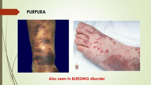
Henoch-Schonlein purpura (HSP) is a disease involving inflammation of small bloodvessels. It most commonly occurs in children. The inflammationcauses blood vessels in the skin, intestines, kidneys, and joints to start leaking. The main symptom is a rash with numerous small bruises, which have a raised appearance, over the legs or buttocks.

Although HSP can affect people at any age, most cases occur in children between the ages of 2 and 11. It is more common in boys than girls. Adults with HSP are more likely to have more severe disease compared to children.

HSP usually ends after four to six weeks — sometimes with recurrence of symptoms over this period, but with no long-term consequences. If organs such as the kidneys and intestines are affected, treatment is often needed and it is important to have regular follow-up to prevent serious complications.

Image result for purpura platelet disorders

Causes and Risk Factors for Henoch-Schonlein Purpura

The exact cause of HSP is not known. The body’s immune system is believed to play a role in targeting the blood vessels involved. An abnormal immune response to an infection may be a factor in many cases. Approximately two-thirds of the cases of HSP occur days after symptoms of an upper respiratory tract infection develop.

Some cases of HSP have been linked to vaccinations for typhoid, cholera, yellow fever, measles, or hepatitis B; foods, drugs, chemicals, and insect bites. Some experts also say that HSP is associated with the colder weather of fall and winter.

Symptoms of Henoch-Schonlein Purpura

The classic symptoms of HSP are rash, joint pain and swelling, abdominal pain, and/or related kidney disease, including blood in urine. Before these symptoms begin, patients may have two to three weeks of fever, headache, and muscular aches and pains. Rarely, other organs, such as the brain, heart, or lungs, may be affected.

Here are some key details about the symptoms of HSP:

Rash. The rash usually appears in all patients with HSP. The initial appearance may resemble hives, with small red spots or bumps on the lower legs, buttocks, knees, and elbows. But these change to appear more like bruises. The rash usually affects both sides of the body equally and does not turn pale on pressing.

Arthritis. Joint inflammation, involving pain and swelling, occurs in approximately three-quarters of cases, particularly affecting the knees and ankles. It usually lasts only a few days and does not cause any long-term, chronic joint problems.

Abdominal pain. In more than half of people with HSP, inflammation of the gastrointestinal tract may cause pain or cramping; it may also lead to loss of appetite, vomiting, diarrhea, and occasionally blood in the stool.

In some cases, patients may have abdominal pain before the rash appears. In rare cases, an abnormal folding of the bowel (intussusception) may cause a bowel blockage, which may require surgery to fix.

Kidney impairment. HSP can cause kidney problems, indicated by such signs as protein or blood in the urine. This is usually only discovered on urine testing, since it does not generally cause any discomfort.

In most patients, the kidney impairment is mild and goes away without any long-term damage. It’s important to monitor the kidney problems closely and make sure they clear up, since about 5% of patients may develop progressive kidney disease. About 1% may go on to develop total kidney failure.

Henoch-Schonlein Purpura Diagnosis and Treatment

The diagnosis of HSP may be clear when the typical rash, arthritis, and abdominal pain are present. A doctor may order some tests to rule out other diagnoses, confirm the diagnosis, and assess its severity.

Occasionally, when the diagnosis is uncertain, particularly if the only symptom is the classic rash, your doctor may perform biopsies of the skin or kidney. Urine and blood tests will likely be done to detect signs of kidney involvement and may need to be repeated during follow-up to monitor any changes in kidney function.

Although there is no specific treatment for HSP, you can use over-the-counter pain medicines, such as acetaminophen or nonsteroidal anti-inflammatory drugs (NSAIDs), such as ibuprofen and naproxen for joint pain. In some cases, corticosteroid medication may be used.

The rash and joint pain will usually go away after four to six weeks without causing any permanent damage. Bouts of the rash may recur in approximately one-third of cases, but they are usually milder, do not involve joint and abdominal symptoms, and they clear up on their own.

Fecal Incontinence: Causes, Symptoms & Treatments

Fecal incontinence is the inability to control bowel movements. It’s a common problem, especially among older adults.

Accidental bowel leakage is usually not a serious medical problem. But it can seriously interfere with daily life. People with bowel incontinence may avoid social activities for fear of embarrassment.

Many effective treatments can help people with bowel incontinence. These include:

  • medicine
  • surgery
  • minimally invasive procedures

Talking to your doctor is the first step toward freedom from bowel incontinence.Image result for fecal incontinence causes, symptoms & treatments

Causes of Bowel Incontinence

The most common cause of bowel incontinence is damage to the muscles around the anus (anal sphincters). Vaginal childbirth can damage the anal sphincters or their nerves. That’s why women are affected by accidental bowel leakage about twice as often as men.

Anal surgery can also damage the anal sphincters or nerves, leading to bowel incontinence.

There are many other potential causes of bowel incontinence, including:

  • Diarrhea (often due to an infection or irritable bowel syndrome)
  • Impacted stool (due to severe constipation, often in older adults)
  • Inflammatory bowel disease (Crohn’s disease or ulcerative colitis)
  • Nerve damage (due to diabetes, spinal cord injury, multiple sclerosis, or other conditions)
  • Radiation damage to the rectum (such as after treatment for prostate cancer)
  • Cognitive (thinking) impairment (such as after a stroke or advanced Alzheimer’s disease)

More than one cause for bowel incontinence is frequently present. It’s also not unusual for bowel incontinence to occur without a clear cause.

Diagnosis of Bowel Incontinence

Discussing bowel incontinence may be embarrassing, but it can provide clues for a doctor to help make the diagnosis. During a physical examination, a doctor may check the strength of the anal sphincter muscle using a gloved finger inserted into the rectum.

Stool testing. If diarrhea is present, stool testing may identify an infection or other cause.

Endoscopy. A tube with a camera on its tip is inserted into the anus. This identifies any potential problems in the anal canal or colon. A short, rigid tube (anoscopy) or a longer, flexible tube (sigmoidoscopy or colonoscopy) may be used.

Surgery may be recommended for people whose bowel incontinence is not helped by noninvasive treatments. The types of surgery include:

Anorectal manometry. Apressure monitor is inserted intothe anus and rectum. This allowsmeasurement of the strength ofthe sphincter muscles.

Endosonography. An ultrasoundprobe is inserted into the anus.This produces images that canhelp identify problems in the anal and rectal walls.

Nerve tests. These tests measure the responsiveness of the nervescontrolling the sphincter muscles. They can detect nerve damage thatcan cause bowel incontinence.

MRI defecography. Magnetic resonance imaging of the pelvis can beperformed, potentially while a person moves her bowels on a specialcommode. This can provide information about the muscles and supportingstructures in the anus, rectum, and pelvis.

Treatments for Bowel Incontinence

Bowel incontinence is usually treatable. In many cases, it can be cured completely.

Recommended treatments vary acc

  • Eat 20 to 30 grams of fiber per day. This can make stool more bulky and easier to control.
  • Avoid caffeine. This may help prevent diarrhea.
  • Drink several glasses of water each day. This can prevent constipation.

Medications. Try these medicines to reduce the number of bowel movements and the urge to move the bowels:

  • Imodium
  • Lomotil
  • Hyoscyamine

Methylcellulose can help make liquid stool more solid and easier to control. For people with a specific cause of diarrhea, such as inflammatory bowel syndrome, other medications may also help.

Exercises. Begin a program of regularly contracting the muscles used to control urinary flow (Kegel exercises). This builds strength in the pelvic muscles and may help reduce bowel incontinence.

Bowel training. Schedule bowel movements at the same times each day. This can help prevent accidents in between.

Biofeedback. A sensor is placed inside the anus and on the abdominal wall. This provides feedback as a person does exercises to improve bowel control.


Dilated cardiomyopathy (DCM): Causes, Symptoms & Treatments

Symptoms:

Many people with dilated cardiomyopathy have no symptoms. Some that do have only minor ones, and live a normal life. Others develop symptoms that may get worse as their heart gets sicker.

Symptoms of DCM can happen at any age and may include:

  • Shortness of breath
  • Swelling of your legs
  • Fatigue
  • Weight gain
  • Fainting
  • Palpitations (fluttering in the chest due to abnormal heart rhythms)
  • Dizziness or lightheadedness
  • Blood clots in the dilated left ventricle because of pooling of the blood. If a blood clot breaks off, it can lodge in an artery and disrupt blood flow to the brain, causing a stroke. A clot can also block blood flow to the organs in the abdomen or legs.
  • Chest pain or pressure
  • Sudden death

Causes :

DCM can be inherited, but it’s usually caused by other things, including:

  • Severe coronary artery disease
  • Alcoholism
  • Thyroid disease
  • Diabetes
  • Viral infections of the heart
  • Heart valve abnormalities
  • Drugs that damage the heart

It can also happen in women after they give birth. That’s called postpartum cardiomyopathy.

Diagnosis

Your doctor will decide if you have DCM after he looks at things like:

  • Your symptoms
  • Your family history
  • A physical exam
  • Blood tests
  • An electrocardiogram
  • A chest X-ray
  • An echocardiogram
  • An exercise stress test
  • Cardiac catheterization
  • A CT scan
  • An MRI

Another test that’s rarely done to find the cause of cardiomyopathy is called a myocardial biopsy, or heart biopsy. A tissue sample is taken from the heart and examined under a microscope.

If you have a relative with dilated cardiomyopathy, ask your doctor if you should be screened for it. Genetic testing may also be available to find abnormal genes.

Treatment

In the case of dilated cardiomyopathy, it’s aimed at making the heart stronger and getting rid of substances in the bloodstream that enlarge the heart and lead to more severe symptoms:

Medications: To manage heart failure, most people take drugs, such as a:

  • Beta blocker
  • ACE inhibitor or an ARB
  • Diuretic

If you have an arrhythmia(irregular heartbeat), your doctor may give you medicine to control your heart rate or make them happen less often. Blood thinners may also be used to prevent blood clots.

Lifestyle changes: If you have heart failure, you should have less sodium, based on your doctor’s recommendations. He may point you toward aerobic exercise, but don’t do heavy weightlifting.

Possible Procedures

People with severe DCM may need one of the following surgeries:

Cardiac resynchronization by biventricular pacemaker: For some people with DCM, stimulating the right and left ventricles with this helps your heart’s contractions get stronger. This improves your symptoms and lets you exercise more.

The pacemaker also will help people with heart block (a problem with the heart’s electrical system) or some bradycardias (slow heart rates).

Implantable cardioverter defibrillators (ICD): These are suggested for people at risk for life-threatening arrhythmias or sudden cardiac death. It constantly monitors your heart’s rhythm. When it finds a very fast, abnormal rhythm, it ”shocks” the heart muscle back into a healthy beat.

Surgery: Your doctor may recommend a surgery for coronary artery disease or valve disease. You may be eligible for one to fix your left ventricle or one that gives you a device to help your heart work better.

Heart transplant: These are usually just for those with end-stage heart failure. You’ll go through a selection process. Hearts that can be used are in short supply. Also, you must be both sick enough that you need a new heart, and healthy enough to have the procedure.


Wernicke- Korsakoff Syndrome: Causes, symptoms & Treatments

What Is Wernicke–Korsakoff Syndrome?

Wernicke-Korsakoff syndrome (WKS) is a type of brain disorder caused by a lack of vitamin B-1. The syndrome is actually two separate conditions that can occur at the same time. Usually, people get the symptoms of Wernicke’s encephalopathy first.

Also called Wernicke’s disease, people with Wernicke’s encephalopathy have bleeding in the lower sections of the brain, including the thalamus and hypothalamus. These areas of the brain control the nervous and endocrine systems. The bleeding causes brain damage that presents symptoms involving your vision, coordination, and balance.

The signs of Korsakoff psychosis tend to follow as the Wernicke’s symptoms decrease. If Wernicke’s disease is treated quickly and effectively, Korsakoff syndrome may not develop. Korsakoff psychosis is the result of chronic brain damage. Korsakoff syndrome affects the areas of your brain that control memory.

Alcoholism, or chronic alcohol abuse, is the most common cause of WKS. WKS can also be linked to diet deficiencies or other medical conditions that impair vitamin B-1 absorption. Vitamin B-1 is also called thiamine.

To diagnose WKS, your doctor will look for clinical signs that point to a vitamin B-1 deficiency. This can include blood tests that measure thiamine levels and your general nutritional health, as well as tests to check your liver function.

Image result for Wernicke-Korsakoff Syndrome

Chronic alcoholism can damage your liver, elevating your liver enzymes. Diagnosis includes a physical examination to assess your:

  • heart rate
  • eye movements
  • reflexes
  • blood pressure
  • body temperature

After diagnosis, your doctor will most likely give you vitamin B-1 intravenously, or through your vein. Fast treatment may reverse many of the neurological symptoms of WKS.

The key to recovery is maintaining adequate vitamin B-1 levels, which means refraining from alcohol abuse if you have WKS. You should also eat a balanced diet.

Risk Factors for WKS;

Risk factors for WKS are related to your diet and lifestyle.

The major risk factors for developing WKS are malnourishment and chronic alcoholism. Other risk factors for WKS include:

  • being unable to afford medical care and proper food
  • undergoing kidney dialysis, which reduces vitamin B-1 absorption
  • AIDS, which makes you more likely to develop conditions that lead to vitamin B-1 deficiency.

Causes of WKS:

The number one cause of WKS is alcoholism.

The less common causes of WKS are conditions that limit nutritional absorption. Eating and nutrient absorption can be restricted by:

  • gastric bypass surgery, which makes it difficult to meet nutritional needs due to limited food portions
  • colon cancer, which can cause pain that causes you to put off eating
  • eating disorders

Alcoholism is the number one cause of WKS because people who are alcoholics generally have a poor diet. Alcohol also prevents vitamin B-1 absorption and storage.

Symptoms of WKS:

Lesions on the brain cause Wernicke’s disease (WD). These lesions are the result of a vitamin B-1 deficiency.

Prominent symptoms of WD are:

  • double vision
  • a drooping upper eyelid
  • up-and-down or side-to-side eye movements
  • loss of muscle coordination
  • a confused mental state

WD can later develop into Korsakoff’s syndrome. People who have WKS have a variety of issues relating to memory. You may suffer from memory loss or be unable to form new memories.

You may also have the following symptoms if you have WKS:

  • amnesia for events that happen after the onset of the disorder
  • difficulty understanding the meaning of information
  • difficulty putting words into context
  • hallucinations
  • exaggerated storytelling, or confabulation
 Diagnosing WKS:

An individual with WKS is often mentally confused. This can make communication with the doctor difficult. Your doctor may overlook the possibility of a physical disorder if you’re confused.

Your doctor may first check for signs of alcoholism. They may check your blood alcohol levels. Sometimes, a doctor will take a liver function test to check for liver damage. Liver damage is a common sign of alcoholism.

Your doctor may also order nutritional tests to make sure you aren’t malnourished. Nutritional tests may include the following:

  • A serum albumin test measures the levels of albumin, which is a protein in the blood. Low levels of albumin may signal nutritional deficiencies as well as kidney or liver problems.
  • A serum vitamin B-1 test is a blood test to check vitamin B-1 levels in the blood. Enzyme activity in the red blood cells can be tested. Low enzyme activity in the red blood cells signals a vitamin B-1 deficiency.

You may also need imaging tests. These tests can help your doctor find any damage that’s characteristic of WKS.

Diagnostic imaging tests for WKS include:

  • an electrocardiogram (EKG) before and after giving vitamin B-1, which can help your doctor find abnormalities
  • a CT scan to check for brain lesions related to WD
  • an MRI scan to look for brain changes related to WD

Your doctor may also use neuropsychological test to determine the severity of any mental deficiencies.

Treatment for WKS :

WKS treatment should begin immediately. Prompt treatment may delay or stop disease progression. Treatments are also able to reverse non-permanent brain abnormalities.

Treatment may first involve hospitalization. At the hospital, you’ll be monitored to ensure your digestive system is absorbing food properly.

The treatment for WKS may include:

  • vitamin B-1 given through an IV in the arm or hand
  • vitamin B-1 given by mouth
  • a balanced diet to keep vitamin B-1 levels up
  • treatment for alcoholism

In a small number of cases, treatment of vitamin B-1 deficiency produces a negative reaction. This is more common in alcoholics.

Negative reactions to receiving vitamin B-1 may vary. Reactions may include alcohol withdrawal symptoms like insomnia, sweating, or mood swings. You may also experience hallucinations, confusion, or agitation.

Dementia: Causes, Symptoms & Treatments

What Is Dementia?

Dementia causes problems with thinking, memory, and reasoning. It happens when the parts of the brain used for learning, memory, decision making, and language are damaged or diseased.

Also called major neurocognitive disorder, it’s not a disease itself. Instead, it’s a group of symptoms caused by other conditions.

Alzheimer’s disease is the most common cause of dementia. Between 60% to 80% of people with dementia have Alzheimer’s. But there are as many as 50 other causes of dementia.

The symptoms of dementia may improve with treatment. But many of the diseases that cause dementia aren’t curable.

Image result for dementia causes, symptoms & treatments

What Causes Dementia?

The most common causes of dementia include:

  • Degenerative neurological diseases. These include Alzheimer’s disease, Parkinson’s disease, Huntington’s disease, and some types of multiple sclerosis. These diseases get worse over time.
  • Vascular disorders. These are disorders that affect the blood circulation in your brain.
  • Traumatic brain injuries caused by car accidents, falls, concussions, etc.
  • Infections of the central nervous system. These include meningitis, HIV, and Creutzfeldt-Jakob disease.
  • Long-time alcohol or drug use
  • Certain types of hydrocephalus, a buildup of fluid in the brain

Types of Dementia

Dementia can be split into two groups based on which part of the brain is affected.

  • Cortical dementias happen because of problems in the cerebral cortex, the outer layer of the brain. They play a critical role in memory and language. People with these types of dementia usually have severe memory loss and can’t remember words or understand language. Alzheimer’s and Creutzfeldt-Jakob disease are two forms of cortical dementia.
  • Subcortical dementias happen because of problems in the parts of the brain beneath the cortex. People with subcortical dementias tend to show changes in their speed of thinking and ability to start activities. Usually, people with subcortical dementia don’t have forgetfulness and language problems. Parkinson’s disease, Huntington’s disease, and HIV can cause these types of dementia.

Some types of dementia affect both parts of the brain.

Are There Treatments for Dementia?

To treat dementia, doctors will treat whatever is causing it. About 20% of the causes of dementia are reversible.

Causes of dementia that may be reversible include:

  • Alcohol or drug abuse
  • Tumors
  • Subdural hematomas, blood clots beneath the outer covering of the brain
  • Normal-pressure hydrocephalus, a buildup of fluid in the brain
  • Metabolic disorders such as a vitamin B12 deficiency
  • Low levels of thyroid hormones, called hypothyroidism
  • Low blood sugar, called hypoglycemia
  • HIV-associated neurocognitive disorders (HAND)

These forms of dementia are partially manageable, but they aren’t reversible and get worse over time:

  • Alzheimer’s disease
  • Vascular dementia
  • Dementia from Parkinson’s disease and similar disorders
  • Dementia with Lewy bodies
  • Frontotemporal dementia (Pick’s disease)
  • Creutzfeldt-Jakob disease

Image result for dementia causes, symptoms & treatments

What Are the Stages of Dementia?

Usually, dementia goes through these stages. But it may vary depending on the area of the brain that is affected.

1) No impairment: Someone at this stage will show no symptoms, but tests may reveal a problem.

2) Very mild decline: You may notice slight changes in behavior, but your loved one will still be independent.

3) Mild decline: You’ll notice more changes in his thinking and reasoning. He may have trouble making plans, and he may repeat himself a lot. He may also have a hard time remembering recent events.

4) Moderate decline: He’ll have more problems with making plans and remembering recent events. He may have a hard time with traveling and handling money.

5) Moderately severe decline: He may not remember his phone number or his grandchildren’s names.He may be confused about the time of day or day of the week. At this point, he will need assistance with some basic day-to-day functions, such as picking out clothes to wear.

6) Severe decline: He’ll begin to forget the name of his spouse. He’ll need help going to the restroom and eating. You may also see changes in his personality and emotions.

7) Very severe decline: He can no longer speak this thoughts. He can’t walk and will spend most of his time in bed.

How Common Is Dementia?

About 5% to 8% of adults over age 65 have some form of dementia. This percentage doubles every 5 years after 65. As many as half of people in their 80s have some dementia.

Patellofemoral Arthritis: Causes, Symptoms & Treatments

Patellofemoral arthritis is where there is degeneration and inflammation of the bone and cartilage at the patella (kneecap). It causes stiffness and pain at the front of the knee (anterior knee pain) and makes activities such as kneeling, climbing stairs, walking on slopes and getting up from sitting difficult and painful. It may develop gradually over time as part of aging or following an injury such as a fall or recurrent dislocations.

Here we will look at the different aspects of patellofemoral arthritis including what is going on in the joint, the common causes and symptoms, how it is diagnosed and the various treatment options, both surgical and non-surgical.
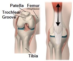
The patella (kneecap) sits in the trochlear groove on the front of the femur (thigh bone). As the knee moves, it glides up and down the groove

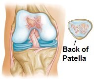
In patellofemoral arthritis, the cartilage on the back of the patella and in the trochlear groove become worn and inflamed. The cartilage thins and frays and if the wear and tear is severe it can expose the underlying bone. In an attempt to protect itself, the body may lay down extra layers of bone, known as bone spurs. The smooth surface is gradually lost leading to bone rubbing on bone. This can be painful and may limit movement.

Patellofemoral arthritis. Left image shows a normal knee, middle image shows loss of joint space between the femur and patella due to arthritis. Right image highlights bone spurs due to arthritis.

In the picture here you can see changes that develop in patellofemoral arthritis. The LEFT image shows a normal knee – note the space between the kneecap and the femur. The MIDDLE image shows the loss of joint space due to arthritis. The RIGHT image highlights the bone spurs that form.

Causes of Kneecap Arthritis

Patellofemoral arthritis may develop for a number of reasons:

With kneecap arthritis there is wear and tear of the cartilage on the back of the patella and in the patella groove on the thigh bone

1) Wear and Tear: Cartilage gradually thins as part of the normal aging process. Degenerative patellofemoral arthritis (known as osteoarthritis) usually affects those over the age of 65. Factors that increase the risk of developing arthritis include obesity, hip angle (which in turn affects knee position), gender (more prevalent in females), genetics and abnormal foot mechanics

2) Kneecap Injuries: Injuries to the kneecap such as fractures or dislocations can damage the cartilage on the patella which can lead to arthritis. Visit the kneecap injury section to find out more

3) Dysplasia: This is when the patella doesn’t sit properly in the trochlear groove, due to either the trochlear or the patellar being a slightly unusual shape, or tightness/laxity in the soft tissues affecting the angle that the patella sits at. As a result, when the knee moves there is excess stress, friction and/or pressure on the bones which can lead to patellofemoral arthritis

4) Medical Conditions: Certain medical conditions such as Gout or Paget’s disease increase the risk of developing patellofemoral arthritis.

Typical Symptoms :

The most common symptom of patellofemoral arthritis is anterior knee pain – pain at the front of the knee. The pain tends to be worse with activities that place pressure through the kneecap such as going up and down stairs, kneeling, squatting and getting up from a chair/bed.

Some people also experience crepitus, where they feel, or sometimes even hear, a cracking, grinding or popping sensation, when they move their knee. In severe cases when there is bone rubbing on bone, people may experience stiffness or the kneecap may get momentarily stuck, known as pseudolocking.

Non-Surgical Treatment Options

Treatment for patellofemoral arthritis usually starts with conservation i.e. non-operative, methods. In most cases, surgery is not required. Non-surgical treatments for patellofemoral arthritis include:

1) Rest and Activity Modification

Avoid activities that aggravate symptoms as much as possible whilst still getting on with daily life. Use the hand rail when going up and downstairs, when sitting down or standing up take your weight through your good leg and if necessary use a stick or crutches when walking.

2) Medication

Non-steroidal anti-inflammatory medication (NSAIDs) such as ibuprofen or Advil may be prescribed to help reduce pain and inflammation.

3) Exercises

Strengthening and stretching exercises help to improve the strength, stability and flexibility of the knee which can help to reduce the pressure on the kneecap and thus reduce pain – they are one of the most effective treatments for patellofemoral arthritis. A physical therapist will be able to give you a rehab programme to follow specific to your needs. Exercises that place pressure on the kneecap, such as squatting, should be avoided. Swimming is a good option as it strengthens the muscles without placing weight through the joint, or opt for cycling over running. Visit the knee exercises section for a whole range of exercises that can help.

4) Heat/Ice

Many people find that using either ice or heat helps to reduce pain and inflammation from patellofemoral arthritis. Ice has the added benefit that it helps to reduce inflammation – visit the ice wraps section to find out more.

5) Weight Loss

Losing weight can make a difference, but is only appropriate if you are overweight. This will help to reduce the pressure going through the kneecap. Talk to your doctor or a dietician before starting on a weight loss programme to ensure it is suitable for you.

6) Corticosteroid Injections:

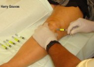
Corticosteroid injections can help to reduce the pain and inflammation associated with patellofemoral arthritis

Corticosteroid injections can help to reduce patellofemoral arthritis pain and inflammation. However, they only help to treat the symptoms of arthritis, not the underlying cause so should be used alongside other treatment methods such as exercises or else the pain is likely to return. Find out more in the knee injectionssection

7) Knee Braces and Taping

Wearing a knee brace or taping the patella can help to support the knee and change the position of the kneecap. Visit the knee braces section to find the right brace for you.

8) Supplements

Glucosamine and chondroitin are popular supplements for reducing knee arthritis pain.

Surgical Options

If moderate to severe symptoms of patellofemoral arthritis persist for more than 3-6 months of non-operative treatment, surgery may be advised. There are a few different options depending on the severity of the arthritis, which parts of the joint are affected and the age of the patient.

1) Chondroplasty

Suitable for mild to moderate patellofemoral arthritis. A chondroplasty is carried out arthroscopically (keyhole surgery). Small incisions are made around the kneecap through which thin surgical instruments and a small camera are inserted. Worn and frayed cartilage is trimmed and bone spurs smoothed out which both help to reduce friction in the joint.

2) Tibial Tuberosity Transfer

This involves shifting the position of the tibial tuberosity – a small bony lump on the tibia (shin bone) where the kneecap attaches via the patella tendon. With this type of surgery, the tibial tuberosity is removed, repositioned and then reattached with anchors or screws. This makes subtle changes to the position of the knee cap improving the tracking of the patella, allowing it to glide more smoothly in the trochlear groove therefore resulting in less stress and friction on the arthritic parts of the patella. Tibial tuberosity transfer tends to be done in cases where patellofemoral arthritis affects only a portion of the kneecap, seeking to offload the arthritic parts.

3) Realignment Surgery

Patella realignment surgery is done when tightness or laxity in the soft tissues means the patella doesn’t sit correctly in the trochlear groove which places excess pressure and friction on certain parts of the bone. With realignment surgery, the soft tissues around the kneecap are tightened or released to change the position of the patella in the trochlear groove. Examples of realignment surgery include lateral patellar retinaculum release, to release tight structures on the outer side of the kneecap, and reattachment or reconstruction of the medial patellofemoral ligament on the inner side of the knee.

4) Cartilage Grafting

With cartilage grafting, the damaged cartilage is replaced with healthy cartilage either from another part of the knee or from a tissue bank. If cartilage is being taken from the person’s own knee, two operations are required. The first is to harvest the cartilage from a non-weight-bearing part of the knee, which is then sent to a laboratory where cells are cultured for 3-6 weeks to increase the number of cartilage cells. In the second operation, the new cartilage cells are transplanted back into the affected parts of the patellofemoral joint. Cartilage grafting is usually done in younger patients in an attempt to delay the need for joint replacement.

5) Patellofemoral Replacement

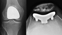
X-ray showing a patellofemoral replacement

With this type of partial knee replacement, the worn and damaged parts of the patellofemoral joint are removed and the surfaces re-lined with prosthetic implants.  The trochlear groove is lined with a thin metal plate, and a plastic “button” is attached to the back of the patella – both components are fixed on using bone cement. Patellofemoral replacement is only suitable if the arthritis is confined to the patellofemoral joint – if other parts of the knee are also affected, a total knee replacement is needed. Patellofemoral replacement surgery tends to be most suitable for younger patients and has the advantage of being less invasive than a total knee replacement. People often report the knee feels more “normal” afterwards, most likely due to the fact that the knee ligaments are all preserved.

6) Patellectomy

With a total or partial patellectomy, part or all of the patella is removed. It is only considered when there are no other options as whilst it may reduce pain, it inhibits the function of the quadriceps resulting in ongoing weakness.
SMA-II: Causes, Symptoms & Treatments

Spinal muscular atrophy type 2 (SMA II) is an inherited condition that affects the muscles. It is characterized primarily by progressive muscle weakness that develops in children between ages 6 and 12 months. Affected children can sit without support; however, they cannot stand or walk unaided. Other signs and symptoms may include a tremor of the fingers, breathing issues, feeding difficulties and skeletal abnormalities (such as scoliosis and hip dislocation). SMA II is caused by mutations in the SMN1 gene and is inherited in an autosomal recessive manner. Treatment is based on the symptoms in each person.

Symptoms:

The signs and symptoms of spinal muscular atrophy type 2 (SMA II) typically become apparent between 6 and 12 months of age. Poor muscle tone may be noticed at birth or within the first few months of life. Affected children may initially slowly gain some motor milestones. However, the highest motor milestone attained is generally the ability to sit independently, and this milestone is often lost by the mid-teens. People with SMA II are not able to stand or walk unaided. Other signs and symptoms may include a tremor of the fingers, breathing issues, feeding difficulties and skeletal abnormalities (such as scoliosis and hip dislocation).

Related image

Related image

Causes:

Spinal muscular atrophy type 2 (SMA II) is caused by changes (mutations) in the SMN1 gene. Extra copies of the SMN2 gene affect how severe the condition is. These genes encode a protein that is important for the normal functioning of certain nerve cells (called motor neurons) which help control muscle movements. Mutations in the SMN1 gene lead to reduced levels of this protein and the death of motor neurons. This in turn causes the characteristic signs and symptoms associated with SMA II.

The protein made by additional copies of SMN2 can compensate for some of the protein lost due to mutations in SMN1. Affected people who have extra copies of SMN2 may, therefore, have milder symptoms and develop the condition later in life.

Diagnosis:

A diagnosis of spinal muscular atrophy (SMA) is first suspected based on the presence of characteristic signs and symptoms. Identifying mutations in the SMN1 gene confirms the diagnosis.[1]

Classifying the type of SMA is based on the age of onset and the maximum function attained. Classisfying SMA as type 2 is based on:

  • onset of muscle weakness usually after age six months; ability to sit independently achieved when placed in a sitting position
  • finger trembling – almost invariably present
  • low muscle tone (flaccidity)
  • absence of tendon reflexes in approximately 70% of individuals
  • average intellectual skills during the formative years and above average by adolescence

The treatment of spinal muscular atrophy type 2 (SMA II) is based on the symptoms present. Physical therapy, occupational therapy and assistive devices (walkers, wheelchairs, etc) are generally recommended to help encourage maximum mobility and independence. These interventions may also prevent or delay scoliosis and abnormal contractions of the muscles and tendons. Some children with SMA II may have difficulty eating enough calories to maintain a normal weight. In these cases, nutritional counseling and/or a feeding tube may become necessary. Breathing problems are also common in people affected by SMA II and may require the use of BiPAP machines or other methods of respiratory support. Some affected children may require surgery to treat scoliosis or severe cases of hip dislocation.

PCOS: Causes, symptoms & treatments

What is polycystic ovarian syndrome?

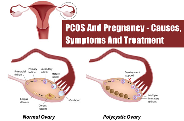
Polycystic ovarian syndrome (PCOS) is a condition in which a woman’s levels of the sex hormones estrogen and progesterone are out of balance. This leads to the growth of ovarian cysts (benign masses on the ovaries). PCOS can affect a women’s menstrual cycle, fertility, cardiac function, and appearance.

What causes PCOS?

While the exact cause of PCOS is unknown, doctors believe that hormonal imbalances and genetics play a role. Women are more likely to develop PCOS if their mother or sister also has the condition.

Overproduction of the hormone androgen may be another contributing factor. Androgen is a male sex hormone that women’s bodies also produce. Women with PCOS often produce higher-than-normal levels of androgen. This can affect the development and release of eggs during ovulation. Excess insulin(a hormone that helps convert sugars and starches into energy) may cause high androgen levels.

Image result for pcos causes, symptoms & treatments

Related image

Symptoms of PCOS

Symptoms of PCOS typically start soon after a woman begins to menstruate for the first time. The type and severity of symptoms varies from person to person. The most common characteristic of PCOS is irregular menstrual periods. Difficulty getting pregnant may be another reason that leads to diagnosis.

Because PCOS is marked by a decrease in female sex hormones, this condition may cause women to develop certain masculine characteristics, such as:

  • excess hair on the face, chest, stomach, thumbs, or toes
  • decrease in breast size
  • deeper voice
  • hair loss

Other symptoms may include:

  • acne
  • weight gain
  • pelvic pain
  • depression
  • infertility

While not symptoms of the disease, many women with PCOS have other concurrent health problems, such as diabetes, hypertension, and high cholesterol. These are linked to the weight gain typical in women with PCOS.

How is PCOS diagnosed?

There is no definitive test for PCOS. To make a diagnosis, your doctor will review your medical history and symptoms and perform tests to rule out other possible conditions. Your doctor will perform a physical and pelvic examination to look for signs of PCOS, such as swollen ovaries or a swollen clitoris.

Blood tests to measure sex hormone levels are typically ordered, as well as:

  • thyroid function tests to determine how much of the thyroid hormone your body produces
  • fasting glucose tests to measure your blood sugar levels
  • lipid level tests to assess the amount of cholesterol in your blood

A vaginal ultrasound allows your gynecologist to create real-time images of your reproductive organs. A pelvic laparoscopy is a surgical procedure in which your doctor makes a small incision in your abdomen and inserts a tiny camera to check for growths on your ovaries. If growths are present, your doctor may take a small tissue sample (biopsy) for further examination.

How is PCOS treated?

PCOS can be treated, but there’s no cure. Treatment focuses on controlling symptoms and managing the condition to prevent complications. The treatment will vary from woman to woman, depending on specific symptoms. Tips for controlling symptoms may include:

  • Eat a healthy diet. A healthy diet and regular exercise are recommended for all women with PCOS, particularly those who are overweight. This can help to regulate your menstrual cycle and lower your blood glucose levels.
  • Take birth control pills if you aren’t planning to become pregnant. Women who don’t want to become pregnant may be prescribed birth control pills. These can help treat acne, regulate the menstrual cycle, and lower levels of male hormones, such as testosterone, in the body. If a woman with PCOS is infertile, fertility drugs may be prescribed to aid in ovulation.
  • Ask your doctor about medications that may help you. Anti-androgens are drugs that reduce male hormone levels. These can help stop excess hair growth and reduce acne. Diabetes medications may also be prescribed to lower blood glucose and testosterone levels.
  • Surgery. Surgery may be recommended for some women with PCOS. Ovarian drilling is a procedure in which your doctor punctures your ovary with a small needle that carries an electric current. This is done in order to destroy part of the ovary. It’s a short-term solution that can promote ovulation and reduce male hormone levels.

Nasal Polyps: Causes, Symptoms & Treatments

Often linked to allergies or asthma, they may cause no symptoms, especially if they’re small and don’t need treatment. Larger ones can block normal drainage from the sinuses. When too much mucus builds up in the sinuses, it can become infected.

Unlike polyps that form in the colon or bladder, nasal ones are rarely cancer. Experts think that long-term inflammation causes them or that they run in families.

Nasal polyps aren’t painful to the touch. Medications or surgery can treat most. They may come back, though.

Image result for nasal polyps causes, symptoms & treatments


Symptoms

If you have any symptoms, they may include:

  • Stuffy or blocked nose
  • Sneezing
  • Postnasal drip
  • Runny nose
  • Facial pain
  • Trouble with sense of smell
  • Loss of taste
  • Itching around the eyes
  • Infections

Most people with nasal polyps have a runny nose, sneezing, and postnasal drip. About 75% have problems with their sense of smell.

Many people also have wheezing, sinus infections, and sensitivity to fumes, odors, dusts, and chemicals. It’s less common, but some people with nasal polyps also have a severe allergy to aspirin and reaction to yellow dyes. If you know you have that allergy, ask your doctor to check for nasal polyps.

Nasal polyps make you more likely to have long-term (chronic) sinusitis. Large ones can even change the shape of your nose.

Who Gets Them?

Anyone can, but they’re most common in adults over age 40 and are twice as likely to affect men as women. Children under age 10 rarely get them. If they do, a doctor will check for signs of cystic fibrosis.

Nasal polyps are linked to allergic rhinitis, asthma, aspirin allergy, sinus infections, acute and chronic infections, something stuck in the nose, and cystic fibrosis. But many times the cause is unknown. Sometimes, people get them before they develop asthma or sinusitis.

Some experts think that symptoms of allergies — including runny nose, sneezing, and itching — make some people more likely to get nasal polyps. But the allergy connection is controversial. Other researchers think that sinus infections are to blame.

Treatments

If you think that you have nasal polyps, your doctor can check. She’ll use a nasal endoscope, which is a tool with a magnifying lens or camera that provides a detailed view of your nose and sinuses. In some cases, your doctor may order more tests or take a small sample (a biopsy) of the polyp.

If you need treatment, you’ll probably start with a nasal corticosteroid spray. In many cases, that can shrink or even get rid of nasal polyps. But some people need to take corticosteroids such as prednisone by mouth for a week.

Unfortunately, nasal polyps tend to come back if the irritation, allergy, or infection continues. So you may need to keep using a corticosteroid spray and get checkups with a nasal endoscope every now and then.

In general, medications such as antihistamines and decongestants aren’t great at managing nasal polyps. But you may need antihistamines to control allergies or antibiotics if you have an infection before you start on steroids.

Surgery:

Sometimes, nasal polyps are so large that corticosteroid nasal sprays don’t work. In such cases, surgery may be an option.

The doctor would likely use a small nasal telescope that removes nasal polyps. You can go home the same day as the surgery.

Surgery helps in most cases. It may be less effective if you have nasal polyps, asthma, and aspirin sensitivity. If that’s you, medication may be more helpful.

Spinocerebellar Ataxia: Causes, Symptoms & Treatments

What is Spinocerebellar Ataxia?

Spinocerebellar ataxia (SCA) is a progressive, degenerative, genetic disease that has multiple types. It is characterized by slowly progressive incoordination of gait, often associated with poor coordination of the hands, speech, and eye movements.Associated behavioral and emotional changes are often present. It is also known as spinocerebellar degeneration or spinocerebellar atrophy.

Structure and Function of the Cerebellum in Brief

The cerebellum is a lobulated structure at the posterior part of the brain, below the cerebral cortex and behind the brainstem. It is divided by a set of large folds into ten smaller lobules. The cerebellum is a prominent feature of the hind brain (rhombencephalon) and due to its unique structure and location, it is easily identifiable in the context of other structures of the brain.

Both clinical observations and experimental evidence have established that the cerebellum is primarily responsible for motor coordination. Every movement of the body parts need to be carefully timed and coordinated. Consequently, the movements are smooth and regulated. These functions are very crucial yet so precise that they are not noticed, except by the trained eye. The importance of the cerebellum becomes evident during disease or injury, when various movement disorders are observed, such as in SCA. Patients having a damaged cerebellum have problem in keeping balance and maintaining muscle coordination.

Related imageWhat are the Causes of Spinocerebellar Ataxia?

SCAs are inherited from parents to offspring. The defective genes are passed on to the children and therefore, these gene mutations are present from birth. The pattern of inheritance is autosomal dominant inheritance.

What are the Symptoms of Spinocerebellar Ataxia?

The symptoms of SCA generally vary with the specific type involved. The symptoms can also vary from patient to patient. Generally, it is characterized by clumsy movements and progressive deterioration in physical ability, although the patient may retain full mental capacity. Although the symptoms generally appear in the third decade of life, they can also appear during childhood or in old age.

What are the Treatment Options for Spinocerebellar Ataxia?

There is currently no specific treatment for SCA, which is a progressive disease that deteriorates over time. However, not all types cause equally severe disability. Specific treatments may become available in the future with further ongoing research.

Currently, the treatment is symptomatic and is aimed at alleviating the symptoms only, without any scope of treating the disease itself. A patient suffering from SCA will likely become wheelchair bound for life and require help for performing daily tasks.

Some of the strategies for managing SCA patients under various circumstances are briefly highlighted below:

  • Impairment of Gait and Posture: Initially, exercises may be recommended for improving balance. Mobility assistive devices such as canes, walkers, and crutches can be used for maintaining balance while walking. If these fail, the only remaining option is a wheelchair.
  • Impairment of Hand and Arm Movements: Provision for assistive devices to help with writing, feeding, and performing self-care activities.
  • Impairment of Speech: Speech therapy and assistive devices may be provided for increasing communication capability.
  • It should be noted that besides ataxia, many patients may exhibit other symptoms as well. These include depressionstiffnesstremorspasticity, and sleep disorders. For these conditions, standard medical treatment can be given to alleviate the symptoms.

    Can Spinocerebellar Ataxia be Prevented?

    Today’s medical technology is not adequately advanced to prevent SCA. However, technology is available to accurately identify the mutated gene in a patient with SCA. The next step is to correct this gene defect, which requires gene therapy. Gene therapy is still in its infancy and primarily confined to the research domain. However, it is hoped that in the near future, it will be possible to replace the defective gene with its healthy counterpart, and this could be done even before the disease manifests.

Pages:123