Global Treatment Services Pvt. Ltd.

Global Treatment Services

Aicardi Syndrome : Causes, symptoms & Treatments

What Is Aicardi Syndrome?

Aicardi syndrome is a rare genetic disorder that interferes with the formation of the corpus callosum, which is the structure that connects the two sides of the brain. In people with Aicardi syndrome, the corpus callosum is either partially or completely missing. The disorder occurs almost exclusively in newborn girls. Medical researchers don’t believe the disorder is passed from the parents to their child.

The exact cause of Aicardi syndrome isn’t known. However, it’s thought to be caused by a first-time mutation in the child’s genes. Since the disorder primarily affects females, researchers believe the mutation specifically occurs on the X chromosome, which is one of the two sex chromosomes. Females have two X chromosomes. Males typically have one X chromosome and one Y chromosome.

People with Aicardi syndrome often have the following:

  • a partial or complete absence of the corpus callosum
  • infantile spasms or seizures that begin during infancy
  • chorioretinal lacunae, or lesions in the light-sensitive layer of tissue at the back of the eye

Not all people with Aicardi syndrome have these features. However, some people may display additional abnormalities in the brain, eyes, and face. The severity of symptoms varies significantly from one person to another. Some have very severe seizures and may not survive past childhood, while others have milder symptoms and may live well into adulthood.

Image result for Aicardi syndrome

What Are the Symptoms of Aicardi Syndrome?

Aicardi syndrome symptoms usually appear in babies between the ages of 2 and 5 months old. Your child may begin jerking or having infantile spasms, a type of seizure that occurs in infants. These seizures can develop into epilepsy later in life. Your child may also develop yellowish spots on their eyes. Lesions on the retina, which is the light-sensitive layer of tissue at the back of the eye, cause these spots.

Other symptoms of Aicardi syndrome include:

  • a coloboma, which is a hole or gap in one of the structures of the eye
  • abnormally small eyes
  • an unusually small head
  • hand deformities
  • intellectual disabilities
  • developmental delays
  • difficulty eating
  • diarrhea
  • constipation
  • gastroesophageal reflux
  • spasticity, which is a condition that causes stiff and rigid muscles

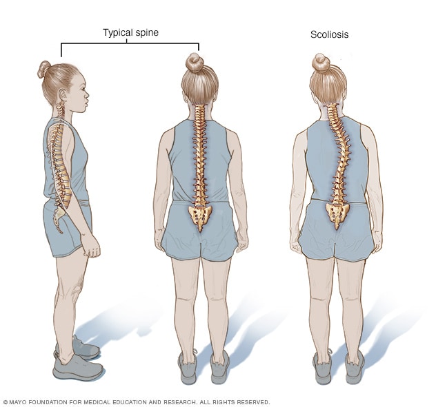
Additional symptoms of Aicardi syndrome include rib and spine abnormalities, such as scoliosis. Children with this disorder may also have unusual facial features, such as a flatter nose, larger ears, or a smaller space between the upper lip and nose. Since Aicardi syndrome can result in poorly developed eyes, children may experience impaired vision or blindness.

Researchers have also found that the brains of children with Aicardi syndrome may have fewer folds and grooves than a normal brain. Fluid-filled cysts may also be present in the brain.

What Causes Aicardi Syndrome?

Aicardi syndrome most often appears in females as well as in boys with Klinefelter’s syndrome, a condition in which a male has an extra X chromosome. For these reasons, researchers believe the disorder is caused by a defect on the X chromosome.

People normally have 46 chromosomes in each cell. Two of the 46 chromosomes are sex chromosomes, known as X and Y. They help determine whether someone will develop male or female sex characteristics. Females have two X chromosomes, while males usually have one X chromosome and one Y chromosome.

During the normal development of an embryo, there should only be one active X chromosome in each cell. This means that one of the two X chromosomes in a female embryo must randomly deactivate during cell formation. In the case of Aicardi syndrome, researchers think that the deactivation of the X chromosomes fails to alternate randomly. As a result, one X chromosome is active in more than half of the body’s cell formations. This is called “skewed X-inactivation.”

Researchers have yet to determine the exact gene that causes skewed X-inactivation, so the cause of the disorder is still not known. This lack of information also makes it difficult to determine the risk factors for Aicardi syndrome.

DIAGNOSIS:

Doctors can usually diagnose Aicardi syndrome based on the symptoms. However, since each child may present different symptoms, additional tests may be needed. Some tests that are used to help doctors make a diagnosis include:

  • eye exams
  • an electroencephalogram (EEG), which evaluates the electrical activity of the brain and detects seizure activity
  • an MRI or CT scan, which provides detailed anatomical images of the head and brain
How Is Aicardi Syndrome Treated?

There’s no cure for Aicardi syndrome at this time. However, some symptoms can be managed with treatment. The most common method of treatment involves controlling the seizures and spasms brought on by the disorder. There are also programs available to help both children and their parents cope with the intellectual disabilities and developmental delays that usually accompany Aicardi syndrome. Your child’s doctor will likely refer you to a pediatric neurologist for further evaluation. A pediatric neurologist is a doctor who specializes in treating nervous system disorders in children. They can help your child with long-term management of Aicardi syndrome. Doctors in these other fields may also be may also be consulted based on the symptoms and severity of the syndrome:

  • neurosurgery
  • ophthalmology
  • orthopedics
  • gastroenterology
  • physical therapy
  • speech therapy
  • occupational therapy

Leg Length Discrepancy: Causes, Symptoms & Treatments

The leg length discrepancy means there is inequality of the length of the lower limbs. It is also called as limb length inequality. The problem is extremely common with 23% of the general population having a discrepancy of 1 cm or more

Leg length discrepancy causes compensatory gait abnormalities, degenerative arthritis of the lower extremity and lumbar spine. In addition to the length inequality, these patients may also have angular and torsional deformities that may influence their functional leg lengths. For example, flexion contractures around the knee and hip can cause apparent shortening of the leg.

Related image

Hip abduction contracture and ankle equinus causes functional lengthening.

Small leg length discrepancy is well compensated by body and hardly requires any treatment. Bigger differences need to be equalized to prevent osteoarthritis , scoliosis and backache in later life.

Causes of Leg Length Discrepancy

The causes can generally be divided into two broad categories: congenital and acquired.

Congenital limb length inequality

  • Longitudinal deficiency of the fibula
  • Hemihypertrophy syndromes
  • Klippel-Trénaunay-Weber syndrome
  • Skeletal dysplasias.
  • Proximal femoral focal deficiency
  • Developmental dysplalsia of hip
  • Unilateral clubfoot
  • Vascular malformation
  • Congenital pseudarthrosis of the tibia
  • Ollier’s disease

Paralytic disorders

  • Spasticity (cerebral palsy)
  • Poliomyelitis
  • Spina bifida
  • Spinal dysraphism

Acquired

  • Physis disruption
    • infection
    • trauma
    • tumor
    • Malunion of fracture

Functional Effects of Limb Length Discrepancy

  • Back pain
    • increased prevalence of back pain
  • Osteoarthritis
    • decreased coverage of femoral head on long leg side leads to osteoarthritis
  • Structural scoliosis
    • Limb length discrepancy increases the incidence of structural scoliosis
  • Inefficient gait

Classification of Limb Length Discrepancy:

  • Static
    • malunion of femur or tibia
  • Progressive
    • Physeal growth arrest
    • Congenital
      • Absolute discrepancy increases
      • Proportion stands the same

Treatment of Leg Length Discrepancy

Goals of treatment are

  • Leg length equality within 1 cm
  • Level pelvis
  • Functional improvement

In certain cases small leg length inequality may be beneficial to the patient especially in neuromuscular disorders to aid in clearance in the swing phase of gait.

Depending on the severity of the difference, different treatments can be used for correction of the inequality.

Treatment of leg length inequality involves many different approaches. Non operative treatment involves use of shoe raises or orthotics.

Surgical approach deals with the problem by

  • Correction of deformity if present
  • Shortening of the normal leg
  • Lengthening of affected leg
  • Combination of shortening and lengthening.

Non-operative Treatment

An inequality < 2cm projected at maturity is generally without any functional problems. Often, surfices. Up to 1 cm can be inserted in the shoe. For larger leg length inequalities, the shoe must be built up.

Leg length inequalities beyond 5 cm are difficult to treat with a shoe lift.

A foot-in-foot prosthesis can be used for larger leg length inequalities but is often not tolerated well. It is often used as temporary measure.

Surgical Treatment

Shortening of long side

Epiphysiodesis of femur, tibia, or both

Epiphysiodesis inhibits the growth of the limb and can be used to correct 2-5 cm of projected limb length discrepancy.

It can be performed only in skeletally immature patients where growthpotential is still present.

Because the procedure effectively shortens the longer leg and is usually done on the uninvolved side, it may be unappealing to the patient and family.

It can be used alone or with limb lengthening of short limb.

Shortening of Mature Limb

This involves ostectomy and removal of bone segment followed by fixation of remaining fragments with desirable fixation device.

Limb lengthening of short side

It is contemplated in cases > 5 cm projected discrepancy. It can be  combined with a shortening procedure (epiphysiodesis, ostectomy) on long side when the discrepancy is large [>8 cmm]

Lengthening is usually done by corticotomy and gradual distraction

The limits of lengthening depend on patient tolerance, bony consolidation, maintenance of range of motion, and stability of the joints above and below the lengthened limb.

Numerous fixation devices are available, such as the ring fixator with fine wires, monolateral fixator with half pins, or a hybrid frame.

What are the Signs and Symptoms of Leg Length Discrepancy (LLD)?

There are multiple signs and symptoms for leg length discrepancy (LLD). Some are obvious and some are subtle. There are differences between individuals on the extent and causes of leg length discrepancy (LLD). The difference in length between the two legs may vary from 3 and a half to 4 percent. These differences often determine the impact that may be experienced by different individuals. Signs and symptoms usually experienced in the patients with leg length discrepancy (LLD) include:

  • If the difference in leg length discrepancy (LLD) is 3 cms and higher, difference will be easily noticed as the person walks.
  • The person affected with leg length discrepancy (LLD) will need to exert more energy to walk.
  • Some people may experience some pain in the lower back, ankle, or hip as they walk. Some studies have collaborated this, while other have refuted this.
  • The person’s posture will be highly affected, causing him/ her to hold the longer leg on the knee for support.
  • The person’s gait will be adversely affected, which will be obviously noticed.

Risk Factors for Leg Length Discrepancy (LLD)

The risk factors for leg length discrepancy (LLD) include:

Patients might have serious dissimilarities in the length of the legs as an effect brought by Leg Length Discrepancy (LLD) due to:

  • Infection.
  • Birth defects.
  • Injuries.
  • Accidents and other causes.

Complications in Leg Length Discrepancy (LLD)

Leg length discrepancy (LLD) with very high difference can become complicated and lead to severe orthopaedic conditions like lower back pain, scoliosis, or arthritis, if left untreated.

  • The patient may experience complications due to contractures of the muscles while having to constantly stand on their toes of the shorter leg for support.
  • Another complication is that the shorter side of their body is often lowered down and suddenly lifted by the longer side, causing them to have abnormal gait that goes up and down.
  • The abnormal gait can cause the child to be highly disturbed psychologically.
  • The patient is often ridiculed and teased by other children, hindering the child’s social life.
  • Most patients with leg length discrepancy (LLD) have to constantly support the shorter leg on the knee while standing, causing them to bend often or they may have to stand on their toes.

Scoliosis: Causes, Symptoms & Treatments

When a person has scoliosis, their backbone curves to the side.

The angle of the curve may be small, large or somewhere in between. But anything that measures more than 10 degrees is considered scoliosis. Doctors may use the letters “C” and “S” to describe the curve of the backbone.

You probably don’t look directly at too many spines, but what you might notice about someone with scoliosis is the way they stand. They may lean a little or have shoulders or hips that look uneven.

What Causes Scoliosis?

In as many as 80% of cases, doctors don’t find the exact reason for a curved spine. Scoliosis without a known cause is what doctors call “idiopathic.”

Some kinds of scoliosis do have clear causes. Doctors divide those curves into two types — structural and nonstructural.

In nonstructural scoliosis, the spine works normally, but looks curved. Why does this happen? There are a number of reasons, such as one leg’s being longer than the other, muscle spasms, and inflammations like appendicitis. When these problems are treated, this type of scoliosis often goes away.

In structural scoliosis, the curve of the spine is rigid and can’t be reversed.

Related image

Causes include:

  • Cerebral palsy
  • Muscular dystrophy
  • Birth defects
  • Infections
  • Tumors
  • Genetic conditions like Marfan syndrome and Down syndrome

Congenital scoliosis begins as a baby’s back develops before birth. Problems with the tiny bones in the back, called vertebrae, can cause the spine to curve. The vertebrae may be incomplete or fail to divide properly. Doctors may detect this condition when the child is born. Or, they may not find it until the teen years.

Family history and genetics can also be risk factors for idiopathic scoliosis. If you or one of your children has this condition, make sure your other kids are screened regularly.

Scoliosis shows up most often during growth spurts, usually when kids are between 10 and 15 years old. About the same number of boys and girls are diagnosed with minor idiopathic scoliosis. But curves in girls are 10 times more likely to get worse and may need to be treated.

Scoliosis diagnosed during the teen years can continue into adulthood. The greater the angle of the spine curve, the more likely it is to increase over time. If you had scoliosis in the past, have your doctor check your back regularly.

Degenerative scoliosis affects adults. It usually develops in the lower back as the disks and joints of the spine begin to wear out as you age.

Treatments:

The main type of surgery for scoliosis involves attaching rods to the spine and doing a spinal fusion. Spinal fusion is used to stabilize and reduce the size of the curve and stop the curve from getting worse by permanently joining the vertebrae into a solid mass of bone.

Other techniques are sometimes used, including instrumentation without fusion, which attaches devices such as metal rods to the spine to stabilize a spinal curve without fusing the spine together. This is only done in very young children when a fusion, which stops the growth of the fused part of the spine, is not desirable. The child usually has to wear a brace full-time after having this surgery.

Pinched Nerve in lower back: Causes, Symptoms & Treatments

A pinched nerve refers to a certain kind of damage to a nerve or group of nerves. It’s caused when a disc, bone, or muscle places increased pressure on the nerve.

It can lead to feelings of:

  • numbness
  • tingling
  • burning
  • pins and needles

A pinched nerve can cause carpal tunnel syndrome, sciatica symptoms (a pinched nerve can’t cause a herniated disc, but a herniated disc can pinch a nerve root), and other conditions.

Some pinched nerves will require professional care to treat. If you’re looking for a way to alleviate mild pain at home, here are nine options you can try. Some of them can be done at the same time. What’s important is to find what works best for you.

TREATMENTS:
I. Laminectomy:
Laminectomy is one of the most common back surgeries. During a laminectomy, a surgeon removes the rear portion of one or more spinal bones (vertebrae). Bone spurs and ligaments that are pressing on nerves may be removed at the same time. Here’s what to expect before, during, and after your laminectomy.
II.  Surgical & Non surgical Spinal Decompression:

What Is Surgical Spinal Decompression?

Surgical spinal decompression is another option for treating certain types of back pain. But it is usually used as a last resort. If other measures don’t work, your doctor may suggest surgical spinal decompression for bulging or ruptured disks, bony growths, or other spinal problems. Surgery may help relieve symptoms from pressure on the spinal cord or nerves, including:

  • Pain
  • Numbness
  • Tingling
  • Weakness

Are There Different Types of Spinal Decompression Surgery?

Your doctor may suggest one or more types of back surgeries to relieve the pressure in your spine. In addition, you may need spinal fusion to stabilize your spine. The following are the more common types of back surgery:

  • Diskectomy: In this procedure, a portion of the disk is removed to relieve pressure on nerves.
  • Laminotomy or laminectomy: A surgeon removes a small portion of bone — a section of bony arch or the entire bony arch — to increase the size of the spinal canal and relieve pressure.
  • Foraminotomy or foraminectomy: A surgeon removes bone and other tissue to expand the openings for nerve roots.
  • Osteophyte removal: During the surgery, bony growths are removed.
  • Corpectomy: This procedure involves removing a vertebral body along with disks between the vertebrae.

What Are the Risks of Spinal Decompression Surgery?

As with any surgery, there are risks. These are some of the more common risks associated with spinal decompression surgery:

  • Infection
  • Bleeding
  • Blood clots
  • Allergic reaction to anesthesia
  • Nerve or tissue damage

Another risk of surgery is that it may not improve back pain much. It can be difficult to determine who will benefit from spinal decompression surgery.

Home remedies:

1. Adjust your posture

You may need to change how you’re sitting or standing to relieve pain from a pinched nerve. Find any position that helps you feel better, and spend as much time in that position as you can.

2. Use a standing workstation

Standing workstations are gaining popularity, and for good reason. Mobility and standing throughout your day are crucial to preventing and treating a pinched nerve.

If you have a pinched nerve or want to avoid one, talk with your human resources department about modifying your desk so that you can stand while working. If you can’t get a standing workstation, be sure to get up and take a walk each hour. Roller balls for tight muscles and an hourly stretching program are a good idea if you use a keyboard frequently. (Wrist braces or supports aren’t recommended as an early treatment strategy.)

3. Rest

No matter where you have a pinched nerve, the best thing is usually to rest as long as possible. Avoid the activity that is causing you pain, such as tennis, golf, or texting.

Rest until the symptoms have completely resolved. When you do start moving that part of your body again, pay attention to how it feels. Stop the activity if your pain returns.

4. Splint

If you have carpal tunnel, which is a pinched nerve in the wrist, a splint can help you rest and protect your wrist. This can be especially helpful overnight so that you don’t curl your wrist in a bad position while you’re sleeping.

5. Stretch

Gentle stretches can help relieve the pressure on your nerve and improve your symptoms. Don’t go too deeply into a stretch. If you begin to feel pain or discomfort, ease up on the stretch. Remember that small movements can have a big impact.

6. Apply heat

You can use heat to relax the muscles that might be tight around a pinched nerve. Heat also increases blood flow, which can help the healing process. You can find heating pads in various sizes at a drugstore.

Hold heat directly onto the pinched nerve for 10–15 minutes at a time.

7. Use ice

Ice reduces swelling and inflammation. Wrap a towel around an ice pack and hold it directly onto the pinched nerve for 10–15 minutes.

8. Elevate your legs

For a pinched nerve in your lower back, try elevating your legs with a 90-degree bend in both your hips and knees.

9. Take an over-the-counter pain reliever

If you want to try a pain reliever, you can take over-the-counter nonsteroidal anti-inflammatory drugs, like ibuprofen (Advil, Motrin IB) or aspirin (Bufferin). Follow the instructions on the bottle, and be sure to check with your doctor before taking any new medications.

Image result for pinched nerve in back causes, symptoms & treatments

When  need to see a doctor:

If your pain is severe, constant, or keeps returning, you should see a doctor. The doctor may run some tests or ask a lot of questions about your lifestyle to determine what’s causing your pinched nerve.

If the pain, tingling, and numbness doesn’t resolve, it’s important that you see your physician. They may prescribe a stronger anti-inflammatory or suggest additional testing, such as an X-ray, to determine exactly where the nerve is pinched. Your doctor may also prescribe physical therapy, which can help to reduce symptoms.

It’s very important that you stop any at-home treatments if they seem to hurt you or make your condition worse. If you have numbness or tingling that isn’t resolving or is getting worse, contact your physician or an orthopedic physical therapist.

See a doctor immediately if you have a pinched nerve that’s:

  • affecting your bowel or bladder
  • causing a whole limb to be weak or give out
  • preventing you from grasping things
  • causing you to drop things

If at-home treatments aren’t helping, or if you continually get pinched nerves, your doctor may prescribe prescription painkillers, physical therapy, or even surgery.

Whisplash: causes, symptoms & treatments

Neck strain is often just called whiplash. Although it’s usually associated with car accidents, any impact or blow that causes your head to jerk forward or backward can cause neck strain. The sudden force stretches and tears the muscles and tendons in your neck.

Neck strain afflicts many amateur and professional athletes. People who play contact sports like football are especially prone to neck strain.

Neck strains are often confused with neck sprains. They’re a bit different. Neck strains are caused by damage to the muscle or the tendons, bands of tissue that connect muscles to bones. Neck sprains are caused by tearing of the ligaments, the tissues that connect the bones to each other.

However, the differences between these strains and sprains probably won’t mean much to you. The causes, symptoms, and treatment of neck sprains and neck strains are usually the same.

Image result for whiplash causes, symptoms & treatments

What Are the Symptoms of Whiplash?

The pain of whiplash is often hard to ignore. The symptoms may include:

  • Pain, decreased range of motion, and tightness in the neck. The muscles may feel hard or knotted.
  • Pain when rocking your head from side to side or backward and forward.
  • Pain or stiffness when moving your head to look over each shoulder.
  • Tenderness.
  • Headaches at the base of the skull that radiate towards the forehead.

Sometimes, the pain of a neck strain is immediate. In other cases, it can take several hours or days before your neck begins to hurt.

The blow that causes neck strain can sometimes cause a concussion, too. Since concussions can be serious, you need to see a doctor right away. You need emergency medical care if you have a headache that worsens or persists, have weakness or trouble talking, or are confused, dizzy, nauseous, excessively sleepy, or unconscious.

To diagnose neck strain, your doctor will give you a thorough examination. You may also need X-rays, CT (computed tomography) scans, and other tests, to rule out other problems.

Physical therapy can be useful in helping to wean a patient from a cervical collar as well as to help strengthen muscles and reduce painful motions. Occupational therapy can be used to help return the patient to the work environment.

If the patient begins to develop psychological symptoms including anger, anxiety or depression following an injury, prompt treatment of the emotional condition is recommended. This can help the patient better understand the good chances for successful recovery and reduce the chances of chronic symptoms.

Here’s the good news: given time, whiplash should heal on its own. To help with recovery, you should:

  • Ice your neck to reduce pain and swelling as soon as you can after the injury. Do it for 15 minutes every 3-4 hours for 2-3 days. Wrap the ice in a thin towel or cloth to prevent injury to the skin.
  • Take painkillers or other drugs, if recommended by your doctor. Non-steroidal anti-inflammatory drugs (NSAIDs), like ibuprofen (Advil, Motrin) or naproxen (Aleve), will help with pain and swelling. However, these medicines can have side effects. Never use them regularly unless your doctor specifically says you should. Check with your doctor before taking them if you take other medicines or have any medical problems. If over the counter medications do not work, prescription painkillers and muscle relaxants may be necessary.
  • Use a neck brace or collar to add support, if your doctor recommends it. However, they are not recommended for long-term use, because they can actually weaken the muscles in your neck.
  • Apply moist heat to your neck — but only after 2-3 days of icing it first. Use heat on your neck only after the initial swelling has gone down. You could use warm, wet towels or take a warm bath.
  • Other treatments, like ultrasound and massage, may also help.

Bulging Disc: causes, Symptoms & Treatments

A bulging disc can have a number of different causes, including trauma from a sports-related injury or a car accident. The condition’s most common cause, however, is the natural aging process. Over time, parts of the spine begin to break down after age-related changes make them less able to support body weight and allow the spine to bend and twist. The spinal discs, which are located between adjacent vertebrae and act as shock absorbers, are prone to shrinkage and water loss as you grow older. This can lead to a loss of elasticity, which can cause the outer layer of a disc to bulge out from between the surrounding vertebrae.

Image result for Bulging disc causes, symptoms & treatments

Symptoms of a bulging disc

If a misshapen disc causes compression of the spinal cord or a spinal nerve root, you may experience symptoms including pain, numbness, tingling and muscle weakness. The exact location of these symptoms will depend on the position of the affected disc in the spinal column:

  • Cervical (upper) spine — nerves send signals to the neck, shoulders, arms and hands
  • Thoracic (middle) spine — nerves here affect the middle back, ribs and abdomen
  • Lumbar (lumbar) spine — nerves travel to the lower back, hips, buttocks, legs and feet

Treating a bulging disc

In a very high number of cases, symptoms from a bulging disc can be relieved through a course of basic conservative treatments. This may include the use of anti-inflammatory or pain medication, appointments with a physical therapist or healthy life choices such as dieting, performing moderate exercise and quitting smoking, among others. Some patients also pursue alternative therapies like restorative yoga, chiropractic manipulation or acupuncture, but be aware that the effectiveness of alternative medicine is the source of debate in the mainstream medical community.

If you are unable to find relief after several weeks or months of conservative treatment or alternative therapy, you may want to consider minimally invasive spine surgery. These procedures are often the clinically appropriate first choice when compared to traditional open spine procedures, offering a reduced risk of infection and no lengthy recovery for our patients.

Bulging disc keyhole surgery is another way to describe a minimally invasive procedure used to alleviate pain and other symptoms associated with spinal nerve compression. A bulging disc causes symptoms when the outer wall of an intervertebral disc compresses or otherwise makes contact with a spinal nerve root or the spinal cord itself. It is called “keyhole,” because the incision used to access the spine is much smaller than the incisions used to perform traditional open neck or back surgery. In most cases, surgery is only recommended if a regimen of conservative treatment proves ineffective after several weeks or months.

The minimally invasive advantage

A smaller incision is not the only defining quality of bulging disc keyhole surgery. There are many advantages to choosing the minimally invasive surgery performed, if you are a candidate, rather than traditional open neck or back surgery. These advantages include:

  • A patient satisfaction score of 96^
  • 81 percent of Laser Spine Institute’s patients return to work within three months
  • An infection rate (0.49 percent^) significantly less than traditional open back surgery (up to 19 percent)

In addition, minimally invasive spine surgery requires no overnight hospital stay, compared to two to five days for traditional open spine surgery. Also, patients generally recover much faster after minimally invasive surgery and there is minimal scarring.

Contact us to schedule your initial consultation and to receive a review of your MRI or CT scan to help determine if are a candidate for one of our procedures.

Valley Fever : Causes, Symptoms & Prevention

Valley fever is a fungal infection caused by coccidioides organisms. It can cause fever, chest pain and coughing, among other signs and symptoms.

Two species of coccidioides fungi cause valley fever. These fungi are commonly found in soil in specific regions. The fungi’s spores can be stirred into the air by anything that disrupts the soil, such as farming, construction and wind.

The fungi can then be breathed into the lungs and cause valley fever, also known as acute coccidioidomycosis. Mild cases of valley fever usually resolve on their own. In more severe cases, doctors prescribe antifungal medications that can treat the underlying infection.

Related image

Symptoms

Valley fever is the initial form of coccidioidomycosis infection. This initial, acute illness can develop into a more serious disease, including chronic and disseminated coccidioidomycosis.

Acute coccidioidomycosis (valley fever)

The initial, or acute, form of coccidioidomycosis is often mild, with few, if any, symptoms. When signs and symptoms do occur, they appear one to three weeks after exposure. They tend to resemble those of the flu, and can range from minor to severe, including:

  • Fever
  • Cough
  • Chest pain
  • Chills
  • Night sweats
  • Headache
  • Fatigue
  • Joint aches
  • Red, spotty rash

The rash that sometimes accompanies valley fever is made up of painful red bumps that may later turn brown. The rash mainly appears on your lower legs, but sometimes on your chest, arms and back. Others may have a raised red rash with blisters or eruptions that look like pimples.

If you don’t become ill from valley fever, you may only find out you’ve been infected when you later have a positive skin or blood test or when small areas of residual infection (nodules) in the lungs show up on a routine chest X-ray. Although the nodules typically don’t cause problems, they can look like cancer on X-rays.

If you do develop symptoms, especially severe ones, the course of the disease is highly variable. It can take months to fully recover, and fatigue and joint aches can last even longer. The severity of the disease depends on several factors, including your overall health and the number of fungus spores you inhale.

Chronic coccidioidomycosis

If the initial coccidioidomycosis infection doesn’t completely resolve, it may progress to a chronic form of pneumonia. This complication is most common in people with weakened immune systems.

Signs and symptoms include:

  • Low-grade fever
  • Weight loss
  • Cough
  • Chest pain
  • Blood-tinged sputum (matter discharged during coughing)
  • Nodules in the lungs

Disseminated coccidioidomycosis

The most serious form of the disease, disseminated coccidioidomycosis, occurs when the infection spreads (disseminates) beyond the lungs to other parts of the body. Most often these parts include the skin, bones, liver, brain, heart, and the membranes that protect the brain and spinal cord (meninges).

The signs and symptoms of disseminated disease depend on which parts of your body are affected and may include:

  • Nodules, ulcers and skin lesions that are more serious than the rash that sometimes occurs with other forms of the disease
  • Painful lesions in the skull, spine or other bones
  • Painful, swollen joints, especially in the knees or ankles
  • Meningitis — an infection of the membranes and fluid surrounding the brain and spinal cord

When to see a doctor

Seek medical care if you are over 60, have a weakened immune system, are pregnant, you develop the signs and symptoms of valley fever, especially if you:

  • Live in or have recently traveled to an area where this disease is common
  • Have symptoms that aren’t improving

Be sure to tell your doctor if you’ve traveled to a place where valley fever is endemic and you have symptoms.

Treatment

Rest

Most people with acute valley fever don’t require treatment. Even when symptoms are severe, the best therapy for otherwise healthy adults is often bed rest and fluids — the same approach used for colds and the flu. Still, doctors carefully monitor people with valley fever.

Antifungal medications

If symptoms don’t improve or become worse or if you are at increased risk of complications, your doctor may prescribe an antifungal medication, such as fluconazole. Antifungal medications are also used for people with chronic or disseminated disease.

In general, the antifungal drugs fluconazole (Diflucan) or itraconazole (Sporanox, Onmel) are used for all but the most serious forms of coccidioidomycosis disease.

All antifungals can have serious side effects. However, these side effects usually go away once the medication is stopped. Possible side effects of fluconazole and itraconazole are nausea, vomiting, abdominal pain and diarrhea.

More serious infection may be treated initially with an intravenous antifungal medication such as amphotericin B (Abelcet, Amphotec, others).

Two newer medications — voriconazole and posaconazole (Noxafil) — may also be used to treat more serious infections.

Antifungals control the fungus, but sometimes don’t destroy it, and relapses may occur. For many people, a single bout of valley fever results in lifelong immunity, but the disease can be reactivated, or you can be reinfected if your immune system is significantly weakened.

Prevention

If you live in or visit areas where valley fever is common, take commonsense precautions, especially during the summer months when the chance of infection is highest. Consider wearing a mask, staying inside during dust storms, wetting the soil before digging, and keeping doors and windows tightly closed

Dermatomycosis: Causes, Symptoms & Treatments

Dermatomycosis is a fungal skin disease. This ailment, also known as trichophytosis, causes fungal bacteria Microsporum or Trichophyton. A person can get infected both from another person, and from an animal. It is important to conduct timely treatment of the disease in order to prevent the development of complications.

Usually pathology develops on:

  • smooth skin;
  • head in the area of ​​hair growth
  • external genitals
  • skin of the feet.

The majority of patients with trichophytosis are children, and inguinal dermatomycosis is a disease most often affected by adults. It is characterized by the appearance of erythematous-type plaques and severe itching.

Ways of infection

Dermatomycosis can be purchased if:

  1.  contacting the patient
  2.  touch objects that the patient uses;
  3.  contact with stray animals
  4.  touch the seat backs in public transport

Trichophytosis of smooth skin is a more rare phenomenon and occurs with close contact with the diseased. The chronic appearance of the disease develops in patients with weakened immunity, and also usually manifests itself in childhood. He does not heal to the end, but proceeds in a latent form. Such patients have flaky areas on the skin, which are usually localized in the region of the temples or occiput.

Classification of ailment

Dermatomycosis can be of the following types:

  • Mycosis on the feet
  • smooth skin dermatomycosis. It is shown by peeling spots from white to brown, which are usually localized on the shoulders, back or neck. They can grow over time;
  • dermatomycosis of feet and hands. Dermatomycosis of the foot is characterized by the appearance of spots and itchy cracks on the legs. This type of disease is characterized by the appearance of sores and blisters
  • inguinal dermatomycosis. The spots are localized on the lower part of the abdomen, hips (especially on the inner surface), buttocks or perineum. Inguinal dermatomycosis involves the presence of spots that have inflammatory areas and are colored in different shades — from pink to brown
  • Mycosis of the scalp. It usually occurs in children and is manifested by the presence of round spots, which are shelled. At the site of their localization the patient’s hair falls out or thinens. When breaking the hair in its place, there remains a stump, which looks like a black dot;
  • onychomycosis. Affects nails. They change their color and become thicker. Affected nails often crumble;
  • face dermatomycosis. It affects the upper or lower lip, as well as the skin on the chin. Characterized by the appearance of nodes that have a bluish tint, which contain a turbid liquid. After the merging of such nodes, the skin becomes rough.

Symptoms:

Symptoms of trichophytosis usually begin to appear one week after contact with a sick person, animal or objects that the infected person used. Symptoms vary depending on the site of the skin lesion

  1.  The superficial lesion is characteristic of the head and smooth skin. In this case, the skin forms circular plaques, which can easily be seen with the naked eye. If the plaque is formed on the site of the hair follicle, then the hair in this place begins to break down or falls out. On the remaining hair there is a plaque of gray color, which is a fungal spore (a characteristic symptom). Smooth plaques are formed on the smooth skin, which itch, and have a rim painted in red;
  2.  inguinal skin lesions are characterized by the appearance of symptoms of inflammation in the groin, buttocks or thighs. It arises because of neglect of the rules of personal hygiene, the wearing of linen from synthetic materials, as well as high sweating
  3.  Onychomycosis promotes the appearance of white or yellow spots on the nail plate.

If you do not notice the symptoms of dermatomycosis of the feet or other part of the body in time, it leads to complications of an infectious nature. On the affected areas, pus can form, which will contribute to worsening of well-being. There are headaches, general weakness and enlarged lymph nodes.

Image result for dermatomycosis causes, symptoms & treatments

Related image

Treatments:

Treatment of pathology involves antifungal therapy. If therapy is started at the first symptoms of the disease, it will be highly effective and will help to completely cure the patient of dermatomycosis. The doctor prescribes ointments, creams or shampoos that contain antifungal agents to treat the patient. These include Exoderyl, Lamisil and other means. Another method of treatment is rubbing the affected areas with iodine.

To get rid of inflammation, the patient should take anti-inflammatory and hormonal drugs.

When the first signs of skin damage appear, the patient should immediately consult a doctor so that he is given the right treatment, because self-medication can lead to various complications that worsen the appearance of the skin and the general condition of a person. During the prescribed course of treatment, the patient should be isolated from contact with others.

Treatment with folk remedies is also effective. There are such popular recipes:

  • grease the cabbage leaf with sour cream and apply to the affected skin
  • mix mustard with vinegar and make a medical compress

Tonsillitis : Causes, Symptoms & Treatments

Tonsillitis is inflammation of the tonsils caused by bacterial or viral infection. Typical symptoms are sore throat, swollen tonsils, difficulty swallowing, fever, and swollen glands in the neck.Treatment will differ depending on whether the cause is bacterial or viral.
Image result for tonsillitis causes symptoms and treatment

Causes and Symptoms of Tonsillitis

Bacterial and viral infections can cause tonsillitis. A common cause is Streptococcus (strep) bacteria. Other common causes include:

  • Adenoviruses
  • Influenza virus
  • Epstein-Barr virus
  • Parainfluenza viruses
  • Enteroviruses
  • Herpes simplex virus

The main symptoms of tonsillitis are inflammation and swelling of the tonsils, sometimes severe enough to block the airways. Other symptoms include:

  • Throat pain or tenderness
  • Redness of the tonsils
  • A white or yellow coating on the tonsils
  • Painful blisters or ulcers on the throat
  • Headache
  • Loss of appetite
  • Ear pain
  • Difficulty swallowing or breathing through the mouth
  • Swollen glands in the neck or jaw area
  • Fever, chills
  • Bad breath

In children, symptoms may also include:

  • Nausea
  • Vomiting
  • Abdominal pain

Treatments for Tonsillitis

Treatment for tonsillitis will depend in part on the cause. To determine the cause, your doctor may perform a rapid strep test or throat swab culture. Both tests involve gently swabbing the back of the throat close to the tonsils with a cotton swab. A lab test can detect a bacterial infection. A viral infection will not show on the test, but may be assumed if the test for bacteria is negative. In some cases, the physical findings are convincing enough to diagnose a probable bacterial infection. In these cases, antibiotics may be prescribed without performing a rapid strep test

If tests reveal bacteria, treatment will consist of antibiotics to cure the infection. Antibiotics may be given as a single shot or taken 10 days by mouth. Although symptoms will likely improve within two or three days after starting the antibiotic, it’s important to take all of the medication your doctor prescribes to make sure the bacteria are gone. Some people need to take a second course of antibiotics to cure the infection.

Periodontitis: Causes, Symptoms & Treatments

What causes bone loss?

Bone loss is a common consequence of loss of teeth and chronic periodontitis. In the case of periodontitis, the bacteria gradually eats away at the underlying jawbone and at the periodontal ligaments that connect the tooth to the bone.

Image result for treatment for bone loss in teeth

The most common cause of bone loss is tooth loss left unreplaced, especially multiple teeth. Jawbone is preserved through the pressure and stimulus of chewing. When that is removed through tooth loss, the bone “resorbs” (reabsorbs) into the body. In the first year after tooth extraction 25% of bone is lost, and this bone loss continues on.

Bone loss

This bone loss occurs in the bone surrounding and supporting the tooth, known as alveolar bone. Alveolar bone forms the ridges in which the teeth are embedded. These ridges atrophy both vertically and horizontally.

Replacing teeth with full or partial dentures doesn’t solve the problem as the dentures exert a very small amount of chewing pressure on the bone compared to natural teeth, as low as 10% or less.

Removal of the molars in the upper jaw can cause additional resorption of the bone due to expansion of the sinus cavity. With no teeth in place, the air pressure in the sinus cavity can cause resorption of the bone lining the sinuses.

Other causes of bone loss

  • Bone loss can also be caused by misaligned teeth, creating a situation where normal chewing doesn’t occur, causing loss of the needed stimulus to the bone.
  • Bone can be lost through infection that damages the bone.
  • A large tumor in the face may require removal of the tumor and some of the jaw.

Bone loss and dentures

Dentures can accelerate bone loss by wearing away at the ridges of bone they are placed on. Every time you bite down or clench your teeth you are placing pressure on the ridge, resulting in its resorption. This is a primary cause of continual problems on getting dentures to fit, of sore spots and difficult or painful chewing.

People who wear dentures can experience another severe consequence of bone loss: collapse of the lower third of their face.

Bone Loss Treatment

The loss of the ridge bone brings your chin closer to the nose, causing your jaw to jut out and your nose appears to stick out further because your upper lip has puckered in. Deep wrinkles appear around the mouth and the cheeks develop “jowls”— sagging skin. This facial collapse can appear to age you by many years!

Preventing bone loss

Bone loss can be prevented by giving the jawbone a replacement tooth with a root that can exert the same or similar pressure as natural teeth. This is done immediately after extraction by replacing single teeth with dental implants, or by using a fixed implant-supported bridge or denture.

A single-tooth implant or a dental bridge with three to four teeth supported by two implants provide a chewing power of 99% of natural bite force. A denture secured with dental implants, such as our Same Day Teeth procedure, provides about 70% to 80% of normal biting force and helps considerably in preventing bone loss.

What about loss of bone density?

When bones loses density it becomes more porous. Density loss is much less common than loss of bone volume, but is something we need to watch out for. Bone can lose density because of a variety of factors, including diet, hormonal imbalance, disease, lifestyle, and even tooth loss.

We can detect low density bone ahead of time using our 3D cone beam CT scanner to capture 3D images. The whiter the color of the bone in the X-ray, the denser it is. We can then avoid these areas of low density, or take precautions by using special implants with a surface that draws the bone to it, which creates denser bone around the implant.

Bone Grafting:

We also use bone grafting to repair damaged and lost bone around teeth that have suffered from severe gum disease.In cases where bone has already been lost, bone grafting might be needed to provide enough bone for dental implant placement. We need enough height of the ridge for any teeth replacement with implants, and when replacing the back teeth (molars), we also need enough width.

A bone graft not only replaces lost bone, it also stimulates the jawbone to regrow and eventually replaces the bone graft with the patient’s own, healthy bone. We use a variety of different types of bone graft material, depending on the patient, including new cutting-edge materials that require less healing time.

Minimally invasive dental implants

The implant unless the bone loss is severe, in which case it might need to be done as a separate procedure. They use minimally invasive methods, including a gentle laser.

Sinus Lifts

When your upper back teeth have been removed, the ridge bone resorbs and the sinus cavity expands, so that eventually the bone separating the sinus cavity and the oral cavity is very thin.

Image result for treatment for bone loss in teeth


Dental implants can’t be placed in such thin bone and in these cases. The sinus is raised by gently pushing up the membrane lining the sinus away from your jaw and packing in bone graft material into the space where the sinus cavity was. Once the bone graft material has fully integrated with the jawbone, we can lace your implants.


When bone loss doesn’t need bone grafting or sinus lifts:

Image result for treatment for bone loss in teeth

The good news is that we can help many patients get implants without any kind of bone grafting through the use of our 3D cone beam CT scanner. This breakthrough technology allows us to see the teeth, jaw and related structures in full 360º view. We can measure the exact width and height of your ridges and assess how much, if any, bone grafting is actually needed. In many cases we can find enough bone to use for implants.

The 3D scanner also permits us to perform procedures such as full-mouth teeth replacement, which can be done using just four implants per arch placed where we can take advantage of the bone available.

Pages:12