Global Treatment Services Pvt. Ltd.

Global Treatment Services

Myocardinal Infarction: Causes, Symptoms and Treatments

“Heart Attack”, technically termed Myocardial Infarction, is a result of total blockage of the blood vessel (coronary artery) which supplies blood to the heart. The blockage is due to the formation of plug composed of cholesterol, blood cells, fibrin and calcium. The heart attack may manifest with a sudden onset of severe central chest pain described as heavy, compressing pain, which can radiate to the left hand, jaw or to the back. It is often associated with profuse sweating and weakness. The presentation can be so sudden that the patient suddenly collapses and becomes unresponsive with profuse sweating and cold peripheries.

A patient with the above mentioned symptoms has to be rushed to the nearest hospital equipped with ECG facilities and a physician / cardiologists on call. Emergency measures include supplemental oxygen through a face mask, pain relief with strong analgesics, intravenous access and initial dose of aspirin and statins (lipid lowering drug). Once the diagnosis of heart attack is confirmed, the treatment is initiated. The important aim of early institution of treatment is to save the heart muscle from damage following lack of blood supply. Blood thinning medications, drugs that improve demand-supply imbalance (beta blockers), drugs that reduce blood cholesterol (statins) and drugs that improve the remodeling of the heart after the attack (ACE inhibitors) form the mainstay of management of heart attacks.

However, the most important component of the treatment consists of Conventional Thrombolytic Therapy -Using clot dissolving medications like Streptokinase, Urokinase or Recombinant TPA to improve blood flow.

Primary Percutaneous Coronary Intervention or Primary Angioplasty (PCI)-An emergency coronary angiogram (to visualize the blockade) followed by balloon Angioplasty with stenting thereby improving the blood flow.

Both methods have got their own merits and demerits. Advantages of primary PCI are that the blockage is visualized, opened and stented, ensuring normal blood flow. The chances of recurrent heart attack, recurrent block and death are less when compared to the clot dissolving medications. Disadvantages include inadequate availability of cathlabs with trained professionals who could perform PCI, and the high cost of the procedure.

Thrombolysis with clot dissolving medications is a time-tested method of treatment. The chief advantage is its universal availability at affordable cost. The disadvantages are the lesser degree of blood flow improvement (54% success rate when compared to 93-98% success with primary PCI), bleeding complications and allergic reactions.

Whatever may be the modality of treatment, the earlier it is instituted the better will be the outcome for the patient. The first one hour is called “The Golden hour” and if treatment as described above is given, the complications and mortality are significantly reduced.

Leg veins: Causes, symptoms and treatments

Leg veins come in many shapes and sizes. Spider veins are generally characterized as smaller blue, red, and purple veins. Varicose veins are characterized as large, ropy, bulging, and sometimes painful veins.

Where did these visible leg veins come from?

There are many different factors that contribute to spider veins and varicose veins including age, occupations that involve long periods of sitting and standing, childbirth, hormones, obesity, history of blood clots, and heredity.

How do I know if I have Varicose Veins?

Visible bulging or ropy veins are usually an indication of varicose veins. Other indications are pain in the legs, ulcers, swelling, leg cramps or pinching sensations, as well as no response to cosmetic vein treatments. An ultrasound is used to determine if varicose veins are present.

What exactly are varicose veins and how are they treated?

The legs are made up of a series of one way valves that carry blood to the heart. If there is reflux present the blood is coming back down instead of going one way. The good news is that varicose veins are treatable and with minimal downtime. Endovenous Laser Therapy is a minimally invasive surgical procedure that allows patients to return to their normal activities directly post treatment. If you don’t have varicose veins and are unhappy with leg vein appearance there is a treatment called sclerotherapy that diminishes the appearance of the veins with just a few injections.

Minimal Access Breast Surgery : Causes, Symptoms & Treatments

Breast cancer develops when one malignant cell starts dividing in an uncontrolled manner from the lining of the breast ducts. As the cells divide, they create a lump which can be felt by the patient if the lump happens to be superficial, if the lump is small or deep, then it is hard to detect with your fingers and must be discovered early with periodic mammograms or ultrasounds.

Causes:

There is no one single factor that causes breast cancer. Breast cancer is multi-factorial disease caused by a combination of stress, abdominal obesity, smoking, lack of exercise, alcohol, pollution, birth control pills, delayed breast feeding, large breasts, advancing age and genetic factors. You do not have to have all these factors get breast cancer but the more factors you have the more likely that you get breast cancer.

Symptoms:

  • A lump in the breast
  • Thickening of the breast
  • Change in shape or look of the breast
  • Skin changes – dimpling or puckering of skin over the breast
  • Swelling, redness or warmth that does not go away
  • Pain in the breast: particularly in one spot: that does not vary with the monthly cycle.
  • Blood or any other discharge from the nipple that starts suddenly and appears in one breast.
  • Any itchy, sore or scaling area on one nipple.

What are the factors that place women at risk?

  • Age: This is the single most important factor. The older a woman gets, the higher the risk, as 80% of all breast cancer occurs in post-menopausal women.
  • Family History: Women with a strong family history of either breast/ovarian cancer, especially among relatives, are more susceptible to breast cancer and they account for 5-10% of all breast cancer patients.
  • Not Having Children: Studies show that women who have borne children are lesser prone to breast cancer.
  • Late First Pregnancy: A woman who has her first child after the age of 30 is more prone to breast cancer.
  • Early Start of Periods: Women who have had their periods at the age of 11 years or so are more prone to breast cancer.
  • Late Menopause: Women, in whom the menopause has started after the age of 55 years.

If you have any of the above signs, be alerted and not alarmed. Consult your doctor.

It is not possible to prevent breast cancer yet, but one can reduce the risk of suffering from the disease.

How can you reduce the risk of breast cancer?

  • Be Breast Aware: Early detection is the aim and therefore one should be aware of the signs of breast cancer and act immediately on noticing any abnormality.
  • Self-Breast Examination: The technique of self-breast examination should be learnt from the doctor and it should be carried out monthly after the age of 20 years. (Technique of self-breast examination is also given in this leaflet.)
  • Clinical Breast Examination: Clinical breast examination is done by a trained doctor and should be done at least every 3 years beginning at the age of 20 years and annually after the age of 40.
  • Mammography: Also known as x-ray of the breast should be done should be done after the age of age of 40.
  • Healthy Well Balanced Diet & Regular Exercise: A low-fat, high fiber diet with plenty of fresh fruit and vegetables, combined with regular exercise is known to enhance overall health and maintain healthy body weight. Remember that post-menopausal and overweight women have an increased risk of breast cancer.

 Prevention & easy detection:

There are now major health programs involved in research and development into prevention, early detection, and new treatments with the aim of reducing the morbidity and mortality from breast cancer

The diagnosis is made by a combination of mammography, breast ultra-sound and increasingly core biopsy and histopathology of the breast lesion rather than the previously used fine needle aspiration and cytological analysis.


Fibroids in females:Causes, symptoms, Treatments

What are Fibroids and How Common are they?

​Fibroids are considered noncancerous tumors of the uterus, and are found in over 80% of women. Although 97% benign, there does exist a malignancy rate of approximately 1-3%.

​Fibroid tumors are the most common uterine pathology and are the primary reason for approximately 80% of the hysterectomies in this country today. Also known as myomas, leiyomomas, fibromas, and fibromyomas, these tumors can have an extensive blood supply from within the uterus.

                            Fibroid Tumors

Fibroid Types

Fibroid tumors usually present in four different types:pedunculated, subserosal,, intramural, submucosal.

  • Pedunculated fibroids are those extending from the uterus on a stalk.This type of tumor may be found within the endometrial cavity or extending outside the uterus into the pelvis. These are the most easily removed tumors, and often the surgical removal can be performed through laparoscopy or hysteroscopy. The symptoms include pain or pressure in a specific area, or bleeding depending on where the tumor is located.
  • Subserosal are those fibroids located just beneath the outer layer of the uterus. These are the tumors that are most easily accessible and are often removed via a simple or laparoscopic myomectomy. If allowed to increase in size, symptoms can include pelvic pain, back pain, urinary frequency, constipation, bloating and indigestion.
  • Intramural tumors are those located deep in the uterus in the main body of the organ. These are the most common and the most difficult to remove, and are responsible for the highest percentage of hysterectomies. These tumors can produce bleeding problems, abdominal pressure, and painful intercourse, in addition to all the symptoms listed above
  • Finally, submucosal fibroids are those directly adjacent to the endometrial lining. These are the tumors primarily responsible for heavy menstrual bleeding. These tumors impinge on the endometrial cavity and can produce long heavy periods, cramps, clots, and cervical
  • pressure.

What Causes Fibroids?

The true etiology of fibroid tumors is unknown. Research has shown significant genetic correlation, as well as a strong cultural component: fibroids are 4-6 times more likely in the African American populations, and yet are rarely seen in the Asian culture. With the introduction of hormones into our general food supply, the incidence of fibroid tumor disease is increasing across the general population regardless
of race or culture.​

We do know that these tumors are directly responsive to estrogen and exhibit their most significant growth spurts just prior to the monthly menses.Women that have a documented estrogen dominance have a much higher rate of tumor
growth, which is also witnessed during pregnancy. Conversely, women in the menopausal phase of life, demonstrate a reduction in the growth of tumors, with some fibroids even shrinking slightly in size.

The Signs and Symptoms of Fibroid Tumors

Signs and symptoms of fibroids are dependent on the size, number and location of the tumors. Many patients complain of menstrual problems like heavy periods, with extended days of flow, cramps and clots.​

However, many women have absolutely no symptoms at all. The following symptoms are known (but not limited)to be associated with fibroids and will increase in severity based on size and number of tumors:

  • heavy bleeding or increased flow
  • clotting
  • cramps
  • abdominal pressure
  • irregular and painful periods
  • bloating
  • indigestion
  • urinary frequency
  • constipation
  • painful intercourse
  • back pain
  • pressure in the legs
  • PMS
  • fatigue

Fibroid Tumor Treatments

For any long term relief, once symptommatic, most fibroids need to be surgically removed. Delaying removal of tumors usually allows the fibroids to grow in size and number. There are multiple treatments available from herbal supplements, birth control pills, IUDs, minimally invasive procedures like UAE to laparoscopic, Robotic and open surgeries.

The type of treatment recommended is often based on the size, number and location of the fibroids and the skill and philosophy of your physician. Please refer to our section on Types of Treatments for more indepth and comparative information and analysis.

 Fertility & Fibroids:

Fertility outcomes with fibroids are dependent on the size, number and location of the tumors. In general, only -12% of fibroids are associated with primary infertilty so most women can have uncomplicated pregnancies with these tumors, and research has shown that if conception takes place only 10-15% of women will have complications with pregnancy.

Submucosal and intracavity tumors are an example of those that can produce complications and even pregnancy loss. Intracavity fibroids are in direct competition for space within the endometrial cavity with a developing fetus. This can create obstacles to development and miscarriage can occur. Submucosal tumors by nature of their location near the lining can also contribute to infertility by inhibiting implanation of the fertilitzed egg. Fundal fibroids are known to block fallopian tubes and are therefore a primary source of infertility by preventing conception.

No matter what type, the longer fibroid tumors are left untreated, the greater the chance of infertility.


Laparoscopic Appendectomy: Symptoms, causes & Treatments

What is the appendix?

If the appendix becomes infected (appendicitis), the infected appendix must be surgically removed (emergency appendectomy) before a hole develops in the appendix (perforation) and spreads the infection to the entire abdominal space (peritonitis).

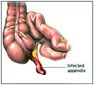
The appendix is a narrow, small, finger-shaped portion of the large intestine that generally hangs down from the lower right side of the abdomen.

Laparoscopic Appendectomy Surgery India offers info on Cost Appendectomy Hospital India, Laparoscopic Abdominal Surgery India

What is appendicitis and what causes it?

Appendicitis is a sudden inflammation of the appendix. Although the appendix does not seem to serve any purpose, it can become diseased and, if untreated, can burst, causing infection and even death.

The cause of appendicitis is usually unknown. Appendicitis may occur after a viral infection in the digestive tract or when the tube connecting the large intestine and appendix is blocked or trapped by stool. It is thought that blockage of the opening of the appendix into the bowel by a hard, small stool fragment causes inflammation and infection of the appendix (appendicitis). The inflammation can cause infection, a blood clot, or rupture of the appendix.

The infected appendix then must be surgically removed (emergency appendectomy) before a hole develops in the appendix and spreads the infection to the entire abdominal space.

What are the symptoms of appendicitis?

  • Abdominal pain – pain may begin in the upper-middle abdomen then develop to sharp localized pain
  • Abdominal pain may be worse when walking or coughing
  • Fever usually occurs within several hours
  • Loss of appetite
  • Nausea
  • Vomiting
  • Constipation
  • Rectal tenderness
  • Chills and shaking

If you have these symptoms, see a doctor immediately! Because of the risk of rupture, which may happen as soon as 48 to 72 hours after symptoms begin, appendicitis is considered an emergency.

How is appendicitis treated?

The best treatment of appendicitis is its surgical removal. Mild appendicitis may sometimes be cured with antibiotics. More serious cases are treated with surgery to remove the appendix, called an appendectomy. Doctors either use an “open” technique or a minimally invasive approach to remove the appendix.

Appendicitis is considered an emergency and anyone with symptoms needs to see a doctor immediately!

What is a laparoscopic appendectomy?

The laparoscopic (minimally invasive) surgical technique involves making several tiny cuts in the abdomen and inserting a miniature camera and surgical instruments. As many as three or four incisions are made. The surgeon then removes the appendix with the instruments, so there is usually no need to make a large incision in the abdomen. The camera projects a magnified image of the area onto a television monitor which helps guide the surgeons as they remove the appendix.
What is the benefit of laparoscopic appendectomy?

Most cases of acute appendicitis can be treated laparoscopically. The main advantages are : -

  • Less post-operative pain
  • Faster recovery and return to normal activity
  • Shorter hospital stay
  • Less post-operative complications
  • Minimally sized incisions/scars

In most cases, patients can be discharged within 24 to 36 hours. By contrast, the hospital stay is typically two to five days for an open procedure.

Can every patient have a laparoscopic appendectomy?

No. Patients with cardiac diseases and COPD would not be good candidates for laparoscopic appendectomy. In addition, laparoscopic appendectomy is not recommended for those with pre-existing disease conditions.

Laparoscopic appendectomy may also be more difficult in patients who have had previous lower abdominal surgery and for obese patients. The elderly may also be at increased risk for complications with general anesthesia. We evaluate every patient to determine the appropriate type of surgery to perform.

Treatment Options:

The appendix is then removed using a camera through the belly button as well as two other small incisions. Depending on how advanced the infection is, the patient is kept on antibiotics. If the infection is very advanced, the operation may be delayed while the patient receives antibiotics for several weeks. After the infection has improved, a laparoscopic appendectomy is performed.

Laparoscopic appendectomy is a safe and effective treatment for appendicitis. However, in the presence of infection, adhesions, or variations in anatomy, this method becomes dangerous and your surgeon may need to make the prudent decision to continue by making the traditional incision to safely complete the operation. This should not be seen as a failure, but as a wise decision by your surgeon to prevent dangerous complications. Other complications, although rare, include bleeding and infection. It is extremely uncommon to require a blood transfusion for this operation.

This operation is generally performed with general anesthesia. An IV line will be placed in your arm for fluids and you will be brought into the operation room. The anesthesiologist and nurses will use monitors to check your heart rate and breathing during the procedure. These may include EKG leads, a blood pressure cuff, an oxygen mask and sleeves on your legs to prevent clots from forming.

Once you are asleep, the operating room team will work together to perform your operation. When your operation is complete, you will be awakened from anesthesia in the operating room but you may not remember this. After a few hours in the recovery room, the nurses will help you out of bed and give you something to drink. It is common to feel groggy and nauseated soon after surgery and medication is available to help with these discomforts. This information is not intended to replace a visit with your physician.

Picturised Procedure of Laparoscopic Appendectomy:

Laparoscopic Appendectomy

Laparoscopic Abdominal Surgery India, Laparoscopic Surgery Training India

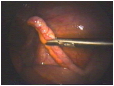
This is a normal appendix in a female patient found to have an infection of the reproductive organs. Since the appendix serves no useful function, it will be removed to save her from ever getting appendicitis in the future

Laparoscopic Appendectomy Surgery India offers info on Cost Appendectomy Hospital India

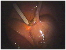
The first step is to free up the appendix down to where it is attached to the large bowel. This requires dividing the mesentery which contains the blood vessels that supply the appendix. Bipolar forceps apply an electric current which seals the blood vessels and prevents bleeding.

Laparoscopic Surgery Training India, Laparoscopic Abdominal Surgery Gurgaon India

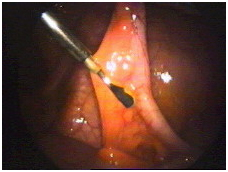
Next, scissors are used to divide the mesentery. Alternating application of the bipolar forceps and scissors allow complete mobilization of the appendix down to its base.


Laparoscopic Appendectomy Surgery India offers info on Cost Appendectomy Hospital India, Laparoscopic Abdominal Surgery India

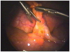
Now the base of the appendix is tied off using a pretied suture which is now being positioned.

Laparoscopic Surgery Training India, Laparoscopic Abdominal Surgery Gurgaon India

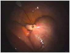
The suture has now been tightened and appears secure. The knot is a fisherman’s knot, which can be tightened, but will not loosen on its own.

Laparoscopic Surgery Training India, Laparoscopic Abdominal Surgery Gurgaon India, Laparoscopic Cancer Surgery India

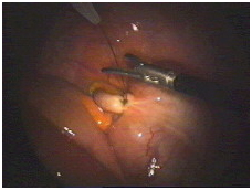
As shown in this picture, the suture is now cut with the scissors.

Laparoscopic Appendectomy Surgery India offers info on Cost Appendectomy Hospital India, Laparoscopic Abdominal Surgery India, Laparoscopic Cancer Surgery India

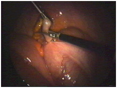
Finally, the appendix is cut free and is ready to be removed.

Laparoscopic Appendectomy Surgery India offers info on Cost Appendectomy Hospital India, Laparoscopic Abdominal Surgery Gurgaon India, Laparoscopic Abdominal Surgery India
The operation is complete, and the area is inspected a final time to make sure there is no bleeding.


Prostatitis: Causes, symptoms & Treatments

Prostatitis is an inflammation or infection of the prostate gland. It affects at least half of all men at some time in their lives. Having this condition does not increase your risk of any other prostate disease.

Symptoms

  • Trouble passing urine or pain when passing urine
  • A burning or stinging feeling when passing urine
  • Strong, frequent urge to pass urine, even when there is only a small amount of urine
  • Chills and high fever
  • Low back pain or body aches
  • Pain low in the belly, groin, or behind the scrotum
  • Rectal pressure or pain
  • Urethral discharge with bowel movements
  • Genital and rectal throbbing
  • Sexual problems and loss of sex drive
  • Blocked urine
  • Painful ejaculation

Prostatitis is not contagious. It is not spread through sexual contact. Your partner cannot catch this infection from you.

Several tests, such as DRE and a urine test, can be done to see if you have prostatitis. Getting the right diagnosis of your exact type of prostatitis is the key to getting the best treatment. Even if you have no symptoms, you should follow your doctor’s suggestion to complete treatment.

Types

There are four types of prostatitis:

Acute bacterial prostatitis

This infection comes on suddenly (acute) and is caused by bacteria. Symptoms include severe chills and fever. There is often blood in the urine.

Treatment: Most cases can be cured with a high dose of antibiotics, taken for 7 to 14 days, and then lower doses for several weeks. You may also need drugs to help with pain or discomfort.

Chronic bacterial prostatitis

Also caused by bacteria, this condition doesn’t come on suddenly, but it can be bothersome. The only symptom you may have is bladder infections that keep coming back. The cause may be a defect in the prostate that lets bacteria collect in the urinary tract.

Treatment: Antibiotic treatment over a longer period of time is best for this type.

Chronic prostatitis or chronic pelvic pain syndrome

This disorder is the most common but least understood form of the disease. Found in men of any age from late teens to elderly, its symptoms go away and then return without warning. There can be pain or discomfort in the groin or bladder area.

Treatment: There are several different treatments for this problem, based on your symptoms. These include antibiotics and other medicines, such as alpha-blockers. Alpha-blockers relax muscle tissue in the prostate to make passing urine easier.

Asymptomatic inflammatory prostatitis

You usually don’t have symptoms with this condition. It is often found when your doctor is looking for other conditions like infertility or prostate cancer. If you have this problem, often your PSA test (see The PSA Test) will show a higher number than normal. It does not necessarily mean that you have cancer.

Treatment: Men with this condition are usually given antibiotics for 4 to 6 weeks, and then have another PSA test.


Propeller flaps for leg reconstruction: How it construct in patients

Reverse sural artery fasciocutaneous flap has become a workhorse for the reconstruction of distal leg soft tissue defects. When its use is not feasible, perforator-based propeller flap offers a better, easier, faster, and cheaper alternative to free flap. We present our experience with two men both aged 34 years who sustained Gustilo 3B injuries from gunshot. The donor area for reversed sural artery flap was involved in the injuries. They had early debridement, external fixation, and wound coverage with perforator-based propeller flaps. The donor sites were covered with skin graft. All flaps survived. There were minor wound edge ulcers due to the pressure of positioning that did not affect flap survival and the ulcers healed with conservative management. Perforator-based propeller flap is a versatile armamentarium for reconstruction of soft tissue defects of the distal leg in resource-constrained settings, especially when the donor area for a reverse flow sural flap artery is involved in the injury.

INTRODUCTION

Reconstruction of soft tissue defects in the distal third of the leg pose challenges for reconstruction as a result of the paucity of donor tissue, bony prominence at the medial and lateral malleolus, and relatively poor blood supply of the region.Random pattern flaps raised in the leg are limited in reliability, size, reach, and arc-of-rotation. Most major injuries of the distal leg are associated with significant soft tissue defects because the tibia is mostly subcutaneous anteromedially. There is also less muscle bulk around its lower third.

Reverse sural artery fasciocutaneous flap has become a workhorse for the reconstruction of distal leg soft tissue defects in settings with limited resources. Its use obviates the need for sophisticated equipment, extended operative time, and super specialized training. Revere sural artery flap is a relatively easy and fast method of transferring a large amount of tissue. It allows for good hemostasis, and it has a short learning curve.

When the vascular supply to this flap is injured as found in our patients, reliable options for reconstruction in the distal third of the leg becomes limited.

Free tissue transfer would have been another modality to choose; however, microsurgical skills, specialized equipment, cost, steep learning curve, and prolonged operating time required for this modality reduces its feasibility in our practice setting and even outside tertiary care centers in more developed countries.

Perforator-based flaps address these problems. They afford speed, simplicity, color and texture match, lower cost, and allow greater arc of movement. Its applicability is possible in most parts of the body. Successful outcomes have been described in the axilla, periolecranon, forearm, lower extremity, hand and trunk. In spite of the versatility of perforator-based flaps, literature search reveals that report of its use is sparse from our practice setting.

We therefore present our experience with the use of three (tibioperoneal system) perforator-based propeller flaps in the reconstruction of distal leg defects following gunshot injuries. Perforator-based propeller flap provides a reliable option for resurfacing soft tissue defects when other options are not feasible.

Anatomy of distal leg perforators

Perforators are small diameter vessels that originate from a main pedicle and perforate the fascia or muscle to reach the skin. The development of flaps based on perforators has followed the understanding of the blood supply from a source artery to the skin.[8,9] Anatomical studies have revealed that they emerge from the crural fascia in four longitudinal rows within the intermuscular septa that border the compartments of the lower leg and the tibia. They are grouped into clusters of perforators with consistent distance proximal to the intermalleolar line. In the leg, there are anterior tibial artery perforators; posterior tibial artery perforators, peroneal artery perforators, and sural artery perforators.[8]

CASE REPORTS

Two men both aged 34–year-old of different addresses at different incidences, were shot in the distal aspect of the left leg.

Case 1

The first patient was attacked by armed robber; he sustained Gustilo 3B open fractures with two soft tissue defects: A lateral one measuring 12 cm × 7 cm and a medial defect 14 cm × 6 cm [Figure 1] with injury to the recurrent sural artery precluding the use of the reversed sural artery flap for wound coverage. The distal part of the tibia and injured tendons were left exposed in the wound bed. He had initial wound debridement and application of external fixators to immobilize his fractures. He subsequently had wound coverage with two perforator-based propeller flaps raised from the medial and lateral aspect of the leg [Figure 2]. The secondary defects were covered with split-thickness skin graft. During the postoperative period, he developed seroma under the lateral flap which was drained and did not have any adverse effect on flap survival. He also developed a Grade 2 pressure ulcer on the lateral edge of the wound due to limb positioning. This healed without surgical intervention. Skin graft take was 100%. Figure 3 shows the surgical outcome six weeks postoperatively.

Figure 1

Figure 1
Preoperative picture before debridement
Figure 2

Figure 2
Immediate postoperative picture
Figure 3

Figure 3
Follow-up picture 6 weeks postoperatively

Case 2

The second patient was shot during an argument; he sustained Gustilo 3B open fractures. The entry wound was quite small, but the exit wound located posteriorly measured 14 cm × 11 cm and a large portion of the Achilles tendon was exposed. The sural artery was found severed in the wound [Figure 4]. He had wound debridement, fracture reduction, and immobilization with an external fixator. A perforator-based propeller flap was raised to cover the wound and split thickness skin graft was applied to the secondary defect [Figure 5]. Flap survived without any area of loss [Figure 6]. The pins of the external fixator became loosened the 10th postoperative day, and the wound had to be manipulated to tighten the loose component. He developed hematoma in the wound after the procedure. This was drained with removal of the staples around the hematoma and the wound healed satisfactorily thereafter. Skin graft take was 100%.

Figure 4

Figure 4
Preoperative picture showing exposed Achilles tendon. The cavitation effect of the gunshot damaged the sural artery
Figure 5

Figure 5
Immediate postoperative picture
Figure 6

Figure 6
Fifth postoperative day

Flap design and surgical technique

Perforators in the proposed donor tissue close to the defect were located preoperatively using handheld Doppler (probe) and marked on the skin using methylene blue [Figure 7]. The flaps were then designed like blades of a propeller adjacent to the defects. The proximal end of the flap to the targeted perforator is made equidistant from the distal end of the defect to the perforator.

Figure 7

Figure 7
Preoperative planning showing the flap outline. Identified perforators were marked as x on the outline

The flap sizes were made to be 0.5 cm more than the size of the defect to be covered to aid closure without tension.

An exploratory skin incision was made and carried through to include the deep fascia. Through this incision, blunt dissection in the subfascial plane was done to isolate the perforator. As soon as the adequacy of the perforator is assured, the other incisions were completed, and other perforators that would not be used were ligated. Fascial strands were released to allow for mobilization of the flaps and to avoid tension on the pedicle during the transfer. The flaps were rotated 180° into place like a propeller blade avoiding kinking of the perforator. Hemostasis was secured with diathermy and tourniquet without exsanguination was used for the first patient, but diathermy and ligatures were used for the second patient. Flaps were inset and secured using skin staples.

The sizes of the secondary defects were reduced by suturing and the remaining defect skin grafted. Primary closure was not possible in all the donor sites due to the size of flaps harvested. Bedside, flap monitoring was achieved via a window created within the dressing to examine the flaps.

Pediatric Stroke : Symptoms, causes & Treatments

Symptoms:

Because people do not think about newborns and children having strokes, they are not often recognized or treated properly. Another challenge with newborns is that they cannot communicate symptoms that are not readily visible.

The common stroke warning signs used to identify stroke in adults can also be used to identify strokes in children.  But, there are some specific symptoms that you should look for in children depending on their age.

In newborns and infants

  • Seizures.
  • Extreme sleepiness.
  • Tendency to use only one side of their body.

In children and teens

  • Severe headaches
  • Vomiting
  • Sleepiness
  • Dizziness
  • Loss of balance or coordination

CAUSES:

While high blood pressure, an irregular heartbeat and a hardening of the arteries are common causes of adult strokes, they are rare in children. Common risk factors for stroke in children include:

  • Congenital heart defects
  • Sickle-cell disease
  • Immune disorders
  • Diseases of the arteries
  • Abnormal blood clotting
  • Head or neck trauma
  • Maternal history of infertility
  • Maternal infection in the fluid surrounding an unborn baby
  • Premature rupture of membrane during pregnancy
  • Pregnancy related high blood pressure in the mother

TREATMENTS:

Children usually recover better from strokes than adults do because their brains are still growing. However, children can experience permanent complications from stroke, such as seizures, weakness, and vision problems.

Treatment for pediatric stroke depends on the specific cause. Some common treatments used with adults are not appropriate for young children and infants. One of the best treatments for ischemic strokes for adults is tPA, or tissue plasminogen activator, a drug that dissolves blood clots.

With children under 18, use of tPA is controversial and currently limited to clinical trials. Further studies on safety and benefits of this treatment are necessary. Rehabilitation methods with pediatric stroke survivors are also not clear and require further clinical trials.

Current treatments for pediatric stroke include:

  • Supportive care to maintain normal body temperature, proper hydration, and normal blood sugar levels.
  • Controlling high blood pressure.
  • Detecting and treating seizures with EEG monitoring and anticonvulsant medication.
  • Managing intracranial pressure.
  • Blood transfusions for children with sickle-cell disease.
  • Antithrombotic therapy, which refers to medications used to prevent blood clots from forming or growing, is used in children but generally not infants.
  • Surgery related to hemorrhagic stroke (and less commonly, ischemic stroke) is sometimes performed to relieve pressure on the brain.


Systemic Lupus Erythematosus (SLE): Symtoms, causes & Treatments

Common symptoms of LUPUS are:
  • severe fatigue.
  • joint pain.
  • joint swelling.
  • headaches.
  • a rash on the cheeks and nose, which is called a “butterfly rash”
  • hair loss.
  • anemia.
  • blood-clotting problems.

What Is Lupus?

Lupus is an autoimmune disease whereby a misdirected immune system leads to inflammation and injury to one’s own body tissues.

Signs of Lupus

Lupus can involve many different parts of the body, including:

  • Skin
  • Joints
  • Heart
  • Lungs
  • Kidneys
  • Brain

There is no specific cure for lupus, but treatments are effective at minimizing lupus damage and improving function. Approximately 80% of lupus patients are women.

Picture of Systemic Lupus Erythematosus 1

Lupus Symptoms: Joint Pain

Though the first signs of the lupus disease can be rash, there are often pains in the muscles and joints. Both sides of the body tend to be affected. Hands, wrists, knees, and feet are commonly affected. The joints can become swollen, warm, and have limited range of motion.

Lupus Symptoms: Butterfly Rash

A classic lupus rash involves the cheeks and over the bridge of the nose. This is referred to as a “butterfly-shaped” lupus rash. It is also common for the skin to be very sensitive to burning and irritation after sun exposure. This is referred to as photosensitivity.

Lupus Symptoms: Nail Changes

Lupus rash can cause a ruddy discoloration of the backs of the hands and fingers. There can also be poor circulation to the nail beds that leads to irregularities of the fingernails. Inflammation at the nail bed can cause swelling and puffiness.

What are risk factors and causes of systemic lupus erythematosus? Is lupus contagious? Is lupus hereditary?

The precise reason for the abnormal autoimmunity that causes lupus is not known. Inherited genes, viruses, ultraviolet light, and certain medications may all play some role.

Lupus is not caused by an infectious microorganism and is not contagious from one person to another.

Genetic factors increase the tendency of developing autoimmune diseases, and autoimmune diseases such as lupus, rheumatoid arthritis, and autoimmune thyroid disorders are more common among relatives of people with lupus than the general population. Moreover, it is possible to have more than one autoimmune disease in the same individual. Therefore, “overlap” syndromes of lupus and rheumatoid arthritis, or lupus and scleroderma, etc., can occur.

Some scientists believe that the immune system in lupus is more easily stimulated by external factors like viruses or ultraviolet light. Sometimes, symptoms of lupus can be precipitated or aggravated by only a brief period of sun exposure.

It also is known that some women with SLE can experience worsening of their symptoms prior to their menstrual periods. This phenomenon, together with the female predominance of SLE, suggests that female hormones play an important role in the expression of SLE. This hormonal relationship is an active area of ongoing study by scientists.

Research has demonstrated evidence that a key enzyme’s failure to dispose of dying cells may contribute the development of SLE. The enzyme, DNase1, normally eliminates what is called “garbage DNA” and other cellular debris by chopping them into tiny fragments for easier disposal. Researchers turned off the DNase1 gene in mice. The mice appeared healthy at birth, but after six to eight months, the majority of mice without DNase1 showed signs of SLE. Thus, a genetic mutation in a gene that could disrupt the body’s cellular waste disposal may be involved in the initiation of SLE.

What is the treatment for systemic lupus?

There is no permanent cure for SLE. The goal of treatment is to relieve symptoms and protect organs by decreasing inflammation and/or the level of autoimmune activity in the body. The precise treatment is decided on an individual basis. Many people with mild symptoms may need no treatment or only intermittent courses of anti-inflammatory medications. Those with more serious illness involving damage to internal organ(s) may require high doses of corticosteroids in combination with other medications that suppress the body’s immune system.

People with SLE need more rest during periods of active disease. Researchers have reported that poor sleepquality was a significant factor in developing fatigue in people with SLE. These reports emphasize the importance for people and physicians to address sleep quality and the effect of underlying depression, lack of exercise, and self-care coping strategies on overall health. During these periods, carefully prescribed exercise is still important to maintain muscle tone and range of motion in the joints.

To protect from sun sensitivity, sunscreens, sun avoidance, and sun protection clothing are used. Certain types of lupus rash can respond to topical cortisone medications.

Nonsteroidal anti-inflammatory drugs (NSAIDs) are helpful in reducing inflammation and pain in muscles, joints, and other tissues. Examples of NSAIDs include aspirin, ibuprofen(Motrin), naproxen (Naprosyn), and sulindac (Clinoril). Since the individual response to NSAIDs varies, it is common for a doctor to try different NSAIDs to find the most effective one with the fewest side effects. The most common side effects are stomach upset, abdominal pain, ulcers, and even ulcer bleeding. NSAIDs are usually taken with food to reduce side effects. Sometimes, medications that prevent ulcers while taking NSAIDs, such as misoprostol (Cytotec), are given simultaneously.

Corticosteroids are more potent than NSAIDs in reducing inflammation and restoring function when the disease is active. Corticosteroids are particularly helpful when internal organs are affected. Corticosteroids can be given by mouth, injected directly into the joints and other tissues, or administered intravenously. Unfortunately, corticosteroids have serious side effects when given in high doses over prolonged periods, and the doctor will try to monitor the activity of the disease in order to use the lowest doses that are safe. Side effects of corticosteroids include weight gain, thinning of the bones and skin, infection, diabetes, facial puffiness, cataracts, and death (necrosis) of the tissues in large joints.

Hydroxychloroquine (Plaquenil) is an antimalarial medication found to be particularly effective for SLE people with fatigue, skin involvement, and joint disease. Consistently taking Plaquenil can prevent flare-ups of lupus. Side effects are uncommon but include diarrhea, upset stomach, and eye-pigment changes. Eye-pigment changes are rare but require monitoring by an ophthalmologist (eye specialist) during treatment with Plaquenil. Researchers have found that Plaquenil significantly decreased the frequency of abnormal blood clots in people with systemic lupus. Moreover, the effect seemed independent of immune suppression, implying that Plaquenil can directly act to prevent the blood clots. This fascinating study highlights an important reason for people and doctors to consider Plaquenil for long-term use, especially for those SLE people who are at some risk for blood clots in veins and arteries, such as those with phospholipid antibodies (cardiolipin antibodies, lupus anticoagulant, and false-positive venereal disease research laboratory test). This means not only that Plaquenil reduces the chance for re-flares of SLE, but it can also be beneficial in thinning the blood to prevent abnormal excessive blood clotting. Plaquenil is commonly used in combination with other treatments for lupus.

For resistant skin disease, other antimalarial drugs, such as chloroquine (Aralen) or quinacrine, are considered and can be used in combination with hydroxychloroquine. Alternative medications for skin disease include dapsone and retinoic acid (Retin-A). Retin-A is often effective for an uncommon wart-like form of lupus skin disease. For more severe skin disease, immunosuppressive medications are considered as described below.

Medications that suppress immunity (immunosuppressive medications) are also called cytotoxic drugs. They are sometimes referred to as chemotherapy because they are also used to treat cancer, generally in much higher doses than those used to treat lupus. Immunosuppressive medications are used for treating people with more severe manifestations of SLE, such as damage to internal organ(s). Examples of immunosuppressive medications include methotrexate (Rheumatrex, Trexall), azathioprine (Imuran), cyclophosphamide (Cytoxan), chlorambucil (Leukeran), and cyclosporine (Sandimmune). All immunosuppressive medications can seriously depress blood-cell counts and increase risks of infection and bleeding. Immunosuppressive medications may not be taken during pregnancy or conception because of risk to the fetus. Other side effects are specific for each drug. For examples, methotrexate can cause liver toxicity, while cyclosporine can impair kidney function.

In recent years, mycophenolate mofetil (CellCept) has been used as an effective medication for lupus, particularly when it is associated with kidney disease. CellCept has been helpful in reversing active lupus kidney disease (lupus renal disease) and in maintaining remission after it is established. Its lower side-effect profile has advantage over traditional immune-suppression medications.

In SLE patients with serious brain (lupus cerebritis) or kidney disease (lupus nephritis), plasmapheresis is sometimes used to remove antibodies and other immune substances from the blood to suppress immunity. Plasmapheresis is a process of removing blood and passing the blood through a filtering machine, then returning the blood to the body with its antibodies removed. Rarely, people with SLE can develop seriously low platelet levels, thereby increasing the risk of excessive and spontaneous bleeding. Since the spleen is believed to be the major site of platelet destruction, surgical removal of the spleen is sometimes performed to improve platelet levels. Other treatments have included plasmapheresis and the use of male hormones.

Plasmapheresis has also been used to remove certain harmful proteins (cryoglobulins) that can lead to vasculitis. End-stage kidney damage from SLE requires dialysis and/or a kidney transplant.

Fibromyalgia: Symptoms, causes & Treatments

Fibromyalgia syndrome affects the muscles and soft tissue. Symptoms include chronic muscle pain, fatigue, sleep problems, and painful tender points or trigger points, which can be relieved through medications, lifestyle changes and stress management.

 What Are the Symptoms of Fibromyalgia?

Symptoms of fibromyalgia include:

  • Chronic muscle pain, muscle spasms, or tightness
  • Moderate or severe fatigue and decreased energy
  • Insomnia or waking up feeling just as tired as when you went to sleep
  • Stiffness upon waking or after staying in one position for too long
  • Difficulty remembering, concentrating, and performing simple mental tasks (“fibro fog”)
  • Abdominal pain, bloating, nausea, and constipation alternating with diarrhea (irritable bowel syndrome)
  • Tension or migraine headaches
  • Jaw and facial tenderness
  • Sensitivity to one or more of the following: odors, noise, bright lights, medications, certain foods, and cold
  • Feeling anxious or depressed
  • Numbness or tingling in the face, arms, hands, legs, or feet
  • Increase in urinary urgency or frequency (irritable bladder)
  • Reduced tolerance for exercise and muscle pain after exercise
  • A feeling of swelling (without actual swelling) in the hands and feet

When it comes to fibromyalgia treatments, there are drugs, alternative remedies, and lifestyle habits that may help decrease pain and improve sleep. Your fibromyalgia specialist may prescribe pain medication or antidepressants to help treat the pain, fatigue, depression, and anxietythat comes with the disease. In addition, your doctor may recommend physical therapy, moist heat, regular aerobic exercise, relaxation, and stress reduction to help you self-manage your symptoms.

There is no one “pill” that treats or cures fibromyalgia. A multidisciplinary approach that uses both medication and alternative or lifestyle strategies seems to work best to treat fibromyalgia symptoms.

Is Fibromyalgia Pain Similar to Arthritis Pain?

Fibromyalgia can cause symptoms similar to arthritis, bursitis, and tendinitis. Consequently, some experts group fibromyalgia with arthritis and related disorders. The pain associated with these other conditions is typically localized to a single area, while the pain and stiffness of fibromyalgia are very widespread and consists of deep muscle pain, morning stiffness, and painful tender points, making it difficult to exercise or be physically active.


related content

SLIDESHOW

A Visual Guide to Acupuncture

Start


How Is Fibromyalgia Fatigue Treated?

Along with deep muscle pain and painful tender points, fatigue is a key symptom of fibromyalgia and it can be debilitating. Not only do you feel exhausted and weak, but bed rest does not seem to help. Many people with fibromyalgia report sleeping eight to 10 hours at night and feeling as if they haven’t slept at all.

Some drugs may help ease the fatigue associated with fibromyalgia. In addition, aerobic exercise can help ease fatigue, minimize pain, improve quality of sleep, and improve mood.


How Does Exercise Help Fibromyalgia Symptoms?

Numerous studies show that exercise is one of the most important treatments for fibromyalgia. Many people with fibromyalgia are are not physically fit. They avoid exercise because they fear increased pain. Yet aerobic or conditioning exercise can actually help relieve pain and depression.

Regular exercise increases the body’s production of endorphins, natural painkillers that also boost mood. Starting slowly and gradually increasing the duration and intensity of exercise can help you enjoy the benefits of exercise without feeling more pain.

Pages:12