Global Treatment Services Pvt. Ltd.

Global Treatment Services

Stroke

A stroke is a medical emergency, and prompt treatment is very important. Early detection & action can reduce brain damage and other complications leading to permanent disability or uncommon death.

What is Stroke?www.gtsmeditour.com (2)

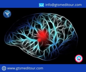
Stroke is a cerebrovascular disease. This means that it affects the blood vessels that feed the brain oxygen. If the brain does not receive enough oxygen, damage may start to occur.

A stroke occurs when the blood supply to part of your brain is interrupted or reduced, preventing brain tissue from getting oxygen and nutrients. Brain cells begin to die in minutes.

Although many strokes are treatable, some can lead to disability or death.

Remember “Be Fast”  when you spot a stroke:www.gtsmeditour.com

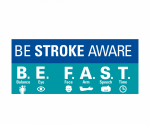
  • Sudden Loss of Balance
  • Loss of vision in one or both Eyes
  • Face drooping
  • Arm weakness
  • Speech difficulty
  • Time to call  medical emergency

Risk factors:

Risk factors for narrowed blood vessels in the brain are the same as those that cause narrowing blood vessels in the heart and heart attack  these includes:

  • High blood pressure/Hypertension
  • High cholesterol
  • Diabetes & Smoking

Symptoms:

The sooner a person having a stroke gets care, the better their outcome is likely to be. For this reason, it’s helpful to know the signs of a stroke so you can act quickly. Stroke symptoms can include:

  • Confusion, including difficulty speaking and understanding speech
  • A Head ache, possibly with altered consciousness or vomiting
  • Numbness or an inability to move parts of the face, arm, or leg, particularly on one side of the body
  • Vision problems in one or both eyes
  • Difficulty walking, including dizziness and a lack of coordination.

What are the different types of strokes?

There are three main types of stroke:

  • transient ischemic attack
  • ischemic stroke
  • hemorrhagic stroke.

Transient ischemic attack (TIA):

Doctors also call a transient ischemic attack (TIA) a warning or ministroke. Anything that temporarily blocks blood flow to your brain causes a TIA. The blood clot and TIA symptoms last for a short period of time.

Ischemic stroke

An ischemic stroke occurs when a blood clot keeps blood from flowing to your brain. The blood clot is often due to atherosclerosis, which is a buildup of fatty deposits on the inner lining of a blood vessel. A portion of these fatty deposits can break off and block blood flow in your brain. The concept is similar to that of a heart attack, where a blood clot blocks blood flow to a portion of your heart.

An ischemic stroke can be embolic, meaning the blood clot travels from another part of your body to your brain. An estimated 15 percent of embolic strokes are due to a condition called atrial fibrillation, where your heart beats irregularly.

A thrombotic stroke is an ischemic stroke caused by a clot forming in a blood vessel in your brain.

Unlike a TIA, the blood clot that causes an ischemic stroke won’t go away without treatment.

Hemorrhagic stroke

A hemorrhagic stroke results when a blood vessel in your brain ruptures or breaks, spilling blood into the surrounding tissues.

There are three main types of hemorrhagic strokes: The first is an aneurysm, which causes a portion of the weakened blood vessel to balloon outward and sometimes rupture. The other is an arteriovenous malformation, which involves abnormally formed blood vessels. If such a blood vessel ruptures, it can cause a hemorrhagic stroke. Lastly, very high blood pressure can cause weakening of the small blood vessels in the brain and result in bleeding into the brain as well.

Diagnosis of stroke

Your doctor will ask you or a family member about your symptoms and what you were doing when they arose. They’ll take your medical history to find out your stroke risk factors. They’ll also:

  • ask what medications you take
  • check your blood pressure
  • listen to your heart

You’ll also have a physical exam, during which the doctor will evaluate you for:

  • balance
  • coordination
  • weakness
  • numbness in your arms, face, or legs
  • signs of confusion
  • vision issues.

Treatment:

Proper medical evaluation and prompt treatment are vital to recovering from a stroke.

You may go through various tests to further help your doctor determine if you’ve had a stroke, or to rule out another condition. These tests include:

Blood tests

Your doctor might draw blood for several blood tests. Blood tests can determine:

  • your blood sugar levels
  • if you have an infection
  • your platelet levels
  • how fast your blood clots

MRI and CT scan

You may undergo either or both a magnetic resonance imaging (MRI) scan and a computerized tomography (CT) scan.

The MRI will help see if any brain tissue or brain cells have been damaged. A CT scan will provide a detailed and clear picture of your brain that shows any bleeding or damage in the brain. It may also show other brain conditions that could be causing your symptoms.

EKG

Your doctor may order an electrocardiogram (EKG), too. This simple test records the electrical activity in the heart, measuring its rhythm and recording how fast it beats. It can determine if you have any heart conditions that may have led to stroke, such as a prior heart attack or atrial fibrillation.

Cerebral angiogram

Another test your doctor may order to determine if you’ve had a stroke is a cerebral angiogram. This offers a detailed look at the arteries in your neck and brain. The test can show blockages or clots that may have caused symptoms.

Carotid ultrasound

A carotid ultrasound, also called a carotid duplex scan, can show fatty deposits (plaque) in your carotid arteries, which supply the blood to your face, neck, and brain. It can also show whether your carotid arteries have been narrowed or blocked.

Echocardiogram

An echocardiogram can find sources of clots in your heart. These clots may have traveled to your brain and caused a stroke.

These stroke types are caused by a blood clot or other blockage in the brain. For that reason, they’re largely treated with similar techniques, which include:

Antiplatelet and anticoagulants

Over-the-counter aspirin is often a first line of defense against stroke damage. Anticoagulant and antiplatelet drugs should be taken within 24 to 48 hours after stroke symptoms begin.

Clot-breaking drugs

Thrombolytic drugs can break up blood clots in your brain’s arteries, which still stop the stroke and reduce damage to the brain.

One such drug, tissue plasminogen activator (tPA), or Alteplase IV r-tPA, is considered the gold standard in ischemic stroke treatment. It works by dissolving blood clots quickly, if delivered within the first 3 to 4.5 hours after symptoms of your stroke began. People who receive a tPA injection are more likely to recover from a stroke, and less likely to have any lasting disability as a result of the stroke.

Mechanical thrombectomy

During this procedure, the doctor inserts a catheter into a large blood vessel inside your head. They then use a device to pull the clot out of the vessel. This surgery is most successful if it’s performed 6 to 24 hours after the stroke begins.

Stents

If your doctor finds where artery walls have weakened, they may perform a procedure to inflate the narrowed artery and support the walls of the artery with a stent.

Surgery

In the rare instances that other treatments don’t work, your doctor may perform surgery to remove a blood clot and plaques from your arteries. This may be done with a catheter, or if the clot is especially large, your doctor may open an artery to remove the blockage.

Hemorrhagic stroke

Strokes caused by bleeds or leaks in the brain require different treatment strategies. Treatments for hemorrhagic stroke include:

Medications

Unlike with an ischemic stroke, if you’re having a hemorrhagic stroke, the treatment goal is to make your blood clot. Therefore, you may be given medication to counteract any blood thinners you take.

You may also be prescribed drugs that can reduce blood pressure, lower the pressure in your brain, prevent seizures, and prevent blood vessel constriction.

Coiling

During this procedure, your doctor guides a long tube to the area of hemorrhage or weakened blood vessel. They then install a coil-like device in the area where the artery wall is weak. This blocks blood flow to the area, reducing bleeding.

Clamping

During imaging tests, your doctor may discover an aneurysm that hasn’t started bleeding yet or has stopped. To prevent additional bleeding, a surgeon may place a tiny clamp at the base of the aneurysm. This cuts off blood supply and prevents a possible broken blood vessel or new bleeding.

Surgery

If your doctor sees that an aneurysm has burst, they may do surgery to clip the aneurysm and prevent additional bleeding. Likewise, a craniotomy may be needed to relieve the pressure on the brain after a large stroke.

Recovery And  Rehabilitation:

It’s important that recovery and rehabilitation from a stroke start as soon as possible. In fact, stroke recovery should begin in the hospital. There, a care team can stabilize your condition, assess the effects of the stroke, identify underlying factors, and begin therapy to help you regain some of your affected skills.

Stroke recovery focuses on four main areas:

Speech therapy

A stroke can cause speech and language impairment. A speech and language therapist will work with you to relearn how to speak. Or, if you find verbal communication difficult after a stroke, they’ll help you find new ways of communication.

Cognitive therapy

After a stroke, many survivors have changes to their thinking and reasoning skills. This can cause behavioral and mood changes. An occupational therapist can help you work to regain your former patterns of thinking and behavior and to control your emotional responses.

Relearning sensory skills

If the part of your brain that relays sensory signals is affected during the stroke, you may find that your senses are “dulled” or no longer working. That may mean that you don’t feel things well, such as temperature, pressure, or pain. A therapist can help you learn to adjust to this lack of sensation.

Physical therapy

Muscle tone and strength may be weakened by a stroke, and you may find you’re unable to move your body as well as you could before. A physical therapist will work with you to regain your strength and balance, and find ways to adjust to any limitations.

Rehabilitation may take place in a rehabilitation clinic, a skilled nursing home, or your own home.

Preventive Measures:

In addition to emergency treatment,Recovery and Rehab  healthcare providers will also advise you on ways to prevent future strokes.

  • Quit smoking.
  • Consume alcohol in moderation.
  • Keep weight in check
  • Regular Checkups

Taking all these measures will help put you in better shape to prevent stroke.

Conclusion/ Take away

If you suspect or experiencing symptoms of a stroke, it’s crucial  you seek emergency medical treatment. Clot-busting medication can only be provided in the first hours after the signs of a stroke begin, (i.e, first 3hrs- 4.5hrs are said to be golden period) and early treatment is one of the most effective ways to reduce your risk for long-term complications and disability.

Prevention is possible, whether you’re preventing a first stroke or trying to prevent a second. Medications can help reduce the risk of blood clots, which lead to strokes. Speakup with your doctor to find a prevention strategy that works for you, including medical intervention and lifestyle changes.


Breast Cancer Awareness

Global Hand Wash Day (13)October is termed as Breast cancer Awareness month across the globe.After skin & lung cancer, breast cancer is the most common cancer diagnosed in women and rarely in men. Breast cancer is cancer that forms in the cells of the breasts.Its treatment depends on the stage of cancer. It may consist of chemotherapy, radiation, hormone therapy and surgery.

On average, 1 in 8 women will develop breast cancer in their lifetimes. About two-thirds of women with breast cancer are 55 or older. Most of the rest are between 35 and 54.

Fortunately, breast cancer is very treatable if you spot it early. Localized cancer (meaning it hasn’t spread outside your breast) can usually be treated before it spreads.

Once the cancer begins to spread, treatment becomes more complicated. It can often control the disease for years.

What is Breast Cancer?

The term “breast cancer” refers to a malignant tumor that has developed from cells in the breast. Usually breast cancer either begins in the cells of the lobules, which are the milk-producing glands, or the ducts, the passages that drain milk from the lobules to the nipple. Less commonly, breast cancer can begin in the stromal tissues, which include the fatty and fibrous connective tissues of the breast.

A tumor can be benign (not dangerous to health) or malignant (has the potential to be dangerous). Benign tumors are not considered cancerous: their cells are close to normal in appearance, they grow slowly, and they do not invade nearby tissues or spread to other parts of the body. Malignant tumors are cancerous. Left unchecked, malignant cells eventually can spread beyond the original tumor to other parts of the body.

Breast cancer is always caused by a genetic abnormality (a “mistake” in the genetic material). However, only 5-10% of cancers are due to an abnormality inherited from your mother or father. Instead, 85-90% of breast cancers are due to genetic abnormalities that happen as a result of the aging process and the “wear and tear” of life in general.

Causes Of Breast Cancer: How Did This Happen?

When you’re told that you have breast cancer, it’s natural to wonder what may have caused the disease. But no one knows the exact causes of breast cancer. Doctors seldom know why one woman develops breast cancer and another doesn’t, and most women who have breast cancer will never be able to pinpoint an exact cause. What we do know is that breast cancer is always caused by damage to a cell’s DNA.

Symptoms:

Different people have different symptoms of breast cancer. Some people do not have any signs or symptoms at all.

Few warning signs of breast cancer are—

  • New lump in the breast or underarm (armpit).
  • Thickening or swelling of part of the breast.
  • Irritation or dimpling of breast skin.
  • Redness or flaky skin in the nipple area or the breast.
  • Pulling in of the nipple or pain in the nipple area.
  • Nipple discharge other than breast milk, including blood.
  • Any change in the size or the shape of the breast.
  • Pain in any area of the breast.

Keep in mind that these symptoms can happen with other conditions that are not cancer.

Stages:

A doctor stage cancer according to the size of the tumor and whether it has spread to lymph nodes or other parts of the body.

There are different ways of staging breast cancer. One way is from stage 0–4, with subdivided categories at each numbered stage. Descriptions of the four main stages are listed below, though the specific substage of a cancer may also depend on other specific characteristics of the tumor, such as HER2 receptor status.

  • Stage 0: Known as ductal carcinoma in situ (DCIS), the cells are limited to within the ducts and have not invaded surrounding tissues.
  • Stage 1: At this stage, the tumor measures up to 2 centimeters (cm) across. It has not affected any lymph nodes, or there are small groups of cancer cells in the lymph nodes.
  • Stage 2: The tumor is 2 cm across, and it has started to spread to nearby nodes, or is 2–5 cm across and has not spread to the lymph nodes.
  • Stage 3: The tumor is up to 5 cm across, and it has spread to several lymph nodes or the tumor is larger than 5 cm and has spread to a few lymph nodes.
  • Stage 4: The cancer has spread to distant organs, most often the bones, liver, brain, or lungs.

    Risk Factors:

    UnControlable:

  • Age. Women over 50 are more likely to get breast cancer than younger women.
  • Race: African American women are more likely than white women to get breast cancer before  menopause.
  • Dense breasts. If your breasts have more connective tissue than fatty tissue, it can be hard to see tumors on a mammogram.
  • Personal history of cancer. Your odds go up slightly if you have certain benign breast conditions. They go up more sharply if you’ve had breast cancer before.
  • Family history. If a first-degree female relative (mother, sister, or daughter) had breast cancer, you’re two times more likely to get the disease. Having two or more first-degree relatives with a history of breast cancer increases your risk at least three times. This is especially true if they got cancer before menopause or if it affected both breasts. The risk can also rise if your father or brother was diagnosed with breast cancer.
  • Genes. Changes to two genes, BRCA1 and BRCA2, are responsible for some cases of breast cancer in families. About 1 woman in 200 has one of these genes. While they make you more likely to get cancer, they don’t mean you definitely will. If you have a BRCA1 or BRCA2 mutation, you have a 7 in 10 chance of being diagnosed with breast cancer by age 80. These genes also raise your odds of ovarian cancer, and they’re linked to pancreatic cancer and male breast cancer. Other gene mutations linked to breast cancer risk include mutations of the PTEN gene, the ATM gen, the TP53 gene, the CHEK2 gene, the CDH1 gene, the STK11 gene, and the PALB2 gene. These carry a lower risk for breast cancer development than the BRCA genes.
  • Menstrual history. Your breast cancer odds go up if:
    • Your periods start before age 12.
    • Your periods don’t stop until after you’re 55.
  • Radiation. If you had treatment for cancers like Hodgkin’s lymphoma before age 40, you have an increased risk of breast cancer.
  • Diethylstilbestrol (DES). Doctors used this drug between 1940 and 1971 to prevent miscarriage. If you or your mother took it, your breast cancer odds go up.

    Controlable:

  • Physical activity The less you move, the higher your chances.
  • Weight and diet. Being overweight after menopause raises your odds.
  • Alcohol. Regular drinking — especially more than one drink a day — increases the risk of breast cancer.
  • Reproductive history.

You have your first child after age 30.

You don’t breastfeed.

You don’t have a full-term pregnancy.

  • Taking hormones. Your chances can go up if you:

Use hormone replacement therapy that includes both estrogen and progesterone during menopause for more than 5 years. This increase in breast cancer risk returns to normal 5 years after you stop treatment.

Use certain birth control methods including birth control pills, shots, implants, IUDS, skin patches, or vaginal rings that contain hormones.

  • Still, most women who are at high risk for breast cancer don’t get it. On the other hand, 75% of women who develop breast cancer have no known risk factors.

Types:

There are several different types of breast cancer, including:

  • Ductal carcinoma: This begins in the milk duct and is the most common type.
  • Lobular carcinoma: This starts in the lobules.

Invasive breast cancer occurs when the cancer cells break out from inside the lobules or ducts and invade nearby tissue. This increases the chance of cancer spreading to other parts of the body.

Noninvasive breast cancer develops when the cancer remains inside its place of origin and has not yet spread. However, these cells can sometimes progress to invasive breast cancer.

Diagnosis:

If you feel a lump or if something shows up on a mammogram, your doctor will begin the breast cancer diagnosis process.

They’ll ask about your personal and family healthy history. Then, they’ll do a breast exam and order tests that include:

Imaging tests. Your doctor will use these to learn more about your breast.

  • Ultrasound. This test uses sound waves to make a picture of your breast.
  • Mammogram. This detailed X-ray gives doctors a better view of lumps and other problems.
  • Magnetic resonance imaging (MRI). This body scan uses a magnet linked to a computer to create detailed images of the insides of your breasts.
  • Biopsy. For this test, the doctor removes tissue or fluid from your breast. They look at it under a microscope to check for if cancer cells and, if they’re there, learn which type they are. Common procedures include:
    • Fine-needle aspiration. This is for easy-to-reach lumps or those that might be filled with fluid.
    • Core-needle biopsy. This type uses a bigger needle to remove a piece of tissue.
    • Surgical (open) biopsy. A surgeon removes the entire lump along with nearby breast tissue.
    • Lymph node biopsy. The doctor removes a part of the lymph nodes under your arm to see if the cancer has spread.
    • Image-guided biopsy. The doctor uses imaging to guide the needle.The doctor can test your biopsy sample for:
      • Tumor features. Is it invasive or in situ, ductal, or lobular? Has it spread to your lymph nodes? The doctor also measures the margins or edges of the tumor and their distance from the edge of the biopsy tissue, which is which is called margin width.
      • Estrogen receptors (ER) and progesterone receptors (PR). This tells the doctor if the hormones estrogen or progesterone make your cancer grow. This affects how likely it is that your cancer will come back and what type of treatment is most likely to prevent that.
      • HER2. This test looks for the gene human epidermal growth factor receptor 2. It can help cancer grow more quickly. If your cancer is HER2-positive, targeted therapy could be a treatment option for you.
      • Grade. This tells the doctor how different the cancer cells look from healthy cells and whether they seem to be slower-growing or faster-growing.
      • Oncotype Dx. This test evaluates 16 cancer-related genes and five reference genes to estimate the risk of the cancer coming back within 10 years of diagnosis.
      • Breast Cancer Index. This test can help your doctor decide how you need endocrine therapy.
      • MammaPrint. This test uses information from 70 genes to predict the risk of the cancer coming back.
      • PAM50 (Prosigna). This test uses information from 50 genes to predict if the cancer will spread.

      You might get blood tests including:

      • Complete blood count (CBC). This measures the number of different types of cells, like red and white blood cells, in your blood. It lets your doctor know if your bone marrow is working like it should.
      • Blood chemistry. This shows how well your liver and kidneys are working.
      • Hepatitis tests. These are sometimes done to check for hepatitis B and hepatitis C. If you have an active hepatitis B infection, you may need medication to fight the virus before you get chemotherapy. Without it, chemo can cause the virus to grow and damage your liver.

Treatment:

Treatment will depend on several factors, including:

  • the type and stage of the cancer
  • the person’s sensitivity to hormones
  • the age, overall health, and preferences of the individual

The main treatment options include:

radiation therapy

  • surgery
  • biological therapy, or targeted drug therapy
  • hormone therapy
  • chemotherapy

Factors affecting the type of treatment a person has will include the stage of the cancer, other medical conditions, and their individual preference.

Surgery

If surgery is necessary, the type will depend on both the diagnosis and individual preference. Types of surgery include:

Lumpectomy: This involves removing the tumor and a small amount of healthy tissue around it.

A lumpectomy can help prevent the spread of the cancer. This may be an option if the tumor is small and easy to separate from its surrounding tissue.

Mastectomy: A simple mastectomy involves removing the lobules, ducts, fatty tissue, nipple, areola, and some skin. In some types, a surgeon will also remove the lymph nodes and muscle in the chest wall.

Sentinel node biopsy: If breast cancer reaches the sentinel lymph nodes, which are the first nodes to which a cancer can spread, it can spread into other parts of the body through the lymphatic system. If the doctor does not find cancer in the sentinel nodes, then it is usually not necessary to remove the remaining nodes.

Axillary lymph node dissection: If a doctor finds cancer cells in the sentinel nodes, they may recommend removing several lymph nodes in the armpit. This can prevent the cancer from spreading.

Reconstruction: Following mastectomy, a surgeon can reconstruct the breast to look more natural. This can help a person cope with the psychological effects of breast removal.

The surgeon can reconstruct the breast at the same time as performing a mastectomy or at a later date. They may use a breast implant or tissue from another part of the body.

Radiation therapy

A person may undergo radiation therapy around a month after surgery. Radiation involves targeting the tumor with controlled doses of radiation that kill any remaining cancer cells.

Chemotherapy

A doctor may prescribe cytotoxic chemotherapy drugs to kill cancer cells if there is a high risk of recurrence or spread. When a person has chemotherapy after surgery, doctors call it adjuvant chemotherapy.

Sometimes, a doctor may choose to administer chemotherapy before surgery to shrink the tumor and make its removal easier. Doctors call this neoadjuvant chemothera

Hormone blocking therapy

Doctors use hormone blocking therapy to prevent hormone sensitive breast cancers from returning after treatment. Hormone therapy may be used to treat estrogen receptor (ER)-positive and progesterone receptor (PR)-positive cancers.

They usually administer hormone blocking therapy after surgery but might sometimes use it beforehand to shrink the tumor.

Hormone blocking therapy may be the only option for people who are not suitable candidates for surgery, chemotherapy, or radiotherapy.

Doctors may recommend a person has hormone therapy for 5–10 years after surgery. However, the treatment will not affect cancers that are not sensitive to hormones.

Examples of hormone blocking therapy medications may include:

  • tamoxifen
  • aromatase inhibitors
  • ovarian ablation or suppression
  • Goserelin, which is a luteinizing hormone-releasing agonist drug that suppresses the ovaries

Hormone treatment may affect fertility.

Biological treatment

Targeted drugs can destroy specific types of breast cancer. Examples include:

  • trastuzumab (Herceptin)
  • lapatinib (Tykerb)
  • bevacizumab (Avastin)

Treatments for breast and other cancers can have severe adverse effects. When deciding on a treatment, people should discuss the potential risks with a doctor and look at ways to minimize the side effects.

Facts to know about Breasts

  1. Breasts contain fat cells, not muscles
  2. Breast size cannot be altered by any excercise. it can only  be altered by painful surgery.
  3. Breast size depends upon a lot of factors such as harmonalchanges, bodyfat, genetics etc.
  4. Most of the time when body gains or losses fat, breast size changes.

Conclusion

There are steps every person can take to help the body stay as healthy as possible, such as eating a balanced diet, maintaining a healthy weight, not smoking, limiting alcohol, and exercising regularly . While these may have some impact on your risk of getting breast cancer, they cannot eliminate the risk.

Developing breast cancer is not your or anyone’s fault. Feeling guilty, or telling yourself that breast cancer happened because of something you or anyone else did, is not productive.

Regular checks and screening can help detect symptoms early. Women should discuss their options with a doctor.There is no way to prevent breast cancer. However, certain lifestyle decisions can significantly reduce the risk of breast cancer as well as other types.