Global Treatment Services Pvt. Ltd.

Global Treatment Services

CHOLECYSTECTOMY, Images, Treatments,

CHOLECYSTECTOMY

INTRODUCTION:
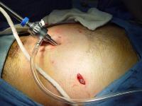
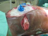
Cholecystectomy is one of the commonest elective surgical procedure performed in India. Most are performed to address symptoms related to biliary colic from cholelithiasis, to treat complications of gallstones (eg, acute cholecystitis, biliary pancreatitis), or as incidental cholecystectomies performed during other open abdominal procedures. Currently, most cholecystectomies are done using the laparoscopic technique in cities; however, the open technique is required in places where facilities or trained staff are not available.
DEFINITION:
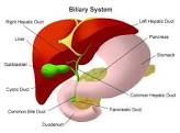
Cholecystectomy implies the surgical resection the gall bladder.
INDIAN INCIDENCE
In India, the incidence of gall stones is around six percent in the total population. It is 10 per cent in women and three per cent in men. In elderly people it may go up to 20 percent.
INDICATIONS:
Asymptomatic patients
Cholecystectomy is not indicated in most patients with asymptomatic stones because only 2-3% of these patients go on to become symptomatic per year. To properly determine the indications for elective cholecystectomy, the risk of the operation (taking into account the age and comorbid factors of the individual patient) must be weighed against the risk of complications and death without operation.
The widespread use of diagnostic abdominal ultrasonography has led to the increasing detection of clinically unsuspected gallstones. This, in turn, has given rise to a great deal of controversy regarding the optimal management of asymptomatic (silent) gallstones.
Patients who are immunocompromised, are awaiting organ allotransplantation, or have sickle cell disease are at higher risk of developing complications and should be treated irrespective of the presence or absence of symptoms.
Additional reasons to consider prophylactic cholecystectomy include the following:
 Calculi >3 cm in diameter, particularly in individuals in geographical regions with a high prevalence of gallbladder cancer
 Chronically obliterated cystic duct
 Nonfunctioning gallbladder
 Calcified (porcelain) gallbladder
 Gallbladder polyp >10 mm or showing rapid increase in size

 Gallbladder trauma
 Anomalous junction of the pancreatic and biliary ducts
 Morbid obesity is associated with a high prevalence of cholecystopathy, and the risk of developing cholelithiasis is increased during rapid weight loss. Routine prophylactic cholecystectomy prior to gastric bypass (RYGB) is controversial, but cholecystectomy should clearly precede or be performed at the time of RYGB in patients with a history of gallbladder pathology.
Symptomatic gallstone disease
Biliary colic with sonographically identifiable stones is the most common indication for elective cholecystectomy.
Acute cholecystitis, when diagnosed within 72 hours from the onset of symptoms, can and usually should be treated by surgery. Once 72 hours pass after the onset of symptoms, inflammatory changes in the surrounding tissues are widely believed to render dissection planes more difficult. Interval cholecystectomy after 4-6 weeks or percutaneous cholecystostomy are other options.
Biliary dyskinesia should be considered in patients who present with biliary colic in the absence of gallstones, and a cholecystokinin–diisopropyl iminodiacetic acid (CCK-DISIDA) scanning should be obtained. The finding of a gallbladder ejection fraction <35% at 20 minutes is considered abnormal and constitutes another indication for cholecystectomy.
Complex gallbladder disease
Gallstone pancreatitis
Cholecystectomy can be safely performed during the same hospitalization after the clinical signs of mild to moderate biliary pancreatitis have resolved. Patients diagnosed with gallstone pancreatitis should first undergo imaging to rule out the presence of choledocholithiasis.
Cholecystectomy should be delayed in cases of acute moderate to severe biliary pancreatitis (5 Ranson criteria).
Choledocholithiasis
The following treatment options are available for patients found to have choledocholithiasis:
Preoperative ERCP with sphincterotomy
Postoperative ERCP with sphincterotomy
Laparoscopic intraoperative cholangiogram with laparoscopic common bile duct (CBD) exploration
Open CBD exploration and T-tube placement
Mirizzi syndrome
P.L. Mirizzi described an unusual presentation of gallstones that, when lodged in either the cystic duct or the Hartmann pouch of the gallbladder, externally compressed the common hepatic duct, causing symptoms of obstructive jaundice.

Although an initial trial of dissection may be performed by an experienced laparoscopic biliary surgeon, one must be prepared for conversion and for biliary reconstruction.
Endoscopic stone fragmentation at ERCP, with papillotomy and stenting, is a viable alternative to operative surgery to treat Mirizzi syndrome in the acute setting.[24 ]Subsequent cholecystectomy may be performed.[25 ]
Cholecystoduodenal fistula
Patients with cholecystoduodenal fistula leading to gallstone ileus should undergo exploratory laparotomy and removal of the stone, followed by exploration of the remainder of the gastrointestinal tract for additional stones. The fistula may be addressed at the time of the initial procedure but is probably better addressed at a second operation (3-4 wk postoperatively) after inflammation has subsided.[25 ]
Cholecystenteric fistula does not represent an absolute contraindication to laparoscopic surgery, although it does require careful visualization of the anatomy and good laparoscopic suturing skills.
Acalculous cholecystitis
A greater proportion of patients with acalculous cholecystitis are too ill to undergo surgery. In these situations, percutaneous cholecystostomy guided by CT or ultrasonography is advised. Ninety percent of these patients demonstrate clinical improvement. Once the patient has recovered, the cholecystostomy tube can be removed, usually at 6 weeks, without sequelae. Interval cholecystectomy is not necessary.[27 ]
Incidental gallbladder cancer
Gallbladder cancer may be an incidental finding at cholecystectomy, with an incidence ranging from 0.3-5.0%.
Uncertainty of diagnosis, uncertainty of the degree of tumor spread, or postoperative identification of cancer on pathological examination of a routine cholecystectomy specimen should engender early reoperation.
Before reoperation, distant metastases should be excluded by a detailed clinical examination including a per-rectal/per-vaginal examination, examination for supraclavicular lymph nodes, and CT/MRI of the chest and abdomen.
Special situations
Children
Cholecystectomy is a safe and effective treatment for most children diagnosed with biliary disease (BD).
Cirrhosis
Cholecystectomy in safe in patients with Child class cirrhosis.
9
Diabetes mellitus
The presence of diabetes mellitus, in and of itself, does not confer sufficient risk to warrant prophylactic cholecystectomy in asymptomatic individuals.
However, consider that acute cholecystitis in a patient with diabetes is associated with a significantly higher frequency of infectious complications such as sepsis.
Pregnancy
The treatment of biliary colic or uncomplicated cholecystitis in a pregnant patient is conservative management followed by elective cholecystectomy. Using antibiotics, analgesics, and antiemetics help most pregnant women avoid surgical intervention. Surgery is generally indicated for patients with recurrent acute cholecystitis who have failed maximal medical therapy.
Classically, the second trimester is considered the safest time for surgery. This is because of the findings of increased risk for spontaneous abortion and teratogenesis during the first trimester, and the increased risk for premature labor and difficulties with visualization in the third trimester.
Pregnancy was formerly considered to be an absolute contraindication to the laparoscopic approach because of concern for potential trocar injury to the uterus and the unknown effects of pneumoperitoneum to the fetal circulation. However, this has not been borne out in the literature, and cholecystectomy is now considered safe.
Recommendations for pregnant patients who undergo laparoscopic cholecystectomy include placing them in the left lateral recumbent position to shift the weight of the gravid uterus off the vena cava and maintenance of insufflation pressures between 10 and 12 mm Hg. In addition, maternal PaCO 2 monitoring must be performed by measuring either arterial blood gases or end-tidal CO 2 , but arterial PaCO 2 may be more accurate.
Other recommendations include avoiding rapid changes in intraperitoneal pressures, avoiding rapid changes in patient position, and using open technique for the umbilical port placement.
Distance from medical centres
In India, another indication can be distance from a treating hospital as long travel that may be needed can lead to complications.
INVESTIGATIONS:
 Haemogram
 Liver Function Tests
 Blood sugar
 Serum creatinine
 Bleeding time, clotting time and prothrombin time
 Xray chest

 ECG
 USG abdomen
 In patients with dilated common bile duct or raised liver functions, MRCP or ERCP may be indicated based on availability. Alternatively, an on table cholangiogram or CBD exploration may be done during surgery. Patient can be referred to another centre in case of non availability of expertise or experience.
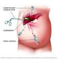
OPERATIVE TECHNIQUES:
Principles of surgery remain the same regardless of whether it is being done by open or laparoscopic technique.
 Safe access to abdomen.
 Clear definition of anatomy in the calots triangle showing either the continuity between cystic duct and gall bladder or junction between cytic duct and common bile duct.
 Avoiding diathermy close to common bile duct.
 Securing cystic artery safely and close to the gall bladder.
 Dissection from liver bed staying close to gall bladder.
 Early conversion to open procedure in case of a difficult laparoscopic surgery.
REFERRAL CRITERIA:
 Common bile duct stones if no facilities for management available.
 Suspected gall bladder cancer.
 Bile duct injury during surgery. Patient should be referred to a tertiary centre as soon as possible.
WHO DOES WHAT?
Doctor:
a) Surgeon: diagnosis & work up
Pre operative planning
Operative procedure
Post operative follow up

b) Anesthetist: PAC, anesthesia, post op ICU management
NURSE:
 Dressing of the wound
 Pre & post operative care
TECHNICIAN:
 Pre op equipment and drugs to be checked and kept ready
 Assist anesthetist in the OT
 Assist the surgeon, positioning of the patient
RESOURCES REQUIRED FOR ONE PATIENT / PROCEDURE (Patient weight 60 Kgs)
Human Resources
Investigations
Drugs/Consumables
Equipment
Surgeon – 1
Medical Officer /
Assistant Surgeon – 1
Anesthetist – 1
Pathologist – 1
Staff Nurse – 1
Technician – 1
Nursing Orderly – 1
Sweeper – 1
Haemogram
LFT
Blood Sugar
S. Electrolytes
KFT
ECG
X-Ray – Chest
USG
Histopathology
Antibiotics
Analgesic
I.V. Fluids
Sutures
Drains
Catheters
Anesthetic drugs
Dressings
OT Table & lights
Instrument trolley
Laparoscopic Surgery set
General Surgery Set
Cautery
Suction
Anesthetic Equipment
Monitors


CHOLECYSTECTOMY-3CHOLECYSTECTOMY-2

General Surgery : Blunt Abdominal Trauma

BLUNT ABDOMINAL TRAUMA
1. Name of the condition: Blunt abdominal trauma
2. When to suspect/ recognize?
a. Introduction: Blunt abdominal trauma (BAT) is an increasingly common problem encountered in the emergency department. The usual causes of BAT include vehicular accident, assault, falls, sports injuries and natural disasters.
b. Case definition: BAT is suspected in any patient involved in above situations and presents with abdominal pain, distention or shock. It should be looked for in patients of polytrauma.
3. Incidence of the condition in our country: One study has reported 2.1% incidence of BAT amongst all surgical patients admitted to a tertiary hospital during 1 year.
4. Differential diagnosis:Abdominal trauma forms adifferential diagnosis of any patient presenting with acute abdomen.
5. Prevention and counseling: Use of appropriate safety measures during various activities associated with BAT can significantly reduce its incidence.
6. Optimal diagnostic criteria, investigations, treatment and referral criteria:
a. Situation 1 At Secondary Hospital/ Non-metro situation: Optimal standard of treatment in situations where resources are limited
I. Clinical diagnosis:This is based on
a. High level of suspicion of intra-abdominal injury
b. Presence of wounds/ bruising on the abdomen
c. Abdominal guarding/ tenderness
d. Presence of free gas/ fluid in the peritoneal cavity
e. Presence of fracture of lower ribs and/ or pelvis increases the likelihood of intra-abdominal injury
f. Note should be made of altered mental state, drug or alcohol intoxication and distracting injuries which may mask the features of BAT
g. Repeated examination increases the accuracy of diagnosis
II. Investigations:
a. All hemodynamically stable patients with suspected BAT should undergo Focused Abdominal Sonography in Trauma (FAST)or Diagnostic Peritoneal Lavage (DPL)
b. Urgent laparotomy is indicated in patients with evidence of BAT who remain hemodynamically unstable despite initial resuscitation
III. Treatment (Standard operating procedure):
a. Inpatient:

i. All patients should have initial cervical stabilization and resuscitation, if required
ii. Initial fluid resuscitation should be done with 2L warmed Ringer Lactate solution infusedrapidly through 2 peripheral lines
iii. A nasogastric tube and a Foley catheter should be put
iv. Laparotomy should be done, if indicated on the basis of clinical features, FASTor DPL
v. Laparotomy should be done through a long midline incision
vi. Bleeding should be controlled by clamping/ packing till definitive control is possible
vii. Hollow viscus should be repaired
viii. In case the intra-abdominal injuries are extensive, patient is very sick and OT facilities/ surgeon’s experience is suboptimal, Damage Control Surgery may be done. Definitive surgery should be done subsequently under improved circumstances or at a higher center.
b. Outpatient: Not indicated
c. Day care: Not indicated
IV. Referral criteria: After Damage Control Surgery if the local facilities are inadequate.
b. Situation 2 At Superspecialty Facility in Metro location where higher end technology is available
1. Clinical diagnosis: Same as 6a
2. Investigations:
a. Same as 6a
b. CECT abdomen is preferred investigation in all hemodynamically stable patients with BAT
c. Angiography and angioembolization may be considered in hemodynamically stable patients with solid organ injury who are suitable for non-operative management
3. Treatment (Standard operating procedure):
a. Inpatient:
i. Same as 6a
ii. In case the intra-abdominal injuries are extensive, patient is very sick and surgeon’s experience is suboptimal, Damage Control Surgery may be done. Definitive surgery should be done subsequently under improved circumstances
iii. Angiography and angioembolization may be considered in hemodynamically stable patients with solid organ injury who are suitable for non-operative management
b. Outpatient: Not indicated
c. Day care: Not indicated
4. Referral criteria: Not indicated
5
7. Who does what and timelines:
a. Doctor: Does initial evaluation, subsequent monitoring, decision regarding investigations and therapeutic intervention.
b. Nurse: Assists in resuscitation, monitoring, investigation and treatment.
c. Technician: Assists in resuscitation, investigation and treatment.
8. Further reading/ references
a. Udeani J, Steinberg SR. Blunt abdominal trauma. URL: http://emedicine.medscape.com/article/433404-print
b. Joint theatre Trauma System Clinical Practice Guideline: Blunt abdominal trauma.

c. M. Swarnkar, P. Singh & S. Dwivedi : Pattern Of Trauma In Central India: An Epidemiological Study With Special Reference To Mode Of Injury. The Internet Journal of Epidemiology.

abdominal truama -laproscopic diagnosis injury by sharp weapon


By

Dr Sanjay Gupta
Professor, Department of Surgery
University College of Medical Sciences, Delhi

General Surgery

The Department of General and Minimally Invasive Surgery is a full-fledged healthcare facility providing complete evaluation, diagnosis, and surgical treatment for a wide variety of disorders. The department uses an integrated

approach to deliver superb care and compassion. In India uses the most advanced minimally invasive surgical innovations and technology to cure the patients.

ct-scanfluoroscopymemmographybone-densitometryx-ray cath-lab

Risk factor and Prevention of Cancer

Risk factor and Prevention of Cancer:

The old saying, “An ounce of prevention is better than a pound of cure”, holds true not only for infectious diseases but also for cancer.

Why cancer prevention is important?

Most cancers are lifestyle-related risk factors. In India, tobacco-related cancers account for 48% of the total cancer cases and cervical cancers form the bulk (36%) of cancers in females.

Primary Prevention

Primary prevention refers to a reduced or to eliminate exposure to carcinogens.

Primary prevention includes:

  • Compulsory education about tobacco related cancers, unhealthy sexual habits and cancer preventive diet.

Modifiable cancer risk factors:

  • Tobacco causes cancers at many sites. Alcohol consumption is associated with oral, oesophageal and other cancers;
  • Obesity associated with increased risk of colon, breast and other cancers.
  • Balanced mixed diet can prevent several cancers.

Chemoprevention

This includes the use of natural or synthetic substances to reduce the risk of developing cancer or its recurrence.

Eg: Selective oestrogen receptor modulators (tamoxifen), NSAIDs, (COX-2 inhibitors) for colon cancer, Retinoids (cis-retinoic acid) for primary cancers of the head and neck and Finasteride, an alpha-reductase inhibitor that can lower the risk of prostate cancer. Vaccines are used to prevent infection by oncogenic agents against hepatitis B for cancers of liver and Human papillomavirus (HPV) vaccine for prevention of Cervical Cancer.Genetic testing with BRCA1, 2 has lead to prophylactic oophorectomy and mastectomy for Breast & Ovarian Cancer, prophylactic colectomy in adenomatous polyposis gene mutation.

Diet and Cancer

Diet is one of the aetiological factors in carcinogenesis. Epidemiological studies (study done by Doll and Peto) have shown that 35% of cancers are associated with diet.

Fat present in red meat modifies DNA synthesis, alters the microflora and induces the production of oestrogens, associated with breast and colon cancers.

Micronutrients and cancer

Micronutrients – vitamins and minerals, non-nutrients such as proteins (legumes), monoterpenes (citrus fruits), polyphenoles (tea and spices) and allium (onion and garlic) have protective effects and prevent cancer. They act through metabolic, antioxidant, differentiation and immune modulation.

Dietary recommendations

  • Increased intake of vegetables and fruits (minimum of five servings per day)
  • Intake of 55% carbohydrates, <30% fats and 20-30g of fibre
  • Avoidance of salt-cured and smoked food
  • Limitation of alcohol.

Secondary prevention includes screening programmes.

Recommendations for the early detection in asymptomatic persons

Screening programmes are:

  • Opportunistic, in which the population seeks medical professionals.
  • Organized, in which medical professionals approach the public.

Nuerosurgery

spine

Neurosurgery (or neurological surgery) is the medical specialty concerned with the prevention, diagnosis, treatment, and rehabilitation of disorders which affect any portion of the nervous system including the brain, spinal cord, peripheral nerves, and extra-cranial cerebrovascular system.

Main divisions of neurosurgery

General neurosurgery involves most neurosurgical conditions including neuro-trauma and other neuro-emergencies such as intracranial hemorrhage. Most level 1 hospitals have this kind of practice.

Specialized branches have developed to cater to special and difficult conditions. These specialized branches co-exist with general neurosurgery in more sophisticated hospitals. To practice these higher specialization within neurosurgery, additional higher fellowship training of 1–2 years is expected from the neurosurgeon. Some of these divisions of neurosurgery are:

  1. Vascular and endovascular neurosurgery
  2. Stereotactic,functional and epilepsy neurosurgery
  3. Oncological neurosurgery
  4. Skull-Butt Surgery
  5. Spine neurosurgery
  6. Peripheral nerve surgery
  7. Pediatric neurosurgery

Neuropathology

The pathology confronted by neurosurgeons could be either congenital, acquired, traumatic, due to infection, or neoplastic or degenerative conditions. Conditions like congenital hydrocephalus, pediatric tumors and myelomeningocele are encountered in children. Trauma with head or spine injury and bleeds due to arteriovenous malformation are encountered in young adults. Degenerative spine disease, aneurysm bleeds and Parkinson’s disease are encountered in much older patients. The science of neuropathology is a well developed branch of pathology.

Neuroanesthesia

Neuroanesthesia is a highly developed science that is linked to neurosurgery. This branch of medicine plays a very important part in day-to-day neurosurgery.

Neurosurgery methods

1. Vascular and endovascular neurosurgery.

2. Stereotactic, functional and epilepsy neurosurgery

3. Oncological neurosurgery

4. Skull- Butt surgery

5. Spine neurosurgery

6. Peripheral Nerve surgery

7.Pediatric neurosurgery


y plays a key role not only in diagnosis but also in the operative phase of neurosurgery.

Neuroradiology methods are used in modern neurosurgery diagnosis and treatment. They include computer assisted imaging computed tomography (CT), magnetic resonance imaging (MRI), positron emission tomography (PET), magnetoencephalography (MEG), and the stereotactic radiosurgery. Some neurosurgery procedures involve the use of intra-operative MRI and functional MRI.

In conventional open surgery the neurosurgeon opens the skull and uses a large opening to access the brain. Techniques of using smaller openings and using microscopes and endoscopes were developed later. With these smaller openings in conjunction with high-clarity microscopic visualization of neural tissue, excellent results can be obtained. However, the open methods are still used in trauma or emergency situations.[10] Principles of Neurosurgery-Rengachari, Ellenbogen, [11] Neurotrauma and Critical Care of the Brain-Jallo, Loftus .’

Microsurgery is utilized in many aspects of neurological surgery. Microvascular techniques are used in EC-IC by-pass surgery and in restoration carotid endarterectomy. The clipping of an aneurysm is performed under microscopic vision. Minimally invasive spine surgery utilizes microscopes or endoscopes. Procedures such as microdiscectomy, laminectomy, and artificial disc replacement rely on microsurgery.[5]

Using Stereotaxy neurosurgeons can approach a minute target in the brain through a minimal opening. This is used in functional neurosurgery where electrodes are implanted or gene therapy is instituted with high level of accuracy as in the case of Parkinson’s disease or Alzheimer’s disease. Using the combination method of open and stereotactic surgery, intraventricular hemorrhages can potentially be evacuated successfully.[6]

Minimally invasive endoscopic surgery is commonly utilized by neurosurgeons when appropriate. Techniques such as endoscopic endonasal surgery are used in pituitary tumors, craniopharyngiomas, chordomas, and the repair of cerebrospinal fluid leaks. Ventricular endoscopy is used in the treatment of intraventricular bleeds, hydrocephalus, colloid cyst and neurocysticercosis. Endonasal endoscopy is at times carried out with neurosurgeons and ENT surgeons working together as a team.

Repair of craniofacial disorders and disturbance of cerebrospinal fluid circulation is done by neurosurgeons who also occasionally team up with maxillofacial and plastic surgeons. Cranioplasty for craniosynostosis is performed by pediatric neurosurgeons with or without plastic surgeons.

Neurosurgeons are involved in Stereotactic Radiosurgery along with Radiation Oncologists in tumor and AVM treatment. Radiosurgical methods such as Gamma knife, Cyberknife and Novalis Shaped Beam Surgery are used as well.

Endovascular Neurosurgery utilize endovascular image guided procedures for the treatment of aneurysms, AVMs, carotid stenosis, strokes, and spinal malformations, and vasospasms. Techniques such as angioplasty, stenting, clot retrieval, embolization, and diagnostic angiography are endovascular procedures.

A common procedure performed in neurosurgery is the placement of Ventriculo-Peritoneal Shunt (VP Shunt). In pediatric practice this is often implemented in cases of congenital hydrocephalus. The most common indication for this procedure in adults is Normal Pressure Hydrocephalus (NPH).

Neurosurgery of the spine covers the cervical, thoracic and lumbar spine. Some indications for spinal coed surgery include spinal cord compression resulting from trauma, arthritis of the spinal discs, or spondylosis. In cervical cord compression, patients may have difficulty with gait, balance issues, and/or numbness and tingling in the hands or feet. Spondylosis is the condition of spinal disc degeneration and arthritis that compresses the spinal canal. This can often result in bone-spurring and disc herniation. Power drills and special instruments are often used to correct any compression problems of the spinal canal. Disk herniations of spinal vertebral discs are removed by Kerrison pitiutary rongeurs. T

his procedure is known aa a discectomy. A laminectomy is the removal of the Lamina portion of the vertebrae of the spine in order to make room for the compressed nerve tissue. Radiology assisted spine surgery is a minimally-invasive procedure and includes the techniques of vertebroplasty and kyphoplasty in which certain types of spinal fractures are managed.

Surgery performed in order to produce spinal analgesia are also often performed by neurosurgeons. Some of these techniques include implantation of deep brain stimulators, spinal cord stimulators and pain pumps.

Surgery of the peripheral nervous system is also possible, and includes the very common procedures of carpal tunnel decompression and peripheral nerve transposition. Numerous other types of nerve entrapment conditions and other problems with the peripheral nervous system are treated as well.


Triplets born to Omani Nationals

Triplets born to Omani Nationals Mr. Khamis & Mrs. Aisha after successful IVF treatment from Lifeline Multispeciality, Hospital, Kerala. Mr. Khamis traveled all the way from Oman to Lifeline to share his overwhelming Joy with Dr. Pappachan and Staff. We congratulate and salute the proud parents for their efforts.

Cosmetic Surgery

Cosmetic Surgery: Definition

The goal of cosmetic surgery is to improve a person’s appearance and, thus, self-esteem by changing the way she or he looks. Cosmetic surgery can be performed on any part of the face and body.

Types

The Face

The Body

Critical Care Unit

critical care unit (CCU):

It is a specially equipped hospital area designed for the treatment of patients with sudden life-threatening conditions. CCUs contain resuscitation and monitoring equipment and are staffed by personnel specially trained and skilled in recognizing and immediately responding to cardiac and other emergencies. See also intensive care unit.

About Critical Care Nursing

Definition of Critical Care Nursing:

Critical care nursing is that specialty within nursing that deals specifically with human responses to life-threatening problems. A critical care nurse is a licensed professional nurse who is responsible for ensuring that acutely and critically ill patients and their families receive optimal care.

Definition of a Critically Ill Patient:

Critically ill patients are defined as those patients who are at high risk for actual or potential life-threatening health problems. The more critically ill the patient is, the more likely he or she is to be highly vulnerable, unstable and complex, thereby requiring intense and vigilant nursing care.

Where Critical Care Nurses Work:

According to “The Registered Nurse Population” study, 56.2% of all nurses work in a hospital setting, and critical care nurses work wherever critically ill patients are found — intensive care units, pediatric ICUs, neonatal ICUs, cardiac care units, cardiac catheter labs, telemetry units, progressive care units, emergency departments and recovery rooms.

Increasingly, critical care nurses work in home healthcare, managed care organizations, nursing schools, outpatient surgery centers and clinics.

What Critical Care Nurses Do:

Critical care nurses practice in settings where patients require complex assessment, high-intensity therapies and interventions and continuous nursing vigilance. Critical care nurses rely upon a specialized body of knowledge, skills and experience to provide care to patients and families and create environments that are healing, humane and caring.

Foremost, the critical care nurse is a patient advocate. AACN defines advocacy as respecting and supporting the basic values, rights and beliefs of the critically ill patient. In this role, critical care nurses:

Respect and support the right of the patient or the patient’s designated surrogate to autonomous informed decision making.
Intervene when the best interest of the patient is in question.
Help the patient obtain necessary care.
Respect the values, beliefs and rights of the patient.
Provide education and support to help the patient or the patient’s designated surrogate make decisions.
Represent the patient in accordance with the patient’s choices.
Support the decisions of the patient or designated surrogate, or transfer care to an equally qualified critical care nurse.
Intercede for patients who cannot speak for themselves in situations that require immediate action.
Monitor and safeguard the quality of care the patient receives.
Act as a liaison between the patient, the patient’s family and other healthcare professionals.

The Roles of Critical Care Nurses

Critical care nurses work in a wide variety of settings, filling many roles including bedside clinicians, nurse educators, nurse researchers, nurse managers, clinical nurse specialists and nurse practitioners. With the onset of managed care and the resulting migration of patients to alternative settings, critical care nurses are caring for patients who are more ill than ever before.

Managed care has also fueled a growing demand for advanced practice nurses in the acute care setting. Advanced practice nurses are those who have received advanced education at the master’s or doctoral level. In the critical care setting, they are most frequently clinical nurse specialists (CNS) or acute care nurse practitioners (ACNP).

A CNS is an expert clinician in a particular specialty — critical care in this case. The CNS is responsible for the identification, intervention and management of clinical problems to improve care for patients and families. They provide direct patient care, including assessing, diagnosing, planning and prescribing pharmacological and nonpharmacological treatment of health problems.

ACNPs in the critical care setting focus on making clinical decisions related to complex patient care. Their activities include risk appraisal, interpretation of diagnostic tests and providing treatment, which may include prescribing medication

Cataracts

What are the different types of cataracts?

Age-related cataract: Most cataracts are related to aging.

Congenital cataract: Some babies are born with cataracts or develop them in childhood, often in both eyes. These cataracts may not affect vision. If they do, they may need to be removed.

Secondary cataract: Cataracts are more likely to develop in people who have certain other health problems, such as diabetes. Also, cataracts are sometimes linked to steroid use.

Traumatic cataract: Cataracts can develop soon after an eye injury, or years later.

What are the different treatments available?

Choose No needle – No stitch – No patch Cataract surgeries by Phacoemulsification with Foldable IOL implantation ,
State of the Art Technology includes;

* Latest Whitestar Signature Phaco system
* Ellips SX Headpiece * HIR – 900 Microscope
* Motorised Slit lamp with EBIOS system
* Micro incision Phaco Surgery with Monofocal / Multifocal / Toric IOL implants

WHITESTAR COLD PHACO Technology of Signature system is clinically proven to produce clearer corneas on post-op day 1. This is one of its types as it can perform Phacoemulsification and Posterior Vitrectomy with the same system.

Pages:12