Global Treatment Services Pvt. Ltd.

Global Treatment Services

Aortic valve stenosis: Causes, Symptoms & Treatments

What is aortic valve stenosis?

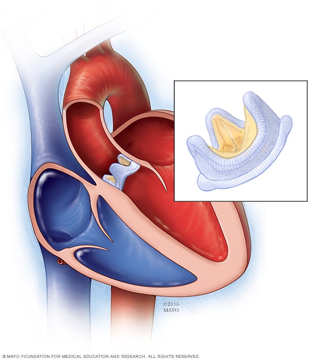
Aortic valve stenosis occurs when the aortic valve narrows and doesn’t open as it should. This limits the amount of blood pumped from your heart into, and then out of, the aorta. This is the main artery of the body. The aortic valve is a key valve in the body’s blood circulation system.

Symptoms:

Aortic valve stenosis generally produces symptoms only once it progresses. You may not have symptoms in the beginning.

You may experience symptoms of severe aortic valve stenosis once the condition has progressed to the symptomatic stage. Symptoms of severe aortic valve stenosis include:

  • chest pain as the heart strains to pump enough blood through the compromised valve
  • feeling tired after exertion, as when you exercise or move
  • feeling short of breath, especially after exertion
  • heart palpitations, or abnormal heartbeats
  • a heart murmur, which is an abnormal swooshing sound produced by the heart as it beats
What are the symptoms of aortic valve stenosis in children and infants?

Infants and children may have different symptoms from adults, or they may not appear to display any symptoms at all. If they do display symptoms, these might include:

  • inability to put on weight
  • not eating well
  • becoming easily fatigued

In severe cases, an infant may have major breathing difficulties that develop within weeks of birth. Mild cases have the potential to worsen as the child gets older.

Causes:

Genetics and certain health conditions may prevent the aortic valve from completing its proper functions. The aortic valve opens and closes every time your heart beats. This continuous action occurs every second of your life.

Genetics

Aortic valve stenosis may be present at birth. When it is, the flaps, or “leaflets,” of the aortic valve are irregularly formed. These leaflets make up the opening of the aortic valve. When functioning properly, they fit together tightly when closed. Children born with aortic valve stenosis usually have one of the following irregularities in their aortic valve leaflets:

  • All three leaflets aren’t present.
  • The leaflets don’t separate properly.
  • The leaflets are too thick to completely open or close.

Rheumatic fever

Rheumatic fever is one of the most common causes of heart valve problems. Rheumatic fever may affect many parts of the body, including the:

  • brain
  • joints
  • heart
  • skin

Rheumatic fever can occur in adults and children who have, or have had, strep throat. Strep throat is a contagious condition caused by Streptococcus bacteria.

Calcification of the valves

Aortic valve stenosis most often occurs in older adults. Age-related aortic valve stenosis is the most common form of the condition, according to the American Heart Association. It’s caused by calcification or scarring of the aortic valve. It usually begins sometime after the age of 60, but people may not begin to experience symptoms until age 70 or 80.

Calcium is an important mineral needed for strong bones. However, the mineral may lead to problems in your heart if it deposits in the aortic valve. Calcium deposits usually affect the leaflets of the aortic valve. They can also prevent the aortic valve from properly opening and closing. These deposits often increase in the aortic valve as we age due to scarring and calcium buildup.

Improperly functioning aortic valve leaflets may also allow blood to leak back into the left ventricle after it enters the aorta. This is called valvular insufficiency, or regurgitation.

The risk factors:

Gender

Men have a higher risk of aortic valve stenosis than women. The condition occurs most often in men between the ages of 30 and 60.

Genetics

Children born with either malformed valve leaflets or fewer than three leaflets will have problems with normal blood flow through the aorta.

Disease

Rheumatic fever can produce significant problems with the valve leaflets. Scar tissue from the disease can make the leaflets hard or even fused. Rheumatic fever damages:

  • heart tissue
  • valves
  • coronary arteries
How is aortic valve stenosis treated?

There are no specific medications to fix aortic valve stenosis. This is because the condition is irreversible once it occurs. Instead, your doctor can prescribe medication to treat the problems caused by the condition, or for the health issues that produced the condition in the first place. Surgery may be able to repair or replace the valve.

Medications

Although medication can’t cure aortic valve stenosis, your doctor may prescribe drugs to manage symptoms or reduce the burden on your heart. Some medications include:

  • Antibiotics. Rheumatic fever requires antibiotics to keep any infection from advancing and causing heart damage.
  • Blood pressure medications. Beta-blockers or calcium channel blockers can help lower your blood pressure.
  • Blood thinners. Blood thinners such as Coumadin may be necessary.
  • Anti-arrhythmics. Medications to manage your heart’s rhythm, called anti-arrhythmics, are sometimes prescribed.

Surgery options include:

Aortic valve repair

Surgeons rarely repair an aortic valve to treat aortic valve stenosis, and generally aortic valve stenosis requires aortic valve replacement. To repair an aortic valve, surgeons may separate valve flaps (cusps) that have fused.

Balloon valvuloplasty

Doctors may conduct a procedure using a long, thin tube (catheter) to repair a valve with a narrowed opening (aortic valve stenosis). In this procedure, called balloon valvuloplasty, a doctor inserts a catheter with a balloon on the tip into an artery in your arm or groin and guides it to the aortic valve. The doctor performing the procedure then inflates the balloon, which expands the opening of the valve. The balloon is then deflated, and the catheter and balloon are removed.

The procedure can treat aortic valve stenosis in infants and children. However, the valve tends to narrow again in adults who’ve had the procedure, so it’s usually only performed in adults who are too ill for surgery or who are waiting for a valve replacement, as they typically need additional procedures to treat the narrowed valve over time.

Aortic valve replacement

Aortic valve replacement is often needed to treat aortic valve stenosis. In aortic valve replacement, your surgeon removes the damaged valve and replaces it with a mechanical valve or a valve made from cow, pig or human heart tissue (biological tissue valve).

Biological tissue valves degenerate over time and may eventually need to be replaced. People with mechanical valves will need to take blood-thinning medications for life to prevent blood clots. Your doctor will discuss with you the benefits and risks of each type of valve and discuss which valve may be appropriate for you.
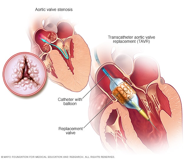
Doctors may perform a less invasive procedure called transcatheter aortic valve replacement (TAVR) to replace a narrowed aortic valve. TAVR may be an option for people who are considered to be at intermediate or high risk of complications from surgical aortic valve replacement.

In TAVR, doctors insert a catheter in your leg or chest and guide it to your heart. A replacement valve is then inserted through the catheter and guided to your heart. A balloon may expand the valve, or some valves can self-expand. When the valve is implanted, doctors remove the catheter from your blood vessel.

Doctors may also conduct a catheter procedure to insert a replacement valve into a failing biological tissue valve that is no longer working properly. Other catheter procedures to repair or replace aortic valves continue to be researched.

Lifestyle and home remedies

You’ll have regular follow-up appointments with your doctor to monitor your condition. You’ll need to continue taking all your medications as prescribed.

Your doctor may suggest you incorporate several heart-healthy lifestyle changes into your life, including:

  • Eating a heart-healthy diet. Eat a variety of fruits and vegetables, low-fat or fat-free dairy products, poultry, fish, and whole grains. Avoid saturated and trans fat, and excess salt and sugar.
  • Maintaining a healthy weight. Aim to keep a healthy weight. If you’re overweight or obese, your doctor may recommend that you lose weight.
  • Getting regular physical activity. Aim to include about 30 minutes of physical activity, such as brisk walks, into your daily fitness routine.
  • Managing stress. Find ways to help manage your stress, such as through relaxation activities, meditation, physical activity, and spending time with family and friends.
  • Avoiding tobacco. If you smoke, quit. Ask your doctor about resources to help you quit smoking. Joining a support group may be helpful.

For women with aortic valve stenosis, it’s important to talk with your doctor before you become pregnant. Your doctor can discuss with you which medications you can safely take, and whether you may need a procedure to treat your valve condition prior to pregnancy.

You’ll likely require close monitoring by your doctor during pregnancy. Doctors may recommend that women with severe valve stenosis avoid pregnancy to avoid the risk of complications.

Long-term outlook

Your health may improve dramatically once you receive treatment. Surgical treatments for aortic valve stenosis have high rates of success. Your outlook depends on a combination of factors:

  • how long you’ve lived with the condition
  • the extent of damage to your heart
  • any complications that may arise from your condition
Managing the symptoms of aortic valve stenosis

Sometimes, aortic valve stenosis isn’t a congenital defect, meaning you weren’t born with the condition. If this is the case, there are healthy lifestyle steps you can take to ease the burden on your heart:

  • Eat a healthy diet low in saturated fat.
  • Exercise regularly.
  • Maintain a healthy weight.
  • Abstain from smoking.
  • Report any abnormal health issues to your doctor.
  • Visit your doctor for any severe sore throat to prevent rheumatic fever.
  • Practice good dental hygiene, as dental infections can travel through the bloodstream and damage the heart valves and muscles.

Ichythyosis: Causes, symptoms & Treatments

Many things can leave your skin dry and flaky, from cold air to the chlorine in swimming pools. In a small number of people, a group of diseases called ichthyosis causes skin dryness and scaling.It can’t be cured, but treatments can relieve the scaling and make you feel more comfortable in your own skin.Ichthyosis is not one disease, but a family of about 20 skin conditions that lead to dry skin. People with this condition lose the protective barrier that keeps moisture in their skin. They also make new skin cells too quickly or shed old cells too slowly. This leads to a buildup of thick, scaly skin.The condition gets its name from the Greek word for fish, because the skin looks like fish scales.

Types of Ichthyosis

Some types only cause dry and scaly skin. Others cause problems inside the body, too.

Most forms of the disease are very rare. The two least rare types are:

  • Ichthyosis vulgaris. It affects about 1 out of every 250 people.
  • X-linked recessive ichthyosis. It affects about 1 out of 6,000 people, but only males.

What Causes Ichthyosis?

It can be inherited or acquired.

Inherited ichthyosis: This is caused by mutations, or changes, to genes.

Genes are the codes that tell your body to make proteins, which determine how your body looks and functions.

Ichthyosis gene mutations affect that proteins that protect your skin and keep it moist. They also affect how quickly skin cells multiply or are shed.

Parents pass these genes to their children. Sometimes children whose parents don’t have ichthyosis get the disease before birth. The gene change happens inside the egg or sperm — or when the egg and sperm meet to make a baby. These are called “spontaneous mutations.”

Some people have the gene mutation but don’t show symptoms. They are called carriers.

Acquired ichthyosis: This starts in adulthood. Doctors don’t know what causes it.

People with the acquired form often have other conditions, such as:

  • Underactive thyroid gland
  • Kidney disease
  • Sarcoidosis, a rare disease that causes patches of inflammation inside the body
  • Cancer such as Hodgkin lymphoma
  • HIV infection

Some medicines might also trigger the condition:

  • Cancer drugs such as vemurafenib (Zelboraf), hydroxyurea (Hydrea, Droxia), and protease inhibitors (a class of drugs used to treat HIVinfections)
  • Nicotinic acid, which is used to treat high cholesterol

Symptoms

In the inherited forms, symptoms appear at birth or in the first few months of life.

Dry, scaly skin is the main symptom. The scales form on only some parts of the body, such as the:

  • Trunk
  • Stomach
  • Buttocks
  • Legs
  • Face and scalp

The scales can be white, gray, or dark brown. They might have thick or thin cracks running through them.

Dryness and scaling get worse in cold, dry weather. They usually improve in warmer weather.

Other signs of ichthyosis are:

  • Skin redness
  • Blisters
  • Peeling
  • Itchiness
  • Pain
  • Lines on the palms of the hands and soles of the feet
  • Tight skin that makes it hard to move

Many people with ichthyosis also have eczema, a red, itchy rash.

Complications

Skin is your body’s barrier. It holds moisture inside and keeps out bacteria and other invaders that might make you sick. When ichthyosis makes pieces of your skin scale off, you lose some of this protective layer.

Scaling can lead to complications such as:

  • Infections
  • Dehydration
  • Blocked sweat glands, which can lead to overheating
  • Slow hair growth from scales on the scalp
  • Burning more calories, because the skin has to work harder to turn over cells

Ichthyosis can cause emotional issues, too. The scales can affect the way you look. People with this condition sometimes feel depressed and have low self-esteem.

Men with ichthyosis are also at increased risk for:

  • Testicular cancer
  • Undescended or hidden testicles
  • Low sperm count

Other complications include:

  • Vision loss from damage to the cornea (the clear layer in the front of the eye)
  • Brain and nervous system problems

Diagnosis

Sometimes it’s so mild that it looks like regular dry skin. If the dryness and scaling bother you, see a dermatologist, a doctor who treats skin problems, for treatment.

She might take a sample of skin to check under a microscope. This is called a biopsy.

She might also test your saliva for gene changes that cause certain types of ichthyosis.

Treatments

It can’t be cured, but treatments can help you manage dry and scaly skin.

Rub cream, lotion, or ointment onto your skin every day to add moisture. Look for rich creams that have any of these ingredients: lanolin, alpha hydroxy acids, urea, or propylene glycol. Products with ceramides or cholesterol also keep skin moist.

Apply the lotion right after you step out of the shower or bath, when your skin is still damp. This will help hold in moisture. Other things you can try:

  • Take baths in salt water.
  • Rub your skin with a pumice stone.
  • Remove dead skin with a product that contains salicylic acid, glycolic acid, or lactic acid.

If the dryness and scaling are severe, your doctor might prescribe an oral retinoid medicine such as acitretin (Soriatane) or isotretinoin (Absorica, Claravis, Sotret, and others). Retinoids can cause side effects such as weakened bones, dry mouth, and upset stomach.

Ichthyosis isn’t life-threatening, but it can be life-changing.

Your dermatologist can recommend treatments to help your skin look and feel better. If you feel depressed or have low self-esteem, talk to a therapist or other mental health experts.

Childhood Ependymoma: Causes, Symptoms & Treatments

Childhood ependymoma is a type of brain tumor. A tumor begins when healthy cells change and grow out of control, forming a mass. A tumor can be cancerous or benign. A cancerous tumor is malignant, meaning it can grow and spread to other parts of the body. A benign tumor means the tumor can grow but will not spread. Ependymoma is a rare, cancerous brain tumor.

Ependymoma starts from radial glial cells, which are a type of cell in the brain. Even though ependymoma can occur in any part of the brain or spine, it most commonly occurs in the cerebellum. The cerebellum is the part of the brain that coordinates the body’s actions.

Symptoms & Causes:

Children with ependymoma may experience the following symptoms or signs. Sometimes, children with ependymoma do not have any of these changes. Or, the cause of a symptom may be another medical condition that is not a tumor.

  • Headaches
  • Nausea and vomiting
  • Changes in vision, such as blurriness
  • Difficulty with walking or balance
  • Jerky eye movements
  • Neck pain
  • Reaching childhood developmental milestones more slowly than expected
  • Seizures/convulsions, which are sudden involuntary movements of a person’s muscles

If you are concerned about any changes your child experiences, please talk with your child’s doctor. Your doctor will ask how long and how often your child has been experiencing the symptom(s), in addition to other questions. This is to help find out the cause of the problem, called a diagnosis.

If a brain tumor is diagnosed, relieving symptoms remains an important part of care and treatment. This may also be called symptom management, palliative care, or supportive care. Be sure to talk with your child’s health care team about symptoms your child experiences, including any new symptoms or a change in symptoms.

Treatments:

option: 1-surgery

or

2- Chemotherapy

or

3- Radiation


Foaming at mouth: Causes, Symptoms & Treatments

Why am I foaming at the mouth?

Foaming at the mouth is a physical symptom. It occurs when an excess of saliva mixes with air or gasses to create a foam.

Frothy saliva is a rare symptom; when you see it, you should be concerned and immediately contact a doctor or 911 for medical assistance.

 Causes:
 
1.overdose

People use recreational drugs because they affect the brain chemistry, causing feelings like euphoria and cravings for more of the drug. Two of the most popular categories of drugs are opioids (painkillers) and stimulants, or “uppers.”

Common opioids are:

  • heroin
  • OxyContin
  • Vicodin

Common stimulants are:

  • Ritalin
  • Adderall
  • methamphetamine

If you take too much of one of these drugs, you could overdose, meaning your body can’t detox the drug before deadly symptoms take effect.

Common signs of opiate or stimulant overdose are:

  • foaming at the mouth or a foam cone
  • loss of consciousness
  • seizures
  • difficulty or stopped breathing

Overdose causes foaming at the mouth because organs like the heart and lungs can’t function properly. Slowed heart or lung movements causes fluids to gather in the lungs, which can mix with carbon dioxide and come out of the mouth like a foam.

Drug overdoses can be fatal. The drug narcan is an antidote to opiate overdose. There is no treatment for stimulant overdose.

2. Seizure

When a person starts convulsing uncontrollably, they are having a seizure. A brain condition called epilepsy can cause seizures. There are also nonepileptic seizures, typically caused by trauma or psychological conditions.

Beyond convulsions, seizures can also cause:

  • loss of consciousness
  • falling
  • frothing at the mouth or drooling
  • biting your tongue
  • incontinence

Foamy saliva can occur during a seizure because the mouth is forced closed, which stimulates the salivary glands and makes you produce extra spit. When the mouth opens again, drool or frothy saliva can come out.

Foaming at the mouth can also occur following a provoked seizure. For example, one case study examined a police officer whose partner accidentally shot him in the head with a Taser while chasing a suspect. The second officer witnessed his colleague lose consciousness, fall to the ground, and begin foaming at the mouth.

Treatments for seizures include antiepileptic medication and brain surgery.

3. Rabies

Rabies is a viral disease that affects the central nervous system. Only warm-blooded animalscan get rabies. Common carriers of the rabies virus are:

  • raccoons
  • foxes
  • skunks
  • bats
  • coyotes
  • wolves

Animals less commonly affected by rabies are:

  • cows
  • dogs
  • cats
  • ferrets
  • humans

The rabies virus is present in the saliva. If an affected animal bites you or licks an open wound or scratch you have, you may get it.

Rabies can only be diagnosed from a brain tissue sample, so it’s important to watch for symptoms of the virus. Foaming at the mouth is the most characteristic symptom of rabies. This happens because the virus affects the nervous system and the animal or person can’t swallow their saliva.

Other symptoms are:

  • fever
  • lost appetite
  • agitation
  • violence and biting
  • convulsions
  • paralysis

There is no way to treat rabies. If you think you have been exposed to rabies, clean your wound with soap and water and put a muzzle on your pet. Then immediately visit an emergency room where you will be given a rabies vaccine.

Treatment for frothing at the mouth

The three causes of foaming at the mouth are very specific and require unique treatment methods:

  • Some drug overdoses can be treated with an injection of narcan.
  • Epileptic seizures can be treated with antiepileptic medication.
  • Nonepileptic seizures can be treated with medication or psychotherapy.
  • Rabies can be prevented with a rabies vaccine and a series of other injections.

Parkinson’s Disease: Causes, Symptoms & Treatments

Parkinson’s disease is a progressive nervous system disorder that affects movement. Symptoms start gradually, sometimes starting with a barely noticeable tremor in just one hand. Tremors are common, but the disorder also commonly causes stiffness or slowing of movement.

In the early stages of Parkinson’s disease, your face may show little or no expression. Your arms may not swing when you walk. Your speech may become soft or slurred. Parkinson’s disease symptoms worsen as your condition progresses over time.

Although Parkinson’s disease can’t be cured, medications might significantly improve your symptoms. Occasionally, your doctor may suggest surgery to regulate certain regions of your brain and improve your symptoms.


Symptoms

Parkinson’s disease signs and symptoms can be different for everyone. Early signs may be mild and go unnoticed. Symptoms often begin on one side of your body and usually remain worse on that side, even after symptoms begin to affect both sides.

Parkinson’s signs and symptoms may include:

  • Tremor. A tremor, or shaking, usually begins in a limb, often your hand or fingers. You may a rub your thumb and forefinger back-and-forth, known as a pill-rolling tremor. Your hand may tremor when it’s at rest.
  • Slowed movement (bradykinesia). Over time, Parkinson’s disease may slow your movement, making simple tasks difficult and time-consuming. Your steps may become shorter when you walk. It may be difficult to get out of a chair. You may drag your feet as you try to walk.
  • Rigid muscles. Muscle stiffness may occur in any part of your body. The stiff muscles can be painful and limit your range of motion.
  • Impaired posture and balance. Your posture may become stooped, or you may have balance problems as a result of Parkinson’s disease.
  • Loss of automatic movements. You may have a decreased ability to perform unconscious movements, including blinking, smiling or swinging your arms when you walk.
  • Speech changes. You may speak softly, quickly, slur or hesitate before talking. Your speech may be more of a monotone rather than with the usual inflections.
  • Writing changes. It may become hard to write, and your writing may appear small.

Causes

In Parkinson’s disease, certain nerve cells (neurons) in the brain gradually break down or die. Many of the symptoms are due to a loss of neurons that produce a chemical messenger in your brain called dopamine. When dopamine levels decrease, it causes abnormal brain activity, leading to symptoms of Parkinson’s disease.

The cause of Parkinson’s disease is unknown, but several factors appear to play a role, including:

  • Your genes. Researchers have identified specific genetic mutations that can cause Parkinson’s disease. But these are uncommon except in rare cases with many family members affected by Parkinson’s disease.However, certain gene variations appear to increase the risk of Parkinson’s disease but with a relatively small risk of Parkinson’s disease for each of these genetic markers.
  • Environmental triggers. Exposure to certain toxins or environmental factors may increase the risk of later Parkinson’s disease, but the risk is relatively small.

Researchers have also noted that many changes occur in the brains of people with Parkinson’s disease, although it’s not clear why these changes occur. These changes include:

  • The presence of Lewy bodies. Clumps of specific substances within brain cells are microscopic markers of Parkinson’s disease. These are called Lewy bodies, and researchers believe these Lewy bodies hold an important clue to the cause of Parkinson’s disease.
  • Alpha-synuclein is found within Lewy bodies. Although many substances are found within Lewy bodies, scientists believe an important one is the natural and widespread protein called alpha-synuclein (a-synuclein). It’s found in all Lewy bodies in a clumped form that cells can’t break down. This is currently an important focus among Parkinson’s disease researchers.

Risk factors

Risk factors for Parkinson’s disease include:

  • Age. Young adults rarely experience Parkinson’s disease. It ordinarily begins in middle or late life, and the risk increases with age. People usually develop the disease around age 60 or older.
  • Heredity. Having a close relative with Parkinson’s disease increases the chances that you’ll develop the disease. However, your risks are still small unless you have many relatives in your family with Parkinson’s disease.
  • Sex. Men are more likely to develop Parkinson’s disease than are women.
  • Exposure to toxins. Ongoing exposure to herbicides and pesticides may slightly increase your risk of Parkinson’s disease.

Complications

Parkinson’s disease is often accompanied by these additional problems, which may be treatable:

  • Thinking difficulties. You may experience cognitive problems (dementia) and thinking difficulties. These usually occur in the later stages of Parkinson’s disease. Such cognitive problems aren’t very responsive to medications.
  • Depression and emotional changes. You may experience depression, sometimes in the very early stages. Receiving treatment for depression can make it easier to handle the other challenges of Parkinson’s disease.You may also experience other emotional changes, such as fear, anxiety or loss of motivation. Doctors may give you medications to treat these symptoms.
  • Swallowing problems. You may develop difficulties with swallowing as your condition progresses. Saliva may accumulate in your mouth due to slowed swallowing, leading to drooling.
  • Chewing and eating problems. Late-stage Parkinson’s disease affects the muscles in your mouth, making chewing difficult. This can lead to choking and poor nutrition.
  • Sleep problems and sleep disorders. People with Parkinson’s disease often have sleep problems, including waking up frequently throughout the night, waking up early or falling asleep during the day.People may also experience rapid eye movement sleep behavior disorder, which involves acting out your dreams. Medications may help your sleep problems.
  • Bladder problems. Parkinson’s disease may cause bladder problems, including being unable to control urine or having difficulty urinating.
  • Constipation. Many people with Parkinson’s disease develop constipation, mainly due to a slower digestive tract.

You may also experience:

  • Blood pressure changes. You may feel dizzy or lightheaded when you stand due to a sudden drop in blood pressure (orthostatic hypotension).
  • Smell dysfunction. You may experience problems with your sense of smell. You may have difficulty identifying certain odors or the difference between odors.
  • Fatigue. Many people with Parkinson’s disease lose energy and experience fatigue, especially later in the day. The cause isn’t always known.
  • Pain. Some people with Parkinson’s disease experience pain, either in specific areas of their bodies or throughout their bodies.
  • Sexual dysfunction. Some people with Parkinson’s disease notice a decrease in sexual desire or performance.

Prevention

Because the cause of Parkinson’s is unknown, proven ways to prevent the disease also remain a mystery.

Some research has shown that regular aerobic exercise might reduce the risk of Parkinson’s disease.

Some other research has shown that people who drink caffeine — which is found in coffee, tea and cola — get Parkinson’s disease less often than those who don’t drink it. However, it is still not known whether caffeine actually protects against getting Parkinson’s, or is related in some other way. Currently there is not enough evidence to suggest drinking caffeinated beverages to protect against Parkinson’s. Green tea is also related to a reduced risk of developing Parkinson’s disease.

Thymectomy for myasthenia gravis: Procedure details

What are the results of thymectomy?

The goal of a thymectomy is to remove the source of abnormal antibody production causing the disease thus leading to resolution of symptoms. The benefits of thymectomy are not realized immediately after surgery, thus patients will continue with there medical regimen after the procedure with the goal of weaning those medications over time. Individual response to thymectomy varies depending on the patient’s age, response to prior medical therapy, the severity of the disease and how long the patient has had myasthenia gravis. In general, 70 percent of patients have complete remission or significant reduction in medication needs within a year of the procedure. The other 30 percent of patients who have thymectomy experience no change in their symptoms.

How does a doctor determine which patients with myasthenia gravis should undergo thymectomy?

Thymectomy is recommended for all patients with thymomas and for patients under 60 who have mild to moderate muscle weakness due to myasthenia gravis. Thymectomy generally is not used for treating patients with myasthenia gravis that affects only their eyes.Thymectomy appears to be most effective when it is performed six to 12 months after the onset of symptoms. It is important to talk to your doctor early in your diagnosis about thymectomy as an option for treatment.

How is thymectomy performed?

Thymectomy can be performed by several different surgical techniques:

Transsternal thymectomy: In this procedure, the incision is made in the skin over the breastbone (sternum), and the breastbone is divided (sternotomy) to expose the thymus. This approach is commonly used for heart surgery. The surgeon removes the thymus through this incision as well as any residual fat in the center of the chest which may harbor extra thymic cells. This approach is commonly used when the patient has a thymoma.

Transcervical thymectomy: In this procedure the incision is made across the lower part of the neck, just above the breastbone(sternum). The surgeon removes the thymus through this incision without dividing the sternum. This is mostly used in patients without thymoma with certain body-types.

Robotic thymectomy and Video-assisted thorascopic thymectomy (VATS): These Minimally invasive techniques use several tiny incisions in the chest. A camera is inserted through one of the incisions and the surgery is performed with video guidance. The surgeon removes the thymus by using special surgical tools inserted into the other incisions. In a robotic-assisted procedure, the surgeon uses robotic arms to perform the surgery. The goal is to provide the same result as the more invasive transsternal approach with less post-operative discomfort and a quicker recovery.

What type of thymectomy is the best?

The transsternal thymectomy is the most commonly performed procedure, however there are no proven differences in outcomes with less invasive approaches. Your neurologist and surgeon will guide you in making a decision about the type of thymectomy you should have. Your surgeon will make a recommendation based on whether a thymoma is present and other factors related to your history and anatomy.There currently is no scientific evidence that proves one type of thymectomy is better than the other in terms of outcomes. To make the best decision for yourself, you should be informed about the different types of thymectomy and consult with your neurologist and surgeon. You also may want to seek a second opinion.

Epiretinal Membrane: Causes, Symptoms & Treatments

What is an Epiretinal Membrane?

The macula is the special area at the center of the retina, which is responsible for clear, detailed vision. The macula normally lies flat against the back of the eye, like film lining the back of a camera. An epiretinal membrane is a condition where a thin sheet of scar tissue grows on the surface of the retina. This scar tissue can contract causing the macula to become wrinkled and may result in swelling of the retina and vision changes. This condition is also known as “macular pucker”, “cellophane maculopathy”, or “premacular fibrosis”.

Parts of the inner eye


What Causes an Epiretinal Membrane and who is at Risk?
Epiretinal membranes typically occur in those over age 50. They are often related to the degeneration of the vitreous, a normal aging process inside of the eye. As the vitreous to pulls away from the retina a scar-like tissue may develop. If this scar-like tissue contracts, it can causes the retina to wrinkle or pucker. Many other eye conditions can be associated with the formation of an epiretinal membrane including trauma, diabetic retinopathy, retinal tears, prior retinal detachment, and ocular inflammation (uveitis). The condition can affect both eyes in approximately 25% of cases.

Pic. showing epiretinal membrane overlying on retina

What are the Symptoms of Epiretinal Membrane?
The condition is mild in most cases with very few symptoms. In more severe cases the vision becomes blurred and distorted, just as one would expect a picture to appear distorted from a camera with wrinkled film. Straight lines, like doorways or telephone poles, often appear wavy. Vision loss can vary from barely noticeable to severe. For most people, vision remains stable and does not get progressively worse.

How is an Epiretinal Membrane Diagnosed?
Your retina specialist can detect macular pucker by examining your retina using a dilated eye exam. A retinal scan called and optical coherence tomography (OCT) is used to document how extensive the scar tissue is and can help provide information about how much retinal damage exists (figure 2). OCT can also be used to follow the growth and contraction of the epiretinal membrane and its response to treatment. A photographic test called a fluorescein angiogram may be done in order to tell the extent of damage to the macula. This technique is also useful for evaluating possible underlying causes of an epiretinal membrane.

How is an Epiretinal Membrane Treated?
Treatment is not necessary if symptoms are mild. When an epiretinal membrane causes vision loss or the symptoms start to interfere with a persons normal daily activities then surgery is recommended. Vitrectomy surgery is the only treatment for effectively removing the membrane and improving the symptoms. During this procedure micro-forceps are used to gently remove the membrane that is wringing the macula. Following removal of the membrane, the retinal architecture usually returns to a more normal state and the symptoms slowly improve (figure 3). Approximately 80-90% of patients have improvement in blurred vision and distortion, though the vision usually does not return all the way to normal.

What is the long-term impact of an Epiretinal Membrane on my vision?
Approximately 90% of epiretinal membranes do not grow or cause progressive blurring or distortion of vision. Mild epiretinal membrane is causing only minimal symptoms typically do not progressively get worse. In the 10% of cases where the epiretinal membranes causes a progressive decrease in vision, the vision loss can be profound and permanent without treatment. Once the epiretinal membrane has been surgically removed the vision is typically stable. It is unusual for an epiretinal membrane to reoccur once it has been removed.

Macular Hole: Causes, Symptoms & Treatments

What is a macular hole?

A macular hole is a small hole in the central part of the retina which can lead to distortion and blurring of central vision. A macular hole normally only occurs in one eye, although they are present in both eyes in 10-15% of cases. They can be detected by a scan of the back of the eye.Macular holes are not related to macular degeneration.

What are the symptoms?

There may be no symptoms in the early stages of a macular hole and clarity of vision can still be good. As a hole progresses, patients may notice distortion and blurring of vision.

Causes

As we age, the vitreous ‘gel’ inside the eye naturally shrinks and pulls away from the retina. Occasionally, the vitreous gel can pull on the retina and create a macular hole. In some cases, the fluid that fills the gap left by the vitreous gel may seep through the hole onto the macula, causing blurring and distortion.

Other conditions that increase risk

The presence of another eye condition may increase your chances of developing a macular hole. These include:

  • Severe short sightedness
  • Epiretinal membranes that progress to the ‘macular pucker’ stage
  • Retinal detachment
  • Diabetic retinopathy
  • Eye injury or trauma


What is the treatment?

Every patient is unique, so we offer a detailed consultation with one of our Consultant Ophthalmic Surgeons to determine suitability, answer all your questions and explain the treatment options.

Vitrectomy: surgical removal of the vitreous gel from the eye, which is then replaced by a bubble of inert gas allowing the hole to close.

 
before surgery
After surgeryOther treatments include injections of ocriplasmin (Jetrea) into the eye to separate the vitreous gel from the retina.

Hirschsprung’s diseases in newborn: Causes, Symptoms & Treatments

It’s a cycle that’s vital to life — we eat food, digest it, and excrete what we can’t use.

Excretion, though often a subject of disgust or humor, is very important. It usually begins on the first day of a baby’s life, when the newborn passes his first stool, called the meconium.

But some babies have trouble. If a baby can’t have a bowel movement, he may have Hirschsprung’s disease, a condition in which there are missing nerve cells in the large intestine.

Hirschsprung’s disease is congenital — that is, it develops during pregnancyand is present at birth. Some congenital conditions happen because of the mother’s diet, or because of an illness she had during her pregnancy. Others are because of genes the parents pass on to the newborn.

Researchers don’t know why some people have Hirschsprung’s disease, though they believe the disease is related to flaws in DNA instructions. Though the disease can be deadly, modern medicine is able to fix the problem with surgery, and children who have been treated can live normal, healthy lives.

What Causes Hirschsprung’s Disease?

It begins to take hold in a baby in the early months of pregnancy.

Usually, as the baby develops, nerve cells are created throughout the entire digestive system, from the esophagus — which leads from the mouth to the stomach — all the way to the rectum. Normally, a person will have up to 500 million of these types of nerve cells. Among other roles, they move food through your digestive system from one end to the other.

In a baby with Hirschsprung’s, the nerve cells stop growing at the end of the large intestine, just before the rectum and anus. In some children, the cells are missing at other places in the digestive system as well.

That means the body can’t sense when waste material reaches a certain point. So the waste material gets stuck and forms a block in the system.

Hirschsprung’s disease affects about one in every 5,000 newborns. Children with other congenital conditions, such as Down syndrome and heart defects, are more likely to have the disease. Parents who carry the code for Hirschsprung’s disease in their genes, especially mothers, may pass it on to their children. Boys get it more than girls.

Symptoms

For the vast majority of people with Hirschsprung’s disease, symptoms usually begin within the first 6 weeks of life. In many cases, there are signs within the first 48 hours.

You may see swelling in your child’s abdomen. Other symptoms you might see include:

No bowel movements: You should be concerned if the newborn does not produce a stool or the meconium in the first couple of days. Older children with Hirschsprung’s may have chronic (ongoing) constipation.

Bloody diarrhea: Children with Hirschsprung’s may also get enterocolitis, a life-threatening infection of the colon, and have bad cases of diarrhea and gas.

Vomiting: The vomit may be green or brown.

In older children, symptoms may include growth issues, fatigue, and severe constipation

Treatments

Hirschsprung’s disease is a very serious condition. But if found quickly, it can almost always be cured by surgery.

Doctors will usually do one of two types of surgery:

Pull-through procedure: This surgery simply cuts out the part of the large intestine with the missing nerve cells. Then the rest of the intestine is connected directly to the anus.

Ostomy surgery: This surgery routes the intestine to an opening made in the body. The doctor then attaches an ostomy bag to the outside of the opening to hold the waste from the intestine. Ostomy surgery is usually a temporary measure until the child is ready for the pull-through procedure.

After surgery, some children may have issues with constipation, diarrhea, or incontinence (lack of control over bowel movements or urination).

A handful may get enterocolitis. Signs include rectal bleeding, fever, vomiting, and a swollen abdomen. If that happens, take your child to the hospital at once.

But with proper care — especially a proper diet and plenty of water — these conditions usually take care of themselves. Within a year of treatment, 95% of children should be cured of Hirschsprung’s disease.

Corneal Disorders: Types, causes, symptoms & treatments

The cornea is your eye’s clear, protective outer layer. Along with the sclera (the white of your eye), it serves as a barrier against dirt, germs, and other things that can cause damage. Fun fact: Your cornea can also filter out some of the sun’s ultraviolet light. But not much, so your best bet to keep it health is to wear a pair of wraparound sunglasses when you’re outdoors.It also plays a key role in vision. As light enters your eye, it gets refracted, or bent, by the cornea’s curved edge. This helps determine how well your eye can focus on objects close-up and far away.If your cornea is damaged by disease, infection, or an injury, the resulting scars can affect your vision. They might block or distort light as it enters your eye.Eye Health - Corneal Rings

Cornea Structure

To understand potential problems, it helps to know the parts of the cornea. Is has three main layers:

Epithelium. The outermost layer. It stops outside matter from getting into your eye. It also absorbs oxygen and nutrients from tears.

troma. The middle and thickest layer lies behind the epithelium. It’s made up mostly of water and proteins that give it an elastic but solid form.Endothelium. This is a single layer of cells on the very back of the stroma. The aqueous humor, a clear fluid in the front chamber of your eye, is in constant contact with this layer. It works like a pump. The stroma absorbs excess liquid and the endothelium pulls it out. Without this function, the stroma would become waterlogged. Your cornea would get opaque and hazy, and so would your vision.

Symptoms of Cornea Problems

The term corneal disease refers to many conditions that affect this part of your eye. These include infections, tissue breakdown, and other disorders you get from your parents.

Your cornea usually heals itself after most minor injuries or infections. But during the healing process, you might notice symptoms like:

  • Pain
  • Blurred vision
  • Tearing
  • Redness
  • Extreme sensitivity to light

These symptoms also come with other eye problems, so they may signal a more serious issue that requires special treatment. If you have them, go to your eye doctor.

What Conditions Can Cause Damage?

Keratitis: This inflammationsometimes occurs after viruses, bacteria, or fungi get into the cornea. They can get in after an injury and cause infection, inflammation, and ulcers. If your contact lenses cause an eye injury, that, too, can lead to keratitis.

Symptoms to look out for:

  • Severe pain
  • Blurred vision
  • Tearing
  • Redness
  • Extreme sensitivity to light
  • Discharge

Treatment usually includes antibiotic or antifungal eyedrops. Some people need antiviral drugs and steroid eyedrops.

Ocular Herpes ( Herpes of the Eye): Like fever blisters, this viral infection can come back again and again. The main cause is the herpes simplex virus I (HSV I), the same one that leads to cold sores. It can also result from the sexually transmitted herpes simplex virus II (HSV II) that causes genital herpes.

This condition creates sores on the cornea. Over time, the inflammation can spread deeper into your cornea and eye.

There’s no cure, but you can often control it with antiviral drugs or steroid eyedrops.Herpes Zoster (Shingles): You can only get it if you had chickenpox. The itchy illness goes away, but the virus that causes it doesn’t leave your body. It stays in your nerves, but it isn’t active. Later in life, it can travel down those nerves and infect specific body parts like your eye. A shingles rash on the face can cause sores on your cornea. They usually heal by themselves, but antiviral medication and topical steroid eyedrops can ease inflammation.Anyone exposed to the chickenpox virus can get shingles, but chances are higher for:

  • Older adults, especially over 80
  • People with a weakened immune system

See your doctor if you have any symptoms. If you’re over 50, get the vaccine.

Corneal Degenerations

There are several types. These diseases can cause problems with the cornea’s structure:

Keratoconus : This disease thins your cornea and changes its shape. It usually starts blurring vision during teenage years and worsens during early adulthood. Changes to the cornea’s curvature can create mild to severe distortion, called astigmatism, and usually nearsightedness. The disease can also cause swelling, scars on your cornea, and vision loss. Your night vision could get so bad you can’t drive after dark.

Causes include:

  • Genetics (you can inherit a tendency for the condition from a parent)
  • Eye trauma (from rubbing your eyes a lot)
  • Eye diseases like retinitis pigmentosa, retinopathy of prematurity, and vernal keratoconjunctivitis, along with Down syndrome, osteogenesis imperfecta, Addison’s disease, Leber’s congenital amaurosis, and Ehlers-Danlos syndrome are associated with keratoconus.

At first, glasses or soft contacts can solve the problem. As the disease goes on, you may need to wear rigid gas permeable lenses. For people with early keratoconus, a procedure called corneal crosslinking can be performed. During the procedure, the doctor instills riboflavin eyedrops and the eyes get exposure to small amounts of UV light. This procedure often prevents worsening of keratoconus and can prevent the need for corneal surgery.

A small number of people with keratoconus will need a cornea transplant. During this procedure, the doctor will replace your damaged cornea with a donated one. This operation is usually successful. But you probably will still need glasses or contacts to see clearly.

Corneal Dystrophies: There are more than 20 of these diseases. They cause structural problems within your cornea. Some of the most common are:

Map-dot-fingerprint dystrophy. This affects the back layer of your epithelium, which separates it from the stroma. It grows irregularly (thick in some places, thin in others). That causes irregularities in your cornea that look like maps, dots, and small fingerprints.

It usually affects adults over 40. It’s usually painless, doesn’t affect your vision, and gets better without treatment. But sometimes the epithelial layer can get worn down and expose the nerves that line your cornea. That causes severe pain, especially when you wake up in the morning. It can also change your cornea’s normal curve and cause astigmatism, nearsightedness, or farsightedness.

As your cornea changes, your vision may get blurry. You may also notice:

  • Moderate to severe pain
  • Increased sensitivity to light
  • Excessive tearing
  • A feeling that something is in your eye

Treatments include an eye patch, a soft contact lens “bandage,” eyedrops, ointments, “tacking it down,” or removing the loose layer. This is a minor procedure your doctor can do in her office.

Fuchs’ dystrophy: This inherited condition causes a slow breakdown of endothelial cells and the swelling of the cornea. This makes it harder to remove water from your stroma. Your eye swells and your vision gets worse. Haze and small blisters may appear on the surface.

Signs of the disease may appear in your 30s or 40s, but it takes about 20 years for it to affect your vision. Women get it more often than men.

An early sign: You wake up with blurred vision that slowly clears during the day. As the disease worsens, swelling becomes more consistent and vision stays blurry.

Treatment includes:

  • Eyedrops/ointments
  • Drying your swollen cornea with a hair dryer (at arm’s length) two or three times a day
  • Corneal transplant (full or partial)

Lattice dystrophy: This is abnormal protein fibers in the stroma. It can happen at any age, but early changes can be seen in childhood.

It gets its name from the clear overlapping lines of proteins. They can make your cornea cloudy and reduce your vision. They could wear down your epithelial layer.Treatments include:

  • Prescription eyedrops
  • Ointments
  • Eye patches
  • Corneal transplant

Symptoms may go away with treatment, but over time you may need a corneal transplant. Results of this surgery are usually good, but the condition can come back.

Pages:12