Global Treatment Services Pvt. Ltd.

Global Treatment Services

SCIATICA

SCIATICA

DEFINITION

The sciatic nerve is the largest nerve in the human body and sciatic is pain which occurs due to irritation of the sciatic nerve

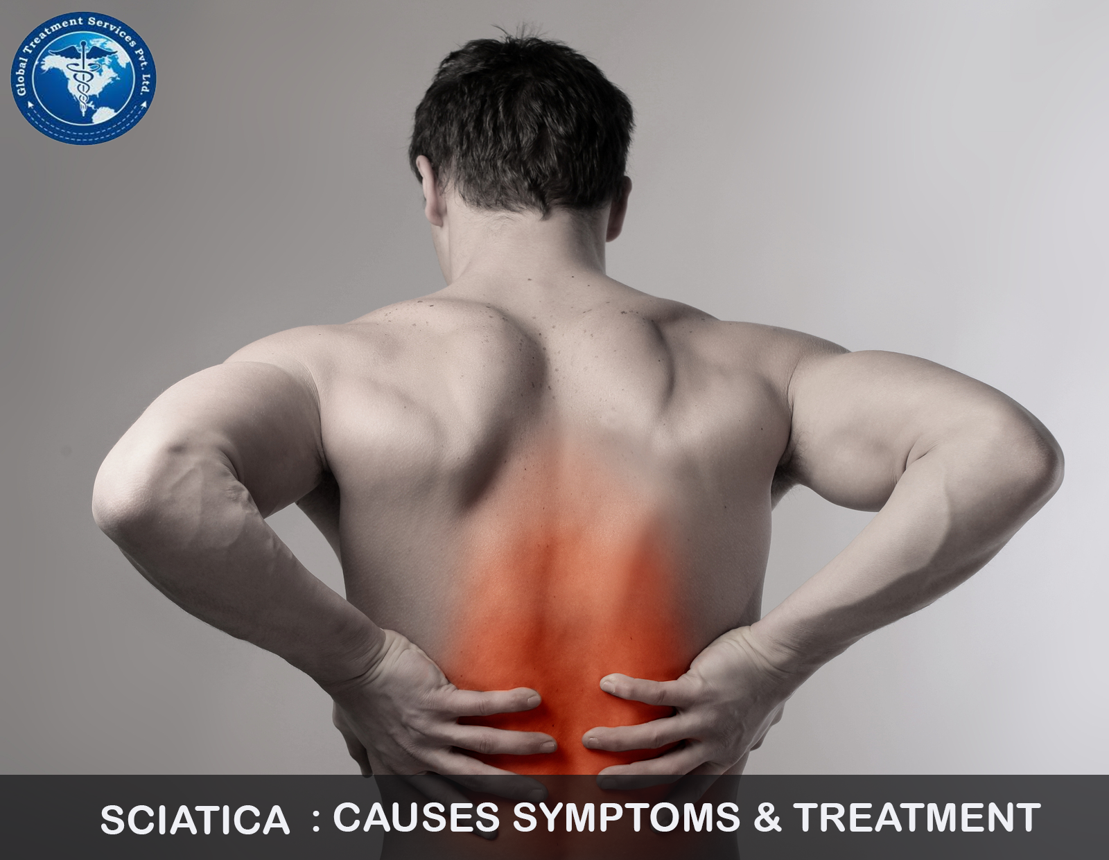
The pain which might feel like a bad leg cramp or can be a shooting pain that makes standing or sitting nearly impossible,is typically felt from the lower back to behind the thigh and radiates down below the knee

CAUSES

  • Herniated or slipped disc:this will cause pressure on the nerve and as a result pain occurs along path of the nerve
  • Piriformis syndrome:which occurs when the small piriformis muscle,which lies deep in the buttocks,becomes tight and spasms,thus putting pressure on and irritating sciatic nerve
  • Spinal stenosis: this results from narrowing of the spinal canal which puts pressure on the nerve
  • Spondylolisthesis: that occurs when one vertebrae slips,so that it is out of line with one above it,narrowing the opening through which sciatic nerve exist

RISK FACTORS

  • Changes in the spine due to advancing age,such as herniated disks and bone spurs
  • being obese which stresses the spine
  • sitting for prolonged period
  • leading a sedentary life style
  • diabetes
  • jobs which require to carry heavy loads,drive for long periods,repeatedly twist your back

SYMPTOMS

  • Lower back pain
  • pain in the rear or leg which worse when sitting
  • hip pain
  • burning or tingling in the leg
  • weakness
  • numbness
  • difficulty in moving the leg or foot
  • shooting pain making difficulty in stand up

COMPLICATIONS

  • Permanent nerve damage
  • loss of feeling in the affected leg
  • weakness of leg
  • loss of bowel or bladder control

INVESTIGATIONS

  • X ray
  • CT
  • MRI

TREATMENT

  • Medications
  • physical therapy:your Doctor may recommend to perform a few back exercises and streches

Exercise:in Sciatica exercises usually the focus on stregthening and streching the spinal column and muscles and tendons

  • Surgery:Micro discectomy is a common surgical approach used to treat sciatica that is caused by a lumbar disc herniation.in this surgery,a small part of the disc material under the nerve root is taken out.while technically an open surgery,a microdiscectomy uses minimal invasive techniques and can be done with relatively small incision and minimal damage

For getting opinion from our network of hospitals visit mvtbooking.com or sent a email to query@gtsmeditour.com

CERVICAL SPONDYLOSIS

cer

DEFINITION

It refers to a degenerative process affecting the vertebral disc and facet joints that gradually develops with age.The condition can narrow the spinal canal that results in the compression of spinal cord and nerve roots

Usually the conditions associated with cervical spondylosis arise from the regular wear and tear of the bones and cartilages located in the neck region

CAUSES

Bone spur: The overgrowth of the bones in the neck or spine area could be due to body trying to grow some extra bone for making the spine stronger.However the growth of extra bones could start pressing on delicate ares of the spine including spinal cord ,nerves and surrounding muscles leading to pain and discomfort

Herniated Discs: This happens due to the development of cracks in the spinal discs.these cracks allow the leakage of the internal material required for cushioning.The material could press on the nerve and spinal coed and would thus,result in symptoms

Dehydrated spinal discs: The presence of the gel like material between the discs of spine bones could dry out with passage of time.this would lead to rub the spinal vertebrae against each other that could result in immense pain

Injury: Injury in neck during some accident or fall

RISK FACTORS

  • Age over 60
  • accident
  • excessive work resulting in wear and tear

SYMPTOMS

  • Pain around shoulder blade
  • pain along entire arm and finger
  • stiffness in the neck muscles
  • constant headaches that usually occur towards the back of head
  • numbness or tingling affecting the arms and shoulder

DIAGNOSIS

While making the diagnosis of cervical spondylosis ,it is essential for the doctor to rule out the possibility of having fibromyalgia

INVESTIGATIONS

  • X-ray
  • CT
  • MRI Scan
  • EMG

COMPLICATIONS

  • Spinal nerve injury
  • Neck Injury
  • Paralysis

TREATMENT

  • Physical therapy
  • Medication
  • Surgery-When the condition becomes severe,then surgery of the affected area remains only option to get relief from pain

ANTERIOR CERVICAL DISKECTOMY AND FUSION

In this the problematic disk and additional bone spur are removed and stabilize the spine through spinal fusion .Typically a plate with screws is added to the front of the spine for added stability

For getting opinion from our network of hospitals visit mvtbooking.com or send a email to    query@gtsmeditour.com


BREAST CANCER

A cancer that forms in the cells of the breast.common between age 40-50.

  1. breast cance

PRECIPITATING FACTORS

  1. Unmarried women
  2. nulliparous women
  3. women with bad obstrectic history
  4. high in oral contraceptives

SYMPTOMS

  1. lumb in the breast
  2. bloody discharge from breast
  3. changes in shape or texture of the nipple or breast

download (13)


CLASSIFICATION

a) intra dochal -within the ducts

b) extra dochal-outside the ducts

CLINICAL TYPES

1.Invasive duct carcinoma-common form ,lumb is very broad &irregular causes drowning of the nipple .

2. Medullary carcinoma-primary tumors is soft and circumscribed and may attain a large size

3. Colloid CA/Tubular CA – Appear as well defined masses common in elderly

4.Inflammatory CA – Seen usually during pregnancy and lactation.Breast is often painful

5. Technical Types

Mastitis Carcinoma:i.e carcinoma + inflammation ;retracted nipple;skin is red ,warm and painful

MANCHESTERS STAGING OF THE BREAST  CANCER

Stage 1 – Growth confined to breast;An area of adherence to the skin smaller than the periphery of the tumor does not affect staging.Even ulceration of the skin in the area defined does not alter it

Stage 2 – same as stage 1 but palpable,mobile,lymph node gathering of growth to the pectoral muscle

Stage 3 – skin involvement larger than tumor. Tumor fixed to pectoral muscle.if the nodes are palpable they must be mobile

Stage 4 –

  • Skin involvement as stage 3
  • Tumor fixed to chest wall
  • Axillary nodes are not mobile
  • Palpable nodes in supra clavicular fossa
  • Secondary deposits in skin
  • Secondary deposits in the opposite breast
  • Distant metastasis

The physician or surgeon can easily notice certain peculiar features in the breast in the conditions of any breast cancer

1. Peu-d-orange

2. Oedema of the arm

3. Browny arm

4. Development of carcinomatous nodules on the affected side of the chest wall .

SPREAD OF CANCER

1.Local spread – From mammary gland to skin muscles and chest wall

2. Lymphatic Spread – Leading to inflammatory of internal mammary lymph nodes,supraclavicular node,axillary lymph nodes etc…

3. Spread  by Blood Stream – To ribs ,lumbar vertebra, femur,thoracic vertebrae, to the skull,may lead to pathological fractures

4. Spread to liver – May lead to jaundice

5. Transcoelomic spread

INVESTIGATIONS

a) Needle biopsy/FNAC

b)Trephine biopsy

c)Open biopsy

d)Frozen section biopsy

e)Mammography

f)Ductography

g)Thermography


TREATMENTS

  1. hormonal
  2. endocrine manipulations
  3. radio therapy
  4. oestrogen blockers
  5. immuno therapy
  6. chemo therapy

At present ,radical mastectomy or extended radical mastectomy offers the prospect of long survival provided the neoplasm categorized as belonging to stage 1 and stage 2 .when stages  3 and 4 are reached,the interests of the patient are best served by simple mastectomy and radiotherapy provided the tumor is hormone stimulated ,with endocrine therapy.

RADICAL MASTECTOMY (halsted)

The breast and associated structures are dissected enblock, and the excised mass is composed of

  1. The whole breast
  2. A large portion of skin ,the centre of which overlies the tumor ,but always included the nipple,where there is much skin involvement more skin must be sacrificed
  3. The  fat and fascia from the tumor border of the clavicle to,including the upper quater of the sheath of the rectus abdominis and from the sternum to the anterior border of the latismus dorsi
  4. pectoralis major,and its fascial sheath
  5. pectoralis minor and sheath
  6. the costo-coracoid membrane
  7. all the fat,fascia and lymph nodes of the axilla
  8. the fascia over and a few fibres of the external oblique,serratus anterior subscapularis ,the latismus dorsi and the upper part of the rectus abdominis

2)Extended radical mastectomy

In this ,the radical mstectomy along with removal of internal mammory lymph glands and second inter costal muscle

For getting opinion from our network hospitals visit mvtbooking.com or send a email to query.gtsmeditour.com


CEREBRAL PALSY

DEFINITION
It is a non progressive neurological disorder of movement ,posture and tone due to a non progressive pathological process in the brain caused by an insult to the developing brain


plasy

Depending on the site and extent of involvement ,it may be associated with

  • visual disturbances
  • seizures
  • mental retardation

Etiology

Both prenatal and perinatal factors are considered.

1. prematurity

2. kernicterus

3. structurally abnormal brain

4. birth asphyxia

5.encephalitis

6.meningitis

Classification

  1. spastic CP
  2. Extra pyramidal CP
  3. Cerebellar or ataxic CP
  4. Mixed CP

Spastic C P

Again classified into

  1. Diplegic C P
  2. Hemiplegic C P
  3. Quadriplegic C P

Diplegic C P

commonest form of C P in preterm

symptoms

  1. spastic lower limbs with increased tendon jerks and extensor plantar  response
  2. scissoring of legs
  3. visual disturbances

Hemiplegic C P

There is paresis of one side of body with upper motor neuron type of spasticity increased reflexes and extensor plantar response

Quadriplegic C P

  1. spasticity in all the four limbs with pseudo bulbar palsy
  2. severe mental retardation
  3. epilepsy

Extra pyramidal C P

Distonia and choreoathetosis in various propotions

  1. Defective coordination of movements
  2. defective regulation of muscle tone,resulting in the inability to execute the intended movements

Ataxic C P

Manifest only in second year of life,before that the child may remain floppy without other cerebellar signs


Investigations

  1. CT
  2. MRI
  3. TORCH screening for associated symptoms

Treatments

Treatment of associated defects and optimum utilization of available motor functions become important

Treatment of associated deficits

  • seizures can be controlled with anticonvulsants
  • early stimulation aimed at functional development of various modalities is a must
  • physiotherapy
  • prevention of contractures -splinting  may prevent this
  • avoid\make use of abnormally persisting reflexes
  • reflex stimulation
  • training in motor activities
  • education
  • Orthopedic & neurosurgical measures
  1.    arthrodesis of the wrist,ankle etc may be considered if it adds to the functional recovery
  2. in established contracture , muscle lengthening to decrease the adduction of hip,flexion of knee etc..
  3. surgical transfer of tendons to the muscles of desired function and release of tendo achilles contracture
  4. peripheral nerve section (obturator nerve) may help to reduce adductor spasm
  5. a more advanced surgery is selective motor fasiculotomy where selected fasicule of a nerve that causes undesirable hyper tones is cut off after identifying them by stimulation studies
  6. surgical implants of electrodes in   selected areas over the dura of spinal cord can be used for spinal stimultaion


For getting opinion from our network of hospitals visit mvtbooking.com or send a email to query@gtsmeditour.com