Global Treatment Services Pvt. Ltd.

Global Treatment Services

Cerebral Venous Thrombosis(CVT): causes, symptoms &treatments

Cerebral venous thrombosis (CVT) is a blood clot of a cerebral vein in the brain. This vein is responsible for draining blood from the brain. If blood collects in this vein, it will begin to leak into brain tissues and cause a hemorrhage or severe brain swelling.

When caught early, CVT can be treated without causing life-threatening complications. 

What are common risk factors of CVT?

Blood clots are more likely to occur in your body when there is an interruption in regular blood flow. While CVT is an uncommon condition, it can be triggered by a number of factors.

Some of the most common risk factors include:

  • birth control or excess estrogen use
  • dehydration
  • ear, face, or neck infection
  • protein deficiencies
  • head trauma or injury
  • obesity
  • cancer
  • tumor

Less common risk factors for CVT include pregnancy and other blood clotting disorders. Both conditions can make blood clot more easily, affecting proper blood flow throughout the body and the brain.

In infants, the most common cause of CVT is infection, specifically in the ear.

In some cases of CVT, the cause is unknown.

If left untreated, CVT can have life-threatening consequences.

Gray488.png

Symptoms of cerebral venous thrombosis

A blood clot in a cerebral vein can cause pressure that leads to brain swelling. This pressure can cause headaches and in more severe cases damage brain tissue.

Symptoms vary depending on where the blood clot occurs in the brain. However, more common symptoms of CVT can include:

  • severe headaches
  • blurred vision
  • nausea
  • vomiting

If you have a more severe case of cerebral venous thrombosis, you may experience stroke-like symptoms. These can include:

  • speech impairment
  • one-sided body numbness
  • weakness
  • decreased alertness

If you begin experiencing any of these symptoms, immediately call 911 or have some someone take you to an emergency room.

Other symptoms from severe CVT include:

  • fainting
  • limited mobility in parts of your body
  • seizures
  • coma
  • death
Diagnosing CVT

When diagnosing cerebral venous thrombosis, doctors will evaluate the symptoms you experience and will also take into account your medical and family history. However, a final diagnosis depends on checking the blood circulation in your brain. To check the blood flow, doctors can use imaging tests to detect blood clots and swelling.

A doctor can misdiagnose a CVT if they use the wrong test. While there are a number of imaging tests available, some aren’t as helpful in diagnosing this condition, such as a simple X-ray of the skull.

The two best imaging tests to help detect CVT are:

  • MRI venogram. An MRI venogram, also referred to as an MRV, is an imaging test that produces images of the blood vessels in the head and neck area. It can help to evaluate blood circulation, irregularities, strokes, or brain bleeds. During this MRI, doctors will inject a special dye into your bloodstream to display blood flow and to help determine if blood is clotting in order to diagnose thrombosis. This test is typically used to clarify images from a CT scan.
  • CT venogram. CT scans use X-ray imaging to show your doctor your bones and arterial vessels. Combined with a venogram, doctors will inject a dye into the veins to produce images of blood circulation and help detect blood clotting.
Cerebral venous thrombosis treatment options

CVT treatment options depend on the severity of the condition. Primary treatment recommendations focus on preventing or dissolving blood clots in the brain.

Medication

Doctors may prescribe anticoagulants, or blood thinners, to help prevent blood clotting and any further growth of the clot. The most commonly prescribed drug is heparin, and it’s injected directly into the veins or under the skin.

Once your doctor thinks you’re stable, they may recommend an oral blood thinner like warfarin as a periodic treatment. This can help to prevent recurrent blood clots, specifically if you have a diagnosed blood clotting disorder.

Other than helping to prevent blood clots, doctors will also address symptoms of CVT. If you’ve experienced a seizure from this condition, doctors will prescribe anti-seizure medication to help control the episode. Similarly, if you begin to experience stroke-like symptoms, a doctor will admit you into a stroke or intensive care unit.

Monitoring

In all cases of CVT, doctors will monitor brain activity. Follow-up venograms and imaging tests are recommended to assess thrombosis and to ensure there are no additional clots. Follow-ups are also crucial to make sure you don’t develop clotting disorders, tumors, or other complications from cerebral venous thrombosis. The doctors will likely run additional blood tests to see if you have any clotting disorders that may have increased your risk of developing CVT.

Surgery

In more severe cases of cerebral venous thrombosis, doctors may recommend surgery to remove the blood clot, or thrombi, and to fix the blood vessel. This procedure is referred to as thrombectomy. In some thrombectomy procedures, doctors may insert a balloon or similar device to prevent blood vessels from closing.


Vertigo: Causes, Symptoms & Treatments

Causes of Vertigo

Vertigo is often caused by an inner ear problem. Some of the most common causes include:

BPPV. These initials stand for benign paroxysmal positional vertigo. BPPV occurs when tiny calcium particles (canaliths) clump up in canals of the inner ear. The inner ear sends signals to the brain about head and body movements relative to gravity. It helps you keep your balance.

BPPV can occur for no known reason and may be associated with age.

Meniere’s disease. This is an inner ear disorder thought to be caused by a buildup of fluid and changing pressure in the ear. It can cause episodes of vertigo along with ringing in the ears (tinnitus) and hearing loss.

Vestibular neuritis or labyrinthitis. This is an inner ear problem usually related to infection (usually viral). The infection causes inflammation in the inner ear around nerves that are important for helping the body sense balance

Less often vertigo may be associated with:

  • Head or neck injury
  • Brain problems such as stroke or tumor
  • Certain medications that cause ear damage
  • Migraine headaches

Symptoms of Vertigo

Vertigo is often triggered by a change in the position of your head.

People with vertigo typically describe it as feeling like they are:

  • Spinning
  • Tilting
  • Swaying
  • Unbalanced
  • Pulled to one direction

Other symptoms that may accompany vertigo include:

  • Feeling nauseated
  • Vomiting
  • Abnormal or jerking eye movements (nystagmus)
  • Headache
  • Sweating
  • Ringing in the ears or hearing loss

Symptoms can last a few minutes to a few hours or more and may come and go.

Treatment for Vertigo

Treatment for vertigo depends on what’s causing it. In many cases, vertigo goes away without any treatment. This is because your brain is able to adapt, at least in part, to the inner ear changes, relying on other mechanisms to maintain balance.

For some, treatment is needed and may include:

Vestibular rehabilitation. This is a type of physical therapy aimed at helping strengthen the vestibular system. The function of the vestibular system is to send signals to the brain about head and body movements relative to gravity.

Vestibular rehab may be recommended if you have recurrent bouts of vertigo. It helps train your other senses to compensate for vertigo.

Canalith repositioning maneuvers. Guidelines from the American Academy of Neurology recommend a series of specific head and body movements for BPPV. The movements are done to move the calciumdeposits out of the canal into an inner ear chamber so they can be absorbed by the body. You will likely have vertigo symptoms during the procedure as the canaliths move.

A doctor or physical therapist can guide you through the movements. The movements are safe and often effective.

Medicine. In some cases, medication may be given to relieve symptoms such as nausea or motion sickness associated with vertigo.

If vertigo is caused by an infection or inflammation, antibiotics or steroids may reduce swelling and cure infection.

For Meniere’s disease, diuretics (water pills) may be prescribed to reduce pressure from fluid buildup.

Surgery. In a few cases, surgery may be needed for vertigo.

If vertigo is caused by a more serious underlying problem, such as a tumor or injury to the brain or neck, treatment for those problems may help to alleviate the vertigo.

Antral Gastritis: Causes, Symptoms & Treatments

Gastritis is a general term for a group of conditions with one thing in common: inflammation of the lining of the stomach. The inflammation of gastritis is most often the result of infection with the same bacterium that causes most stomach ulcers. Regular use of certain pain relievers and drinking too much alcohol also can contribute to gastritis.

Gastritis may occur suddenly (acute gastritis), or appear slowly over time (chronic gastritis). In some cases, gastritis can lead to ulcers and an increased risk of stomach cancer. For most people, however, gastritis isn’t serious and improves quickly with treatment.

Symptoms

The signs and symptoms of gastritis include:

  • Gnawing or burning ache or pain (indigestion) in your upper abdomen that may become either worse or better with eating
  • Nausea
  • Vomiting
  • A feeling of fullness in your upper abdomen after eating

Gastritis doesn’t always cause signs and symptoms.

When to see a doctor

Nearly everyone has had a bout of indigestion and stomach irritation. Most cases of indigestion are short-lived and don’t require medical care. See your doctor if you have signs and symptoms of gastritis for a week or longer. Tell your doctor if your stomach discomfort occurs after taking prescription or over-the-counter drugs, especially aspirin or other pain relievers.

If you are vomiting blood, have blood in your stools or have stools that appear black, see your doctor right away to determine the cause.

Causes

Gastritis is an inflammation of the stomach lining. Weaknesses or injury to the mucus-lined barrier that protects your stomach wall allows your digestive juices to damage and inflame your stomach lining. A number of diseases and conditions can increase your risk of gastritis, including Crohn’s disease and sarcoidosis, a condition in which collections of inflammatory cells grow in the body.

Risk factors

Factors that increase your risk of gastritis include:

  • Bacterial infection. Although infection with Helicobacter pylori is among the most common worldwide human infections, only some people with the infection develop gastritis or other upper gastrointestinal disorders. Doctors believe vulnerability to the bacterium could be inherited or could be caused by lifestyle choices, such as smoking and diet.
  • Regular use of pain relievers. Common pain relievers — such as aspirin, ibuprofen (Advil, Motrin IB, others) and naproxen (Aleve, Anaprox) — can cause both acute gastritis and chronic gastritis. Using these pain relievers regularly or taking too much of these drugs may reduce a key substance that helps preserve the protective lining of your stomach.
  • Older age. Older adults have an increased risk of gastritis because the stomach lining tends to thin with age and because older adults are more likely to have H. pylori infection or autoimmune disorders than younger people are.
  • Excessive alcohol use. Alcohol can irritate and erode your stomach lining, which makes your stomach more vulnerable to digestive juices. Excessive alcohol use is more likely to cause acute gastritis.
  • Stress. Severe stress due to major surgery, injury, burns or severe infections can cause acute gastritis.
  • Your own body attacking cells in your stomach. Called autoimmune gastritis, this type of gastritis occurs when your body attacks the cells that make up your stomach lining. This reaction can wear away at your stomach’s protective barrier.Autoimmune gastritis is more common in people with other autoimmune disorders, including Hashimoto’s disease and type 1 diabetes. Autoimmune gastritis can also be associated with vitamin B-12 deficiency.
  • Other diseases and conditions. Gastritis may be associated with other medical conditions, including HIV/AIDS, Crohn’s disease and parasitic infections.

Diagnosis:

Although your doctor is likely to suspect gastritis after talking to you about your medical history and performing an exam, you may also have one or more of the following tests to pinpoint the exact cause.

  • Tests for H. pylori. Your doctor may recommend tests to determine whether you have the bacterium H. pylori. Which type of test you undergo depends on your situation. H. pylori may be detected in a blood test, in a stool test or by a breath test.For the breath test, you drink a small glass of clear, tasteless liquid that contains radioactive carbon. H. pylori bacteria break down the test liquid in your stomach. Later, you blow into a bag, which is then sealed. If you’re infected with H. pylori, your breath sample will contain the radioactive carbon.
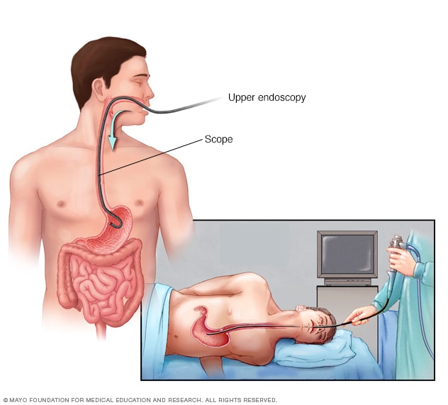
  • Using a scope to examine your upper digestive system (endoscopy). During endoscopy, your doctor passes a flexible tube equipped with a lens (endoscope) down your throat and into your esophagus, stomach and small intestine. Using the endoscope, your doctor looks for signs of inflammation.If a suspicious area is found, your doctor may remove small tissue samples (biopsy) for laboratory examination. A biopsy can also identify the presence of H. pylori in your stomach lining.
  • X-ray of your upper digestive system. Sometimes called a barium swallow or upper gastrointestinal series, this series of X-rays creates images of your esophagus, stomach and small intestine to look for abnormalities. To make the ulcer more visible, you may swallow a white, metallic liquid (containing barium) that coats your digestive tract.
 Treatment

Treatment of gastritis depends on the specific cause. Acute gastritis caused by nonsteroidal anti-inflammatory drugs or alcohol may be relieved by stopping use of those substances.

Medications used to treat gastritis include:

  • Antibiotic medications to kill H. pylori. For H. pylori in your digestive tract, your doctor may recommend a combination of antibiotics, such as clarithromycin (Biaxin) and amoxicillin (Amoxil, Augmentin, others) or metronidazole (Flagyl), to kill the bacterium. Be sure to take the full antibiotic prescription, usually for seven to 14 days.
  • Medications that block acid production and promote healing. Proton pump inhibitors reduce acid by blocking the action of the parts of cells that produce acid. These drugs include the prescription and over-the-counter medications omeprazole (Prilosec), lansoprazole (Prevacid), rabeprazole (Aciphex), esomeprazole (Nexium), dexlansoprazole (Dexilant) and pantoprazole (Protonix).Long-term use of proton pump inhibitors, particularly at high doses, may increase your risk of hip, wrist and spine fractures. Ask your doctor whether a calcium supplement may reduce this risk.
  • Medications to reduce acid production. Acid blockers — also called histamine (H-2) blockers — reduce the amount of acid released into your digestive tract, which relieves gastritis pain and encourages healing. Available by prescription or over-the-counter, acid blockers include ranitidine (Zantac), famotidine (Pepcid), cimetidine (Tagamet HB) and nizatidine (Axid AR).
  • Antacids that neutralize stomach acid. Your doctor may include an antacid in your drug regimen. Antacids neutralize existing stomach acid and can provide rapid pain relief. Side effects can include constipation or diarrhea, depending on the main ingredients.

Complications

Left untreated, gastritis may lead to stomach ulcers and stomach bleeding. Rarely, some forms of chronic gastritis may increase your risk of stomach cancer, especially if you have extensive thinning of the stomach lining and changes in the lining’s cells.

Tell your doctor if your signs and symptoms aren’t improving despite treatment for gastritis.

Prevention

Preventing H. pylori infection

It’s not clear how H. pylori spreads, but there’s some evidence that it could be transmitted from person to person or through contaminated food and water. You can take steps to protect yourself from infections, such as H. pylori, by frequently washing your hands with soap and water and by eating foods that have been cooked completely.

Hallux Valgus: Symptoms & Treatments

Hallux valgus is the commonest forefoot deformity, with an estimated prevalence of 23% to 35%. It causes symptoms on the medial edge of the foot, the sole, and the small toes. Non-operative treatment may alleviate symptoms but does not correct the deformity of the big toe. Surgery is indicated if the pain persists. The correct operation must be selected from a wide variety of available techniques.

Symptoms

Hallux valgus causes pain particularly in the bunion on the inner side of the foot, on loading under the foot and in the smaller toes

Hallux valgus causes symptoms in three particular ways. First and foremost is pain in the bunion, the pressure-sensitive prominence on the medial side of the head of the first metatarsal. It hurts to wear a shoe. Furthermore, the valgus deviation of the great toe often results in a lack of space for the other toes. They become displaced, usually upwards, leading to pressure against the shoe. This is termed hammer toe or claw toe. Finally, normal function of the forefoot relies heavily on the great toe pressing down on the ground during gait. Since the valgus deformity stops this happening to a sufficient degree, metatarsal heads II–V are overloaded. The resulting pain is referred to as transfer metatarsalgia.

An external file that holds a picture, illustration, etc.<br /><br />
Object name is Dtsch_Arztebl_Int-109-0857_001.jpg

Conservative treatment

Only while the skeleton is still growing can the position of the great toe be improved with lasting effect. A night splint can be prescribed to move the great toe to medial. After the end of growth, adequate correction is no longer possible and conservative treatment is restricted to alleviation of symptoms.

By the time patients consult a physician, most of them have already resorted to softer and wider shoes to alleviate pain from pressure on their bunion. Ring pads and other dressings tend to increase the prominence of the bunion and are usually unsuccessful. Antiphlogistic salves can be administered locally, nonsteroidal anti-inflammatory drugs systemically.

Pain in the smaller toes can be alleviated with pads and toe straighteners. Wide, soft shoes are helpful if they give the toes enough space. Once hammer toes or claw toes have developed, however, surgery is necessary.

Conservative treatment

Pain on standing and walking can be alleviated by inlays under the forefoot. Permanent improvement of the position of the great toe is not possible.

In our experience, insoles are effective for alleviation of metatarsalgia. They must feature a pad that pushes the metatarsals upward proximal to the pressure-sensitive heads. It often suffices to advise the patient to wear shoes with soft soles and without excessively high heels (no more than 4 cm). The malposition of the great toe, of course, cannot be corrected with insoles alone.

Surgical procedures

The different surgical procedures are based on various underlying principles, e.g., correction osteotomy, resection arthroplasty, or arthrodesis.

Spinal Hemangioma: Causes, Symptoms & Treatments

What is a Spinal Hemangioma?

A spinal hemangioma is a tumor that mostly occurs in the thoracic and the lumbar spine. Also referred to as vertebral hemangioma, it is non-cancerous and has few symptoms. This is why most people do not realize that they have this condition unless an imaging test for another condition reveals its presence. The vertebral body is the most commonly affected by this tumor; however, it may also affect the muscles around the spine. Pain is the most common symptom and is reported by around 20% of the patients. Let’s have a brief look at the causes and treatments of spinal hemangioma pain.

Causes of Vertebral Hemangioma Pain:

The causes of spinal hemangioma are not much known, however, it is believed that there may be a genetic cause involved. Studies have also found that a high amount of estrogen present after birth may also increase the chances of this tumor. This is also considered to be a likely cause because the condition is more common in females than in men.

As mentioned above, there are rarely any symptoms when this tumor is present. In fact, the tumor is often found in 10-12% autopsies, which indicates that a person can actually go through life without even experiencing any symptom. However, some individuals may experience pain. A likely cause of this pain is a spinal hemangioma that is quite large and involves the entire vertebral body. There may also be a collapse and loss of height in the vertebrae if the tumor is extensive. A severe collapse means that the spinal cord and the nerves emanating from it will get compressed causing pain, numbness or weakness in the legs, and even loss of function in the bowel or bladder.

Hemangioma diagnosis

If a hemangioma is suspected, the doctor will order an x-ray to check for a specific pattern on the bone, called a trabecular pattern. Trabecular, or cancellous, bone is a lattice-shaped structure within the bone.

A CT scan may also show a polka dot appearance in the bone. If this appears, an MRI will be ordered to show whether the tumor has expanded into the spinal column or spinal canal, or encroached on the spinal cord. MRI can also show the extent of nerve damage in the spine and can assist in planning surgical treatment.

Hemangioma symptoms

Most hemangiomas are symptom-free, but symptoms may include:

  • Back pain
  • Pain that radiates along a nerve due to inflammation or irritation of the nerve root
  • Spinal cord compression

Treatments:

It is also possible that the spinal hemangioma may extend beyond the vertebrae. This could be a cause of back pain in some individuals. Trauma may also occur and lead to a compression fracture of the vertebrae causing hemorrhage or bleeding. Extra pressure due to a pregnancy may put extra pressure on the vertebrae and cause or increase the back pain.

Once the tumor is discovered, these patients need to go for regular checkups to diagnose the presence of compression fractures, neurological dysfunction, or the development of a soft tissue mass. If pain and other symptoms are present due to a hemorrhage or another reason, the treatment will depend on the extent of the bleeding and the severity of the neurological symptoms. The size and location of the hemangioma will also determine the treatment administered.

Embolization is one of the treatment options available. In this procedure, a tiny catheter or tubing is inserted and a specific material, such as polyvinyl alcohol foam, is injected. This material travels to the bleeding site. As it builds up at the site of the hemorrhage, the flow of blood stops.

The most common treatment for these painful lesions is radiotherapy, in which high energy X-rays are used to kill the tumor cells. Studies have found that high doses administered to patients with pain associated with spinal hemangioma are quite successful in achieving complete pain relief.

Excision or surgical removal of the spinal hemangioma may be required if spinal cord compression is present causing pain, numbness, and weakness. In cases where, a partial removal of the tumor is performed, radiotherapy is required as an additional treatment. It is also the most common treatment for children since they are more prone to the harmful effects of radiation.Radiation therapy is effective in the treatment of pain caused by hemangiomas. Ethanol injections with fluoroscopic guidance have also proven effective in treating pain. Another treatment option is embolization followed by removal of the small bones that make up a vertebra (laminectomy) or removal of the vertebra (vertebrectomy).

The presence of spinal hemangioma pain and neurological symptoms are the likely reasons that treatment is required. If the symptoms are worsening rapidly, surgical procedures are recommended. However, if the symptoms develop slowly, radiotherapy or embolization are considered to be better options.

Gangrene: Causes, symptoms & treatments

Gangrene is a condition that occurs when body tissue dies. It is caused by a loss of blood supply due to an underlying illness, injury, and/or infection. Fingers, toes, and limbs are most often affected, but gangrene can also occur inside the body, damaging organs and muscles. There are different types of gangrene and all require immediate medical attention.

Gangrene Causes

Blood plays a very important role in your health. Not only does it transport oxygen and nutrients throughout your body to feed cells, it delivers disease-fighting antibodies that protect your body from infection. When blood cannot travel freely throughout the body, your cells cannot survive, infection can develop, and tissue can die from gangrene. Any condition that affects blood flow increases your risk of gangrene, including:

  • Diabetes
  • Atherosclerosis
  • Peripheral arterial disease
  • Smoking
  • Trauma or serious injury
  • Obesity
  • Raynaud’s phenomenon (a condition in which the blood vessels that supply the skin become intermittently narrowed)
  • Weakened immune system
Gangrene Types
There are two main types of gangrene:Dry gangrene: More common in people with blood vessel disease,  diabetes, and autoimmune diseases, dry gangrene usually affects the hands and feet. It develops when blood flow to the affected area is impaired, usually as a result of poor circulation. In this type, the tissue dries up and may be brown to purplish-blue to black in color and often falls off. Unlike other types of gangrene, infection is typically not present in dry gangrene. However, dry gangrene can lead to wet gangrene if it becomes infected.Wet gangrene: Unlike dry gangrene, wet gangrene almost always involves an infection. Injury from burns or trauma where a body part is crushed or squeezed can rapidly cut off blood supply to the affected area, causing tissue death and increased risk of infection. The tissue swells and blistersand is called “wet” because of pus. Infection from wet gangrene can spread quickly throughout the body, making wet gangrene a very serious and potentially life-threatening condition if not treated quickly.

Types of wet gangrene include:Internal gangrene: If gangrene occurs inside the body due to blocked blood flow to an internal organ, then it is referred to as internal gangrene. This is usually related to an infected organ such as the appendix or colon.Gas gangrene: Gas gangrene is rare but dangerous. It occurs when infection develops deep inside the body, such as inside muscles or organs, usually as a result of trauma. The bacteria that causes gas gangrene, called clostridia, release dangerous toxins or poisons that wreak havoc throughout the body, along with gas which can be trapped within body tissue. As the condition progresses, the skin may become pale and gray, and make a crackling sound when pressed, due to the gas within the tissue. Gas gangrene warrants immediate medical treatment. Without treatment, death can occur within 48 hours.

Fournier’s gangrene: Also a rare condition, Fournier’s gangrene is caused by an infection in the genital area. Men are affected more often than women. If the infection gets into the bloodstream, a condition called sepsis, it can be life-threatening.

Gangrene Symptoms

You may notice the following symptoms at the site of the dry gangrene:

  • Dry and shriveled skin that changes color from blue to black and eventually sloughs off
  • Cold and numb skin
  • Pain may or may not be present

Symptoms of wet gangrene may include:

  • Swelling and pain at the site of infection
  • Change in skin color from red to brown to black
  • Blisters or sores that produce a bad-smelling discharge (pus)
  • Fever and feeling unwell
  • A crackling noise that comes from the affected area when pressed

Internal gangrene usually is painful in the area of the gangrene. For example, a person with gangrene of the appendix or colon would be expected to have severe abdominal pain in the vicinity of the gangrene.

Gangrene Warning

If infection from gangrene gets into the blood, you may develop sepsis and go into septic shock. This can be life-threatening if not treated immediately. Symptoms of sepsis may include:

  • Low blood pressure
  • Rapid heartbeat
  • Shortness of breath
  • Change in body temperature
  • Light-headedness
  • Body pain and rash
  • Confusion
  • Cold, clammy, and pale skin

If you think you or a loved one may have sepsis, go to the emergency room immediately.

Gangrene Treatments

Treatment for gangrene involves removing the dead tissue, treating and preventing the spread of infection, and treating the condition that caused gangrene to develop. The sooner you receive treatment, the better your chance of recovery. Depending on the type of gangrene, treatment may include:

Surgery. Also called debridement, the dead tissue is surgically removed to prevent the spread of infection. In some situations, amputation (removal of the affected limb, finger or toe) may be required.

Maggot therapy. Believe it or not, maggots still play a role in modern medicine. Maggots provide a non-surgical way to remove dead tissue. When used to treat gangrene, maggots from fly larvae (specially bred in a laboratory so they are sterile) are placed on the wound, where they consume the dead and infected tissue without harming healthy tissue. They also help fight infection and speed up healing by releasing substances that kill bacteria.

Antibiotics. Antibiotics are used to treat and prevent infections. These are usually given by intravenous injection into a vein.

Oxygen therapy. Hyperbaric oxygen therapy may be used in some cases of wet gangrene or ulcers related to diabetes or peripheral artery disease. During treatment, the patient is put in a specially designed chamber filled with oxygen at a higher pressure than oxygen found in the outside air. The theory is that this high level of oxygen saturates the blood and encourages healing of the dying tissue. Oxygen therapy may also reduce the growth of bacteria that cannot thrive in an oxygen-rich environment.

In order to prevent gangrene from occurring again, the cause of any blood supply blockage must be determined so that the underlying condition can be treated. Often vascular surgery, such as bypass surgery or angioplasty, is needed to restore blood flow. Medication to prevent blood clots may be used in some cases.

Gangrene is a serious medical condition that requires immediate medical treatment. While most people with dry gangrene recover fully with treatment, gangrene that involves an infection can be life-threatening. The sooner you get treatment, the better your chances of recovery. If you have ongoing and unexplained pain in any area in your body, fever, a wound that is slow to heal, or you notice a change in skin color, make an appointment to see your doctor right away or go to the emergency room.

Dyspepsia: Causes, symptoms & Treatments

Dyspepsia, also known as indigestion, is a term that describes discomfort or pain in the upper abdomen. It is not a disease.

The term refers to a group of symptoms that often include bloating, discomfort, nausea, and burping.

In the majority of cases, indigestion is linked to eating or drinking. It can also be caused by infections or the use of certain medications.

Fast facts on dyspepsia

  • Dyspepsia is the same as indigestion, and it is not a disease. It is the name for a group of symptoms that cause discomfort in the abdomen.
  • Overeating, consuming greasy or spicy foods, and hiatus hernia can all cause indigestion.
  • An endoscopy can be used to identify the root cause.
  • Dietary choices and medication are used to manage dyspepsia symptoms.

What is dyspepsia?

Dyspepsia is a group of symptoms rather than a specific condition.

Most people with indigestion feel pain and discomfort in the stomach or chest. The sensation generally occurs soon after consuming food or drink. It may make a person feel full or uncomfortable during a meal, even if they have not eaten a large amount of food.

Dyspepsia symptoms

Indigestion is not a condition but a group of symptoms that affect digestion.

The following symptoms of dyspepsia are also common:

  • nausea
  • belching
  • pain
  • a feeling of fullness, or satiety
  • feeling bloated

In very rare cases, indigestion may be a symptom of stomach cancer.

Mild dyspepsia rarely needs further investigation, and should not be a cause for concern. A visit to a doctor is only needed if symptoms continue for more than 2 weeks.

Seek emergency treatment if the pain is severe and any of the following also occurs:

  • loss of appetite or weight loss
  • vomiting
  • inability to swallow
  • black stools
  • yellow coloring in the eyes and skin
  • chest pain during exertion
  • shortness of breath
  • sweating
  • chest pain that spreads to the jaw, arm, or neck

Heartburn and dyspepsia are often confused for one another, but they are two separate conditions despite regularly occurring at the same time. Heartburn is a symptom of acid reflux, described as a burning feeling behind the breastbone that usually occurs after eating.

Causes

Consuming too much caffeine is one of the many possible causes of dyspepsia.

Indigestion is usually caused by the lifestyle of an individual and the foods they eat. It can also be related to an infection or other digestive conditions.

The symptoms are normally triggered by stomach acid coming into contact with the mucosa. Stomach acids break down the mucosa, causing irritation and inflammation. This triggers the uncomfortable symptoms of indigestion.

Common causes of indigestion include:

  • eating too much or too rapidly
  • eating fatty, greasy, or spicy foods
  • drinking too much caffeine or alcohol
  • consuming too much chocolate or soda
  • emotional trauma
  • gallstones
  • gastritis, or inflammation of the stomach
  • hiatus hernia
  • infection, especially with a bacteria called Helicobacter pylori (H. pylori)
  • nervousness
  • obesity
  • pancreatitis, or inflammation of the pancreas
  • peptic ulcers
  • smoking
  • certain medications, such as antibiotics and non-steroidal anti-inflammatory drugs (NSAIDs)
  • stomach cancer

When a doctor cannot find a cause for indigestion, the individual may have functional dyspepsia. This is a type of indigestion without any structural or metabolic disease to explain the symptoms. It may be caused by an impairment of the stomach that prevents it from accepting and digesting food in a normal way.

Diagnosis

Dyspepsia is mild and infrequent for most people with symptoms. In such cases, no treatment is needed.

People who experience regular indigestion or severe abdominal pain should see a primary care physician.

A doctor will ask the person experiencing dyspepsia about their symptoms. They will also find out about their medical and family histories and examine the chest and stomach. This may involve pressing down on different areas of the abdomen to find out whether any are sensitive, tender, or painful under pressure.

If the doctor suspects an underlying cause, they can use the following diagnostic tests to identify any underlying health problems:

  • Blood test: If the person with indigestion also has any symptoms of anemia, the doctor may order a blood test.
  • Endoscopy: People who have not responded to previous treatment for dyspepsia may be referred for a more detailed examination of the upper gastrointestinal (GI) tract. A long thin tube with a camera at the end is inserted through the mouth and into the stomach. This produces clear images of the mucosa. The doctor can also perform a biopsy during this procedure to test for cancer.
  • Tests to diagnose H. pylori infection: These may include a urea breath test, a stool antigen test, and a blood test. An endoscopy would also identify H. pylori as well as any peptic ulcers that are present. Peptic ulcers are often caused by H. pylori.
  • Liver function test: If the doctor suspects a problem with the bile ducts in the liver, they may request a blood test to assess how the liver is working.
  • X-rays: X-ray images are taken of the esophagus, stomach, and small intestine.
  • Abdominal ultrasound: High-frequency soundwaves make images showing movement, structure, and blood flow in the abdomen. A gel is applied to the abdomen and a hand-held device pressed against the skin. The device gives off sound waves, and the doctor can see a detailed picture of the inside of the abdomen on a monitor.
  • Abdominal CT scan: This may involve injecting a dye into the veins. The dye shows up on the monitor. The CT scan takes a series of X-ray images to produce a 3D image of the inside of the abdomen.

Further examinations are often only used in severe cases.

Complications

Dyspepsia is mild and infrequent in most cases. However, severe indigestion can occasionally cause the following complications.

Esophageal stricture: Acid reflux can cause indigestion. This is a condition in which stomach acids leak back up into the esophagus and irritate the sensitive lining of the stomach, known as the mucosa. The irritation can scar the esophagus, which then becomes narrow and constricted.

People with esophageal stricture may start to find swallowing difficult. Food can get stuck in the throat, causing chest pain. Esophageal dilatation is sometimes needed to widen the esophagus.

Pyloric stenosis: This occurs when stomach acid causes long-term irritation of the lining of the digestive system. The pylorus is the passage between the stomach and the small intestine. In pyloric stenosis, it becomes scarred and narrowed. As a result, food is not properly digested.

Surgery may be required to widen the pylorus.

Treatments

Treatment for indigestion depends on the cause and severity of symptoms.

If symptoms are mild and infrequent, lifestyle changes will probably ease them. This usually involves consuming fewer fatty and spicy foods and less caffeine, alcohol, and chocolate. Sleeping for at least 7 hours every night may also help to ease mild indigestion.

Exercising regularly and quitting smoking are also important lifestyle changes in treating indigestion.

Medications

In severe or frequent cases of indigestion, a doctor may prescribe medication.

Antacids: These counter the effects of stomach acid. Examples include Alka-Seltzer, Maalox, Rolaids, Riopan, and Mylanta. These are over-the-counter (OTC) medicines that do not need a prescription. A doctor will usually recommend an antacid medication as one of the first treatments for dyspepsia.

H-2-receptor antagonists: These reduce stomach acid levels and last longer than antacids. However, antacids act more quickly. Examples include Zantac, Tagamet, Pepcid, and Axid. Some of these are OTC, while others are only available on prescription.

Some people may experience nausea, vomiting, constipation, diarrhea, and headaches after taking these. Other side effects may include bruising or bleeding.

Proton pump inhibitors (PPIs): Examples include Aciphex, Nexium, Prevacid, Prilosec, Protonix, and Zegerid. PPIs are highly effective for people who also have gastroesophageal reflux disease (GERD). They reduce stomach acid and are stronger than H-2-receptor antagonists.

Speak to a doctor about possible side effects.

Dyspepsia diet

Banana chopped

Foods high in fiber, such as fruit, can help prevent indigestion.

A high-fiber diet is a good way to manage digestive health. It has the effect of clearing out the intestine and making digestion a smoother, cleaner process.

Fruits, nuts, legumes, and wholegrain foods are packed with fiber and an excellent choice for protecting against indigestion. Many yogurts and cereals have also been fortified with fiber.

Eating a balanced diet that excludes spicy or greasy foods is key. Be sure to consume fluids with every meal, as this helps to move food through the digestive tract.

Consuming four or five smaller meals in a day as opposed to three larger ones can also help the digestive system.


Stoke in Brain (CVA): causes, types, symptoms& treatments

A stroke occurs when a blood vessel (an artery) that supplies blood to a certain area of the brain bursts or becomes clogged by a blood clot. Within minutes, the nerve cells  from that that area are affected and are lacking oxygen and they die within minutes – hours (depending on the severity of  obstruction). As a result, that part of the body that is controlled by the stroke affected area of the brain can not function properly.
If you notice any symptoms of a stroke you must know that it is a medical emergency and immediate treatment is required, just as in the case of a myocardial infarction (a part of the heart muscle dies due to the lack of blood supply to that area). If medical treatment is started soon after the onset of stroke symptoms, the fewer brain cells will die or be affected by the lack of oxygen.

Stroke Causes

Ischemic stroke is caused by a blood clot that blocks the blood flow to the brain, the blood clot can develop in a narrowed artery that supplies blood  to the brain or may reach the cerebral circulation after it migrated from the heart or  from any other region of the body. Blood clots usually occur as a result of other disorders that cause modifications to the normal blood flow, such as:

  • Hardening of the arteries also known as atherosclerosis and it is caused by high blood pressure, diabetes and high cholesterol blood levels
  • Atrial fibrillation, or other cardiac arrhythmias (irregular heart rhythms)
  • Certain heart valve disorders, such as the presence of an artificial heart valve, a repaired heart valve, valvular heart disease and mitral valve stenosis (narrowing of valvular orifice)
  • Heart valve infection (endocarditis)
  • The presence of foramen ovale, which is a congenital heart defect (a hole through which blood can enter the left atrium from the right atrium
  • Vessel clogging disorders
  • Inflammation of blood vessels (vasculitis)
  • Myocardial infarction.

Though in rare rare cases low blood pressure (hypotension) may also be the cause of an ischemic stroke. Low blood pressure leads to reduced blood flow to the brain, can be caused by a narrowing of the arteries or damage of heart attack, massive blood loss or a severe infection.
Some surgeries (such as endarterectomy) or other procedures (such as angioplasty) used to treat narrowed carotid arteries may lead to a blood clot at the site where it occurred, causing a subsequent stroke.

Stroke

Causes Of Hemorrhagic Stroke

Hemorrhagic stroke is caused by rupture of blood vessels and bleeding inside the brain (also called intracerebral hemorrhage) or into the space around the brain (called subarachnoid hemorrhage). Elevated values of the blood pressure for long periods can be the cause of a stroke. Bleeding in the space around the brain can be caused by a ruptured aneurysm or blood pressure that was not kept under control.
Other  less common  causes of hemorrhagic stroke,are

    • Inflammation of blood vessels, which may occur in syphilis or tuberculosis
    • Hemophilia
    • Injurys to the neck or head that can affect the blood vessels that can be found in these areas
    • Radiation therapy treatments used for treating neck or brain cancers
    • Cerebral amyloid angiopathy (a disorder in which blood vessels degenerate).

Stroke Risk Factors

The stroke riks factors are changable risk factors and unchangeble risk factors. Certain conditions are proven to increase the risk of stroke, if these conditions can be kept under control, the risk of stroke may decrease.

The stroke risk factors that can be controlled are:

  • High blood pressure (hypertension) is the second most important risk factor after age
  • Diabetes. About a quarter of people with diabetes die from a stroke. The presence of diabetes doubles the risk of stroke due to damage to the blood vessels that occurs in this disease.
  • Elevevated bad blood cholesterol levels (HDL) can lead to coronary artery disease and myocardial infarction which damages the heart muscle (myocardium) which may increase the risk of a stroke
  • Coronary artery disease that can lead to a heart attack and a stroke
  • Other heart disease such as atrial fibrillation, endocarditis, heart valve disease, cardiomyopathy, patent foramen ovale
  • Smoking, including passive smoking
  • Physical inactivity
  • Obesity
  • Certain drugs such as oral contraceptives – especially in women who smoke or who have other bleeding disorders – and anticoagulants or corticosteroids. It seems that women in menopause that follow a hormone replacement treatment have a lower risk of stroke
  • Increased consumption of alcohol, because  people who drink alcohol excessively, especially those with acute alcohol intoxication have a higher risk of stroke.
  • Cocaine usage or other illicit drugs.

Uncontraollabe risk factors:

  • Age. The risk of stroke increases with age, each ten years double the stroke risk after the age of 55. At least 66 percent of all people with stroke were aged 65 or more.
  • Race. Black and Hispanic Americans have a higher risk than people of other races. Compared with whites, young black Americans, both women and men have a risk of 2 to 3 times more likely to make a stroke and die from this cause.
  • Sex. Stroke is more common in men than in women up to 75 years, but after this age women are more affected. At all ages, more women than men will die from a stroke.
  • Family history. The risk of stroke is greater if a parent, brother or sister had a stroke or transient ischemic attack present in the past.

Stroke Symptoms

Keep in mind that these symptoms of stroke require immediate medical care:

  • State of numbness, weakness or paralysis of the face, arm or leg, usually on one side
  • Blurred vision in one eye or in both eyes, spots, double vision (diplopia) or blindness
  • Confusion, trouble speaking or understanding words spoken by others
  • Gait, dizziness, loss of balance or coordination
  • Severe headache
  • The ischemic stroke symptoms differ from those of hemorrhagic. Symptoms also depend on the location of blood clot or bleeding and expansion of the affected region.
  • Symptoms of ischemic stroke (caused by a clot that has blocked a blood vessel) usually occur in the opposite side of the body. For example, a stroke in the right brain gives symptoms in the left side of the body.
  • Symptoms of a hemorrhagic stroke (caused by bleeding in the brain) may be similar to those produced by a ischemic stroke, but differ in symptoms related to high blood pressure in the brain, such as severe headaches, nausea and vomiting, stiff neck , dizziness, convulsions, irritability and confusion
  • Symptoms of a stroke may progress over several minutes, hours or days, often gradually. For example, mild weakness may progress to an inability to move the arm and leg on one side.

If the stroke is caused by a large blood clot (ischemic) symptoms appear suddenly, within a few seconds, but if an artery already narrowed by atherosclerosis  is blocked, symptoms usually develop gradually, raging from a few minutes to hours or less, in a few days.
In time, if  there are more small strokes, the person may experience a gradual change of thinking, behavior, balance or movement (multi-infarct dementia). The symptoms of a small stroke are not always easily recognized. They can be attributed to wrong conditions that can mimic the stroke symptoms.

Stroke (CVA)

Stroke Treatment

Initial treatment of a stroke differs because it depends on the cause – whether it was caused by a blood clot (ischemic stroke) or bleeding in the brain (hemorrhagic stroke). Before starting the treatment a head computerized tomography (CT), or a magnetic resonance imaging (MRI) to see the type of stroke. Other tests can determine the location of the clot or bleeding to assess the extent of the brain lession. While determining treatment options, blood pressure will be carefully monitored and oxygen will be administrated if necessary. Initial treatment focuses on restoring the blood flow (for ischemic stroke) or hemorrhage control (in hemorrhagic stroke). As with heart attack, permanent damage due to a stroke often develops in the first few hours.

Ischemic stroke

Emergency treatment for an ischemic stroke depends on the location and cause of clot formation. Measures to stabilize vital signs, including drugs are taken. If the stroke is diagnosed within 3 hours after the onset of symptoms, drugs that dissolve the clots are administrated called tissue plasminogen activator (t-PA) drugs, which could increase the chances of survival and recovery. However, t-PA can not be safely administered to any patient. If the stroke is hemorrhagic, the use of t-PA is extremely dangerous. Choosing to use or not to use t-PA must be evaluated in the emergency room quickly. Aspirin alone or in combination with another antiplatelet drugs can be administrated. However, aspirin is not recommended within 24 hours after administration of t-PA. Other drugs to control blood glucose levels (glucose) or fever and convulsions may be administrated. Generally hypertension is not treated immediately unless systolic pressure is greater than 220 (mm Hg) and diastolic is above 120.

Hemorrhagic stroke

Initial treatment for hemorrhagic stroke is very difficult, efforts are being made to control bleeding, to lower intracranial pressure and stabilize vital signs, especially blood pressure values. Several drugs are administered in the case of a hemorrhagic stroke. Drugs to control blood pressure, decreased brain swelling, blood sugar levels, fever or seizures. Signs of increased intracranial pressure, psychomotor restlessness, confusion, difficulty to perform commands and headaches are closely monitored. Usually surgery is not recommended for controlling mild or moderate bleeding. However, if there was a large amount of bleeding and if the person’s general condition deteriorates rapidly, surgery may be needed to remove the accumulated blood in the brain and decrease the intracranial pressure . If the bleeding is caused by a rupture of an aneurysm, surgery to repair the aneurysm is the only option.

  • A metal clip for clipping the aneurysm in order to prevent a new bleeding
  • Endovascular embolization, a procedure which involves inserting a small coil inside the aneurysm and block it

Ssurgery decision depends on the location and general condition of the person after stroke.

Stroke

Ongoing treatment

After receiving emergency treatment and the general condition has stabilized, a treatment aimed at recovery and preventing another stroke is started. It is important to control stroke risk factors such as high blood pressure, atrial fibrillation, elevated cholesterol levels and diabetes. Aspirin or other antiplatelet drugs are recommended. In the case of an ischemic stroke (caused by a blood clot), anticoagulants to prevent another stroke are needed. Also drugs such as statins to lower cholesterol or drugs to control blood pressure are needed.

Drugs that lower blood pressure include:

  • Inhibitors of angiotensin converting enzyme (ACE)
  • Angiotensin II receptor blockers
  • Beta blockers
  • Diuretic
  • Calcium channel blockers.