Overview
Eyelid myokymia makes your eyelids twitch or involuntary muscle movement can be seen on skin and if you don’t know why, it’s natural to feel confused or concerned. This condition is extremely common, especially under certain circumstances. But fortunately, it’s also usually nothing to worry about. And even when it’s due to more serious conditions, the twitching is usually very treatable. It is often triggered by stress, fatigue, poor nutrition, or excessive caffeine intake. While typically self-limiting, lasting a few days or weeks, persistent or severe cases may warrant medical attention.Myokymia is usually a minor, temporary concern. Most cases go away within days or weeks without any treatment. But sometimes, eyelid myokymia becomes more than just an annoyance and starts interfering with your work or other parts of your daily routine. Treatment may also be an option if you have myokymia that happens consistently for at least three months.
Symptoms
The main symptom of eyelid myokymia is a twitching you can feel (and you can probably see it if you look in a mirror while it happens). Myokymia twitches usually last only seconds to minutes, but they can last hours for some people. In rare cases, they can become constant.
The twitches are usually:
- Slow
- Constant
- Gentle
- Rippling (almost like waves on water)
Myokymia usually affects just one eye at a time, but it can affect both. You can have it in your upper or lower eyelids, but lower is more common. In rare cases, it will affect both the upper and lower eyelids of the same eye. Myokymia can sometimes also cause nystagmus (when the eyelid twitching also makes your eyeball move).
Causes
Some of the most common causes of myokymia include:
- Being tired or sleep-deprived
- Caffeine intake (especially if you have too much)
- Nicotine use
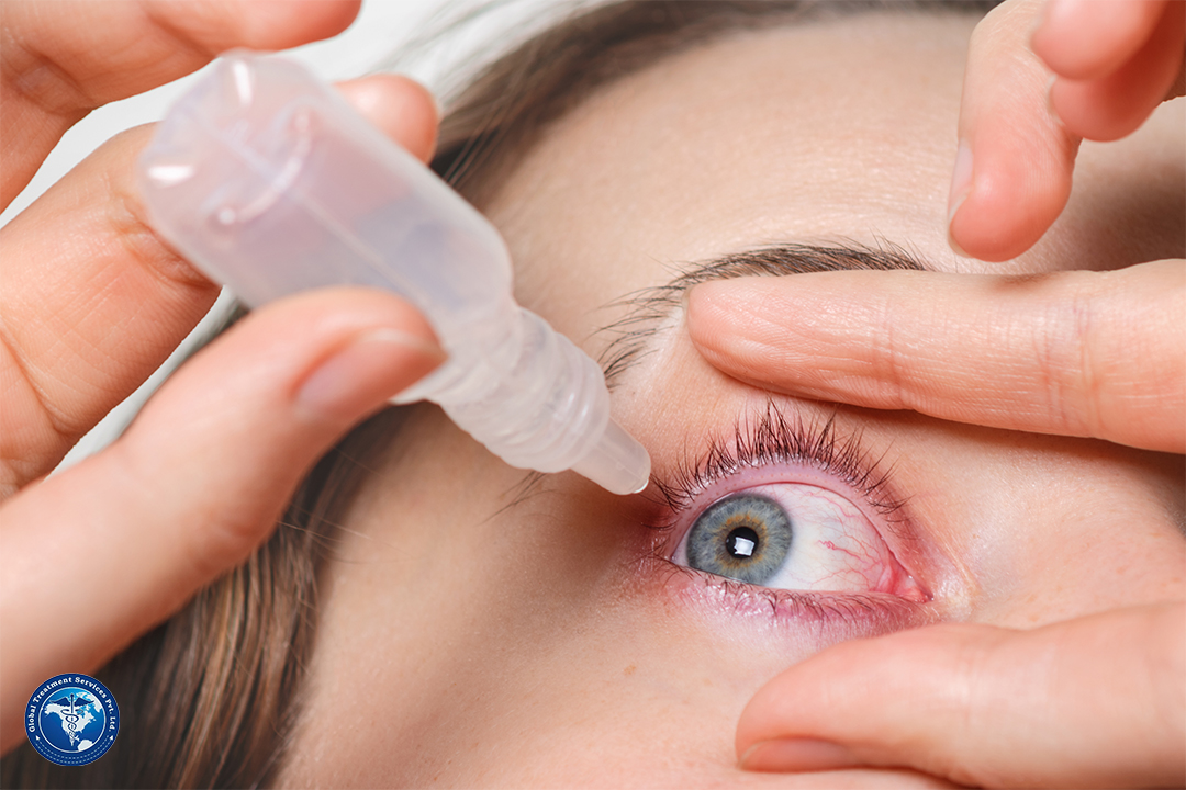
- Dry eyes
- Severe fatigue or overexerting yourself
- Stress
Diagnosis
An eye care specialist/Optholmologist or other provider can usually diagnose eyelid myokymia with a simple physical exam and a neurological exam. These exams let your provider watch the twitching happen and diagnose it or rule out other causes based on what they see.
When myokymia is longer-lasting, more disruptive or starts spreading and getting worse, your eye specialist or other provider will likely recommend other tests. These are usually imaging tests like CT scans or MRIs.
Treatment
The most common treatment approaches include:
- Changing things that could cause or contribute to your myokymia. Some simple examples include limiting how much caffeine you consume, managing your stress or making sure to get enough quality sleep. Limiting alcohol intake and reducing or quitting nicotine (including smoking and vaping, or smokeless forms like chewing tobacco or snuff) may also help. These can also reduce your risk of developing myokymia in the first place.
- Medication changes. If you’re having myokymia because of a medication, your provider may recommend changing to another.
- Medication injections. The most common medication treatments for myokymia are neurotoxins like onabotulinumtoxinA (Botox®). They temporarily block nerve signals traveling to your eyelids. The injection points are all around your eye, just underneath your skin, and this doesn’t involve any injections into your eye itself. These medications paralyze the related muscles, keeping the twitching from happening entirely until the effect wears off.
Eyelid myokymia is usually nothing to worry about. For most people, it’s a minor condition and isn’t enough to affect their usual routines and activities. The twitching from myokymia often lasts only a few seconds to minutes, and it’ll probably go away if you resolve the potential causes or contributing factors like lack of sleep or caffeine intake.
But if eyelid myokymia doesn’t go away after a few weeks or it’s disrupting your life, it’s a good idea to talk to an eye care specialist or your primary care provider. They can help figure out what might be causing your myokymia or refer you to a provider who can diagnose and treat it.
If you have any big concerns related to health you can connect us via email : query@gtsmeditour.com.


:max_bytes(150000):strip_icc()/GettyImages-523913021-59563bd55f9b5815d93c266d.jpg)