Global Treatment Services Pvt. Ltd.

Global Treatment Services

Cerebal Palsy

Overview

Cerebral palsy is a disorder of movement, muscle tone or posture that is caused by damage that occurs to the immature, developing brain, most often before birth.

Signs and symptoms appear during infancy or preschool years. In general, cerebral palsy causes impaired movement associated with abnormal reflexes, floppiness or rigidity of the limbs and trunk, abnormal posture, involuntary movements, unsteady walking, or some combination of these.

People with cerebral palsy may have problems swallowing and commonly have eye muscle imbalance, in which the eyes don’t focus on the same object. People with cerebral palsy also may suffer reduced range of motion at various joints of their bodies due to muscle stiffness.

Cerebral palsy’s effect on functional abilities varies greatly. Some affected people can walk while others can’t. Some people show normal or near-normal intellectual capacity, but others may have intellectual disabilities. Epilepsy, blindness or deafness also may be present.

Image result for cerebral palsy

www.gtsmeditour.com


Symptoms

Signs and symptoms can vary greatly. Movement and coordination problems associated with cerebral palsy may include:

  • Variations in muscle tone, such as being either too stiff or too floppy
  • Stiff muscles and exaggerated reflexes (spasticity)
  • Stiff muscles with normal reflexes (rigidity)
  • Lack of muscle coordination (ataxia)
  • Tremors or involuntary movements
  • Slow, writhing movements (athetosis)
  • Delays in reaching motor skills milestones, such as pushing up on arms, sitting up alone or crawling
  • Favoring one side of the body, such as reaching with only one hand or dragging a leg while crawling
  • Difficulty walking, such as walking on toes, a crouched gait, a scissors-like gait with knees crossing, a wide gait or an asymmetrical gait
  • Excessive drooling or problems with swallowing
  • Difficulty with sucking or eating
  • Delays in speech development or difficulty speaking
  • Difficulty with precise motions, such as picking up a crayon or spoon
  • Seizures

The disability associated with cerebral palsy may be limited primarily to one limb or one side of the body, or it may affect the whole body. The brain disorder causing cerebral palsy doesn’t change with time, so the symptoms usually don’t worsen with age. However, muscle shortening and muscle rigidity may worsen if not treated aggressively.

Brain abnormalities associated with cerebral palsy also may contribute to other neurological problems. People with cerebral palsy may also have:

  • Difficulty with vision and hearing
  • Intellectual disabilities
  • Seizures
  • Abnormal touch or pain perceptions
  • Oral diseases
  • Mental health (psychiatric) conditions
  • Urinary incontinence

Causes

Cerebral palsy is caused by an abnormality or disruption in brain development, usually before a child is born. In many cases, the exact trigger isn’t known. Factors that may lead to problems with brain development include:

  • Mutations in genes that lead to abnormal brain development
  • Maternal infections that affect the developing fetus
  • Fetal stroke, a disruption of blood supply to the developing brain
  • Infant infections that cause inflammation in or around the brain
  • Traumatic head injury to an infant from a motor vehicle accident or fall
  • Lack of oxygen to the brain (asphyxia) related to difficult labor or delivery, although birth-related asphyxia is much less commonly a cause than historically thought

Risk factors

A number of factors are associated with an increased risk of cerebral palsy.

Maternal health

Certain infections or health problems during pregnancy can significantly increase cerebral palsy risk to the baby. Infections of particular concern include:

  • German measles (rubella). Rubella is a viral infection that can cause serious birth defects. It can be prevented with a vaccine.
  • Chickenpox (varicella). Chickenpox is a contagious viral infection that causes itching and rashes, and it can cause pregnancy complications. It too can be prevented with a vaccine.
  • Cytomegalovirus. Cytomegalovirus is a common virus that causes flu-like symptoms and may lead to birth defects if a mother experiences her first active infection during pregnancy.
  • Herpes. Herpes infection can be passed from mother to child during pregnancy, affecting the womb and placenta. Inflammation triggered by infection may then damage the unborn baby’s developing nervous system.
  • Toxoplasmosis. Toxoplasmosis is an infection caused by a parasite found in contaminated food, soil and the feces of infected cats.
  • Syphilis. Syphilis is a sexually transmitted bacterial infection.
  • Exposure to toxins. Exposure to toxins, such as methyl mercury, can increase the risk of birth defects.
  • Zika virus infection. Infants for whom maternal Zika infection causes microcephaly can develop cerebral palsy.
  • Other conditions. Other conditions may increase the risk of cerebral palsy, such as thyroid problems, intellectual disabilities or seizures.

Infant illness

Illnesses in a newborn baby that can greatly increase the risk of cerebral palsy include:

  • Bacterial meningitis. This bacterial infection causes inflammation in the membranes surrounding the brain and spinal cord.
  • Viral encephalitis. This viral infection similarly causes inflammation in the membranes surrounding the brain and spinal cord.
  • Severe or untreated jaundice. Jaundice appears as a yellowing of the skin. The condition occurs when certain byproducts of “used” blood cells aren’t filtered from the bloodstream.

Other factors of pregnancy and birth

While the potential contribution from each is limited, additional pregnancy or birth factors associated with increased cerebral palsy risk include:

  • Breech births. Babies with cerebral palsy are more likely to be in a feet-first position (breech presentation) at the beginning of labor rather than headfirst.
  • Complicated labor and delivery. Babies who exhibit vascular or respiratory problems during labor and delivery may have existing brain damage or abnormalities.
  • Low birth weight. Babies who weigh less than 5.5 pounds (2.5 kilograms) are at higher risk of developing cerebral palsy. This risk increases as birth weight drops.
  • Multiple babies. Cerebral palsy risk increases with the number of babies sharing the uterus. If one or more of the babies die, the chance that the survivors may have cerebral palsy increases.
  • Premature birth. A normal pregnancy lasts 40 weeks. Babies born fewer than 37 weeks into the pregnancy are at higher risk of cerebral palsy. The earlier a baby is born, the greater the cerebral palsy risk.
  • Rh blood type incompatibility between mother and child. If a mother’s Rh blood type doesn’t match her baby’s, her immune system may not tolerate the developing baby’s blood type and her body may begin to produce antibodies to attack and kill her baby’s blood cells, which can cause brain damage.

Complications

Muscle weakness, muscle spasticity and coordination problems can contribute to a number of complications either during childhood or later during adulthood, including:

  • Contracture. Contracture is muscle tissue shortening due to severe muscle tightening (spasticity). Contracture can inhibit bone growth, cause bones to bend, and result in joint deformities, dislocation or partial dislocation.
  • Malnutrition. Swallowing or feeding problems can make it difficult for someone who has cerebral palsy, particularly an infant, to get enough nutrition. This may cause impaired growth and weaker bones. Some children may need a feeding tube for adequate nutrition.
  • Mental health conditions. People with cerebral palsy may have mental health (psychiatric) conditions, such as depression. Social isolation and the challenges of coping with disabilities can contribute to depression.
  • Lung disease. People with cerebral palsy may develop lung disease and breathing disorders.
  • Neurological conditions. People with cerebral palsy may be more likely to develop movement disorders or worsened neurological symptoms over time.
  • Osteoarthritis. Pressure on joints or abnormal alignment of joints from muscle spasticity may lead to the early onset of painful degenerative bone disease (osteoarthritis).
  • Osteopenia. Fractures due to low bone density (osteopenia) can stem from several common factors such as lack of mobility, nutritional shortcomings and antiepileptic drug use.
  • Eye muscle imbalance. This can affect visual fixation and tracking; an eye specialist should evaluate suspected imbalances.

Prevention

Most cases of cerebral palsy can’t be prevented, but you can lessen risks. If you’re pregnant or planning to become pregnant, you can take these steps to keep healthy and minimize pregnancy complications:

  • Make sure you’re vaccinated. Vaccination against diseases such as rubella may prevent an infection that could cause fetal brain damage.
  • Take care of yourself. The healthier you are heading into a pregnancy, the less likely you’ll be to develop an infection that may result in cerebral palsy.
  • Seek early and continuous prenatal care. Regular visits to your doctor during your pregnancy are a good way to reduce health risks to you and your unborn baby. Seeing your doctor regularly can help prevent premature birth, low birth weight and infections.
  • Practice good child safety. Prevent head injuries by providing your child with a car seat, bicycle helmet, safety rails on beds and appropriate supervision.

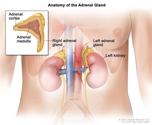
Adrenalectomy

Overview

Image result for adrenalectomy

Adrenal glands

One adrenal gland sits above each of your kidneys. Your two adrenal glands produce various hormones that help regulate your metabolism, immune system, blood pressure, blood sugar and other essential functions.

Most adrenal tumors are noncancerous (benign). You may require surgery to remove an adrenal gland if the tumor is producing excess hormones or is large in size (more than 2 inches or 4 to 5 centimeters). You may also require surgery if the tumor is cancerous (malignant) or suspected to be cancerous. This surgery is called adrenalectomy (uh-dree-nul-EK-tuh-me).You may also need an adrenalectomy to remove cancer that has spread (metastasized) from another location, such as the kidney or lung.

If both adrenal glands are removed, you will need to take supplemental hormone medications. If only one gland is removed, the other will take over full function without the need for ongoing medications.

Types of adrenalectomy

Surgeons may perform an adrenalectomy through minimally invasive (laparoscopic) or traditional open surgery, or use cryoablation. The procedure they recommend depends on the size and type of tumor or the condition affecting your adrenal gland.

  • Minimally invasive surgery. Endocrine surgeons at Mayo Clinic are often able to use minimally invasive (laparoscopic) surgery for tumors of the adrenal gland because the gland is relatively small. Laparoscopic surgery has many benefits, including smaller scars, less pain and a shorter recovery period than traditional open surgery.

    An alternative approach to laparoscopic surgery is a posterior retroperitoneoscopic adrenalectomy (PRA), in which surgeons make small incisions in your back. Sometimes surgeons use the robotic da Vinci Surgical System to perform laparoscopic adrenalectomies.

  • Open surgery. Doctors usually reserve open surgery for large or cancerous (malignant) tumors. They perform open surgery using traditional instruments and incisions.
  • Cryoablation. In addition, Mayo Clinic doctors may use cryoablation to treat adrenal tumors. This procedure uses CT imaging to guide the insertion of a probe that freezes and destroys adrenal tumors. Interventional radiologists may use cryoablation as a treatment option for small tumors that have spread to the adrenal gland (metastasis), particularly when surgery carries a high risk.

Your doctor can explain your treatment options and discuss whether an adrenalectomy is the most appropriate treatment for you.

Autism Spectrum Disorder

Overview

Autism spectrum disorder is a condition related to brain development that impacts how a person perceives and socializes with others, causing problems in social interaction and communication. The disorder also includes limited and repetitive patterns of behaviour. The term “spectrum” in autism spectrum disorder refers to the wide range of symptoms and severity.

Autism spectrum disorder includes conditions that were previously considered separate — autism, Asperger’s syndrome, childhood disintegrative disorder and an unspecified form of pervasive developmental disorder. Some people still use the term “Asperger’s syndrome,” which is generally thought to be at the mild end of autism spectrum disorder.

Autism spectrum disorder begins in early childhood and eventually causes problems functioning in society — socially, in school and at work, for example. Often children show symptoms of autism within the first year. A small number of children appear to develop normally in the first year, and then go through a period of regression between 18 and 24 months of age when they develop autism symptoms.

While there is no cure for autism spectrum disorder, intensive, early treatment can make a big difference in the lives of many children.

Image result for autism spectrum disorder

Symptoms

Some children show signs of autism spectrum disorder in early infancy, such as reduced eye contact, lack of response to their name or indifference to caregivers. Other children may develop normally for the first few months or years of life, but then suddenly become withdrawn or aggressive or lose language skills they’ve already acquired. Signs usually are seen by age 2 years.

Each child with autism spectrum disorder is likely to have a unique pattern of behaviour and level of severity — from low functioning to high functioning.

Some children with autism spectrum disorder have difficulty learning, and some have signs of lower than normal intelligence. Other children with the disorder have normal to high intelligence — they learn quickly, yet have trouble communicating and applying what they know in everyday life and adjusting to social situations.

Because of the unique mixture of symptoms in each child, severity can sometimes be difficult to determine. It’s generally based on the level of impairments and how they impact the ability to function.

MICRO Vascular Decompression

Overview

Microvascular decompression (MVD) is a surgery to relieve abnormal compression of a cranial nerve causing trigeminal neuralgia, glossopharyngeal neuralgia, or hemifacial spasm. MVD involves opening the skull (craniotomy) and inserting a sponge between the nerve and offending artery triggering the pain signals. Medications often provide initial relief, but when drugs become ineffective or cause side effects, MVD is an option.

What is microvascular decompression (MVD)?

MVD is a surgical procedure to relieve the symptoms (pain, muscle twitching) caused by compression of a nerve by an artery or vein. Surgery involves opening the skull (craniotomy) and exposing the nerve at the base of the brainstem to insert a tiny sponge between the compressing vessel and the nerve. This sponge isolates the nerve from the pulsating effect and pressure of the blood vessel.

Trigeminal neuralgia is an irritation of the fifth cranial nerve causing severe pain that usually affects one side of the face, normally in the forehead, cheek, jaw, or teeth (Fig. 1). To treat trigeminal neuralgia, a sponge is placed between the trigeminal nerve and the superior cerebellar artery or a branch of the petrosal vein. By removing the compression, the painful symptoms are relieved.

Figure 1. Trigeminal neuralgia can be caused by an artery or vein compressing the trigeminal nerve root as it originates at the brainstem

What happens during surgery?

Patients are admitted to the hospital the morning of the procedure. You will meet with a nurse who will ask your name, date of birth, what procedure you’re having, and the side of your facial pain. They will explain the pre-op process and discuss any questions you may have. An anesthesiologist will meet with you and explain the effects of anesthesia and its risks. They will place an intravenous (IV) line in your arm and then transport you to the operating room.

There are 5 steps of the procedure. The operation generally takes 2 to 3 hours.

Step 1: prepare the patient
In the OR room, general anesthesia is administered while you lie on the operating table. Once asleep, your body is rolled over on its side and your head is placed in a 3-pin skull fixation device, which attaches to the table and holds your head in position during the procedure. Next, the area behind your ear is prepped with antiseptic. A hair-sparing technique may be used, where only a 1/4-inch wide area along the proposed skin incision is shaved.

Step 2: perform a craniectomy
A 3-inch curved skin incision is made behind the ear. The skin and muscles are lifted off the bone and folded back. Next, a 1-inch opening is made in the occipital bone with a drill (Fig. 2). The bone is removed to expose the protective covering of the brain called the dura. The dura is opened with surgical scissors and folded back to expose the brain.

Figure 2. A skin incision is made behind the ear (dashed line) and a 1-inch craniectomy (solid line) is made in the skull.

Step 3: expose the nerve
Retractors placed on the brain gently open a corridor to the trigeminal nerve at its origin with the brainstem. The surgeon exposes the trigeminal nerve and identifies any offending vessel causing compression (Fig. 3). The vessel and nerve are often restricted by thickened connective tissue that must be dissected free with scissors and knife.

Figure 3. The superior cerebellar artery is adherent to the trigeminal nerve causing compression and painful trigeminal neuralgia attacks.

Step 4: insert a sponge
The surgeon cuts an appropriate size of teflon sponge and inserts it between the nerve and the vessel (Fig. 4). Sometimes a vein is adherent to the nerve and causing compression. In these cases, the vein is cauterized and moved away.

Figure 4. A sponge is inserted between the nerve and the blood vessel causing compression.

Step 5: closure
Once the sponge is in place, the retractor is removed and the brain returns to its natural position. The dura is closed with sutures and made watertight with tissue sealant. Since the bone opening is very small, it is not replaced. Instead, a titanium plate covers the skull opening and is secured with tiny screws (Fig. 5). The muscles and skin are sutured back together. A soft adhesive dressing is placed over the incision.


Figure 5. A circular titanium plate secured with screws covers the craniectomy made in the skull.

Mastectomy Surgery (Breast Cancer)

A mastectomy is surgery to remove all breast tissue from a breast as a way to treat or prevent breast cancer.

For those with early-stage breast cancer, a mastectomy may be one treatment option. Breast-conserving surgery (lumpectomy), in which only the tumor is removed from the breast, may be another option.

Deciding between a mastectomy and lumpectomy can be difficult. Both procedures are equally effective for preventing a recurrence of breast cancer. But a lumpectomy isn’t an option for everyone with breast cancer, and others prefer to undergo a mastectomy.

Newer mastectomy techniques can preserve breast skin and allow for a more natural breast appearance following the procedure. This is also known as skin-sparing mastectomy.

Surgery to restore shape to your breast — called breast reconstruction — may be done at the same time as your mastectomy or during a second operation at a later date.

 Image result for mastectomy pictures

Why it’s done

A mastectomy is used to remove all breast tissue if you have breast cancer or are at very high risk of developing it. You may have a mastectomy to remove one breast (unilateral mastectomy) or both breasts (bilateral mastectomy).

Mastectomy for breast cancer treatment

A mastectomy may be a treatment option for many types of breast cancer, including:

  • Ductal carcinoma in situ (DCIS), or noninvasive breast cancer
  • Stages I and II (early-stage) breast cancer
  • Stage III (locally advanced) breast cancer — after chemotherapy
  • Inflammatory breast cancer — after chemotherapy
  • Paget’s disease of the breast
  • Locally recurrent breast cancer

Your doctor may recommend a mastectomy instead of a lumpectomy plus radiation if:

  • You have two or more tumors in separate areas of the breast.
  • You have widespread or malignant-appearing calcium deposits (microcalcifications) throughout the breast that have been determined to be cancer after a breast biopsy.
  • You’ve previously had radiation treatment to the breast region and the breast cancer has recurred in the breast.
  • You’re pregnant and radiation creates an unacceptable risk to your unborn child.
  • You’ve had a lumpectomy, but cancer is still present at the edges (margin) of the operated area and there is concern about cancer extending to elsewhere in the breast.
  • You carry a gene mutation that gives you a high risk of developing a second cancer in your breast.
  • You have a large tumor relative to the overall size of your breast. You may not have enough healthy tissue left after a lumpectomy to achieve an acceptable cosmetic result.
  • You have a connective tissue disease, such as scleroderma or lupus, and may not tolerate the side effects of radiation to the skin.

Mastectomy to prevent breast cancer

You might also consider a mastectomy if you don’t have breast cancer, but have a very high risk of developing the disease.

A preventive (prophylactic) or risk-reducing mastectomy involves removing both of your breasts and significantly reduces your risk of developing breast cancer in the future.

A prophylactic mastectomy is reserved for those with a very high risk of breast cancer, which is determined by a strong family history of breast cancer or the presence of certain genetic mutations that increase the risk of breast cancer.

PAROTID GLAND SWELLING

Your salivary glands make as much as a quart of saliva each day. Saliva is important to lubricate your mouth, help with swallowing, protect your teeth against bacteria, and aid in the digestion of food. The three major pairs of salivary glands are:

  • parotid glands on the insides of the cheeks
  • submandibular glands at the floor of the mouth
  • sublingual glands under the tongue

There are also several hundred minor salivary glands throughout the mouthand throat. Saliva drains into the mouth through small tubes called ducts.

When there is a problem with the salivary glands or ducts, you may have symptoms such as salivary gland swelling, dry mouth, pain, fever, and foul-tasting drainage into the mouth.

Causes of Salivary Gland Problems

Many different problems can interfere with the function of the salivary glands or block the ducts so they can’t drain saliva. The following are some of the more common salivary gland problems:


Mumps_MED_ILL_EN

Salivary stones, or sialoliths. The most common cause of swollen salivary glands, salivary stones are buildups of crystallized saliva deposits. Sometimes salivary stones can block the flow of saliva. When saliva can’t exit through the ducts, it backs up into the gland, causing pain and swelling. Pain is usually off and on, is felt in one gland, and gets progressively worse. Unless the blockage is cleared, the gland is likely to become infected.

Salivary gland infection, or sialadenitis. Bacterial infection of the salivary gland, most commonly the parotid gland, may result when the duct into the mouth is blocked. Sialadenitis creates a painful lump in the gland, and foul-tasting pus drains into the mouth.

Sialadenitis is more common in older adults with salivary stones, but it can also happen in babies during the first few weeks after birth. If not treated, salivary gland infections can cause severe pain, high fevers, and abscess (pus collection).

Infections. Viral infections such as mumps, flu, and others can cause swelling of the salivary glands. Swelling happens in parotid glands on both sides of the face, giving the appearance of “chipmunk cheeks.”

Salivary gland swelling is commonly associated with mumps, happening in about 30% to 40% of mumps infections. It usually begins approximately 48 hours after the start of other symptoms such as fever and headache.