Global Treatment Services Pvt. Ltd.

Global Treatment Services

Gastroparesis: Causes, Symptoms &Treatments

  • Uncontrolled diabetes
  • Gastric surgery with injury to the vagus nerve
  • Medications such as narcotics and some antidepressants
  • Parkinson’s disease
  • Multiple sclerosis
  • Rare conditions such as: amyloidosis (deposits of protein fibers in tissues and organs) and scleroderma (a connective tissue disorder that affects the skin, blood vessels, skeletal muscles, and internal organs).

What Are the Symptoms of Gastroparesis?

There are many symptoms of gastroparesis, including:

  • Heartburn or GERD
  • Nausea
  • Vomiting undigested food
  • Feeling full quickly when eating
  • Abdominal bloating
  • Poor appetite and weight loss
  • Poor blood sugar control

What Are the Complications of Gastroparesis?

Some of the complications of gastroparesis include:

  • Food that stays in the stomach too long can ferment, which can lead to the growth of bacteria.
  • Food in the stomach can harden into a solid collection, called a bezoar. Bezoars can cause obstructions in the stomach that keep food from passing into the small intestine.
  • People who have both diabetes and gastroparesis may have more difficulty because blood sugar levels rise when food finally leaves the stomach and enters the small intestine, making blood sugar control more of a challenge.

How Is Gastroparesis Diagnosed?

To diagnose gastroparesis, your doctor will review your symptoms and medical history. He or she will also give you a physical exam and may order certain blood tests, including blood sugar levels. Other tests used to diagnose and evaluate gastroparesis may include:

  • Barium X-ray : You drink a liquid (barium), which coats the esophagus, stomach, and small intestine and shows up on X-ray. This test is also known as an upper GI (gastrointestinal) series or a barium swallow.
  • Radioisotope gastric-emptying scan (gastric scintigraphy): You eat food that contains a very small amount of radioisotope (a radioactive substance), then lie under a scanning machine; if the scan shows that more than 10% of food is still in your stomach 4 hours after eating, you are diagnosed with gastroparesis.
  • Gastric manometry: A thin tube that is passed through your mouthand into the stomach measures the stomach’s electrical and muscular activity to determine the rate of digestion.
  • Electrogastrography: This test measures electrical activity in the stomach using electrodes placed on the skin.
  • The smart pill: This is a small electronic device that is swallowed. It sends back information about how fast it is traveling as it moves through the digestive system.
  • Ultrasound : This is an imaging test that uses sound waves to create pictures of body organs. Your doctor may use ultrasound to eliminate other diseases.
  • Upper endoscopy : This procedure involves passing a thin tube (endoscope) down the esophagus to examine the lining of the stomach.

What Is the Treatment for Gastroparesis?

Gastroparesis is a chronic (long-lasting) condition. This means that treatment usually doesn’t cure the disease. But there are steps you can take to manage and control the condition.

Some patients may benefit from medications, including:

  • Reglan (metoclopramide): You take this drug before eating and it causes the stomach muscles to contract and move food along. Reglan also decreases the incidence of vomiting and nausea. Side effects include diarrhea, drowsiness, anxiety, and, rarely, a serious neurological disorder.
  • Erythromycin: This is an antibiotic that also causes stomach contractions and helps move food out. Side effects include diarrhea and development of resistant bacteria from prolonged exposure to the antibiotic.
  • Antiemetics: These are drugs that help control nausea.

People who have diabetes should try to control their blood sugar levels to minimize the problems of gastroparesis.

Dietary Modifications for Gastroparesis

One of the best ways to help control the symptoms of gastroparesis is to modify your daily eating habits. For instance, instead of three meals a day, eat six small meals. In this way, there is less food in the stomach; you won’t feel as full, and it will be easier for the food to leave your stomach. Another important factor is the consistency of food; liquids and low residue foods are encouraged (for example, applesauce should replace whole apples with intact skins).

You should also avoid foods that are high in fat (which can slow down digestion) and fiber (which is difficult to digest).

Other Treatment Options for Gastroparesis

In a severe case of gastroparesis, a feeding tube, or jejunostomy tube, may be used. The tube is inserted through the abdomen and into the small intestine during surgery. To feed yourself, put nutrients into the tube, which go directly into the small intestine; this way, they bypass the stomach and get into the bloodstream more quickly.

Using an instrument through a small incision, botulinum toxin (such as Botox) can be injected into the pylorus, the valve that leads from the stomach to the small intestine. This can relax the valve, keeping it open for a longer period of time to allow the stomach to empty.

Another treatment option is intravenous or parenteral nutrition. This is a feeding method in which nutrients go directly into the bloodstream through a catheter placed into a vein in your chest. Parenteral nutrition is intended to be a temporary measure for a severe case of gastroparesis.

Gastritis: Causes, Symptoms & Treatments

What Is Gastritis?

What Causes Gastritis?

Gastritis can be caused by irritation due to excessive alcohol use, chronic vomiting, stress, or the use of certain medicationssuch as aspirin or other anti-inflammatory drugs. It may also be caused by any of the following:

  • Helicobacter pylori (H. pylori): A bacteria that lives in the mucous lining of the stomach; without treatment, the infection can lead to ulcers, and in some people, stomach cancer.
  • Bile reflux: A backflow of bile into the stomach from the bile tract (that connects to the liver and gallbladder)
  • Infections caused by bacteria and viruses

If gastritis is left untreated, it can lead to a severe loss of blood and may increase the risk of developing stomach cancer.

What Are the Symptoms of Gastritis?

Symptoms of gastritis vary among individuals, and in many people there are no symptoms. However, the most common symptoms include:

  • Nausea or recurrent upset stomach
  • Abdominal bloating
  • Abdominal pain
  • Vomiting
  • Indigestion
  • Burning or gnawing feeling in the stomach between meals or at night
  • Hiccups
  • Loss of appetite
  • Vomiting blood or coffee ground-like material
  • Black, tarry stools

How Is Gastritis Diagnosed?

To diagnose gastritis, your doctor will review your personal and family medical history, perform a thorough physical evaluation, and may recommend any of the following tests:

  • Upper endoscopy. An endoscope, a thin tube containing a tiny camera, is inserted through your mouth and down into your stomach to look at the stomach lining. The doctor will check for inflammation and may perform a biopsy, a procedure in which a tiny sample of tissue is removed and then sent to a laboratory for analysis.
  • Blood tests. The doctor may perform various blood tests, such as checking your red blood cell count to determine whether you have anemia, which means that you do not have enough red blood cells. He or she can also screen for H. pylori infection and pernicious anemia with blood tests.
  • Fecal occult blood test (stool test). This test checks for the presence of blood in your stool, a possible sign of gastritis.

What Is the Treatment for Gastritis?

Treatment for gastritis usually involves:

  • Taking antacids and other drugs (such as proton pump inhibitors or H-2 blockers) to reduce stomach acid
  • Avoiding hot and spicy foods
  • For gastritis caused by H. pylori infection, your doctor will prescribe a regimen of several antibiotics plus an acid blocking drug (used for heartburn)
  • If the gastritis is caused by pernicious anemia, B12 vitamin shots will be given.
  • Eliminating irritating foods from your diet such as lactose from dairy or gluten from wheat

Once the underlying problem disappears, the gastritis usually does, too.

You should talk to your doctor before stopping any medicine or starting any gastritis treatment on your own.

Leg Fatty Tumors: Causes and Treatments

Leg Fatty TumorsTt

Fatty tumors in legs come in two different types–benign and malignant. There are three different kinds of benign fat tissue tumors–lipomas, the most common; lipoblastomas, occurring in infants and young children; and hibernomas, which are like lipomas but much less common. A malignant or cancerous soft tissue tumor is called a sarcoma. There are two types: osteosarcomas, which develop from bone, and soft tissue sarcomas which can develop from fat, muscle, nerves, fibrous tissues, blood vessels or deep skin tissues. They can be found in any part of the body, but most occur in the arms or legs. Sarcomas are not common and most are called carcinomas.


Causes, Diagnosis and Treatment of Benign Fatty Tumors

A lipoma is a slow-growing, fatty lump usually situated between your skin and muscle layer. It moves easily with finger pressure, is doughy to the touch and generally not tender. Lipomas can become painful if they grow and press on nerves. Most are detected during middle age. The exact cause of lipomas is not known, but they do tend to run in families, indicating perhaps a genetic predisposition. Diagnosis of this type tumor is usually done with a physical exam or a tissue sample (biopsy) for lab findings. Sometimes an ultrasound or MRI or CT scan is used if the tumor is large, has unusual features or seems to go deeper than the fatty tissue. In a small number of cases the lipoma is actually a form of cancer called a liposarcoma, but these are usually very painful. There is usually no treatment for a lipoma unless it bothers you, is painful or starts to grow. Surgical removal is the most common form of treatment. Steroid injections will shrink the tumor but usually doesn’t eliminate it. Liposuction uses a needle and syringe to remove the lump, but it is difficult to remove the entire lipoma with this method.

Endocrine Disorders:Causes & Types

The endocrine system is a network of glands that produce and release hormones that help control many important body functions, including the body’s ability to change calories into energy that powers cells and organs. The endocrine system influences how your heart beats, how your bones and tissues grow, even your ability to make a baby. It plays a vital role in whether or not you develop diabetes, thyroid disease, growth disorders, sexual dysfunction, and a host of other hormone-related disorders.

Glands of the Endocrine System

Each gland of the endocrine system releases specific hormones into your bloodstream. These hormones travel through your blood to other cells and help control or coordinate many body processes.

Endocrine glands include:

  • Adrenal glands: Two glands that sit on top of the kidneys that release the hormone cortisol.
  • Hypothalamus: A part of the lower middle brain that tells the pituitary gland when to release hormones.
  • Ovaries: The female reproductive organs that release eggs and produce sex hormones.
  • Islet cells in the pancreas: Cells in the pancreas control the release of the hormones insulin and glucagon.
  • Parathyroid: Four tiny glands in the neck that play a role in bone development.
  • Pineal gland: A gland found near the center of the brain that may be linked to sleep patterns.
  • Pituitary gland: A gland found at the base of brain behind the sinuses. It is often called the “master gland” because it influences many other glands, especially the thyroid. Problems with the pituitary gland can affect bone growth, a woman’s menstrual cycles, and the release of breast milk.
  • Testes: The male reproductive glands that produce sperm and sexhormones.
  • Thymus: A gland in the upper chest that helps develop the body’s immune system early in life.
  • Thyroid: A butterfly-shaped gland in the front of the neck that controls metabolism.

Even the slightest hiccup with the function of one or more of these glands can throw off the delicate balance of hormones in your body and lead to an endocrine disorder, or endocrine disease.

Endocrine disorders are typically grouped into two categories:

  • Endocrine disease that results when a gland produces too much or too little of an endocrine hormone, called a hormone imbalance.
  • Endocrine disease due to the development of lesions (such as nodules or tumors) in the endocrine system, which may or may not affect hormone levels.

The endocrine’s feedback system helps control the balance of hormones in the bloodstream. If your body has too much or too little of a certain hormone, the feedback system signals the proper gland or glands to correct the problem. A hormone imbalance may occur if this feedback system has trouble keeping the right level of hormones in the bloodstream, or if your body doesn’t clear them out of the bloodstream properly.

Increased or decreased levels of endocrine hormone may be caused by:

  • A problem with the endocrine feedback system
  • Disease
  • Failure of a gland to stimulate another gland to release hormones (for example, a problem with the hypothalamus can disrupt hormone production in the pituitary gland)
  • A genetic disorder, such as multiple endocrine neoplasia (MEN) or congenital hypothyroidism
  • Infection
  • Injury to an endocrine gland
  • Tumor of an endocrine gland

Most endocrine tumors and nodules (lumps) are noncancerous. They usually do not spread to other parts of the body. However, a tumor or nodule on the gland may interfere with the gland’s hormone production.

Types of Endocrine Disorders

There are many different types of endocrine disorders. Diabetes is the most common endocrine disorder diagnosed in the U.S.

Other endocrine disorders include:

Adrenal insufficiency. The adrenal gland releases too little of the hormone cortisol and sometimes, aldosterone. Symptoms include fatigue, stomachupset, dehydration, and skin changes. Addison’s disease is a type of adrenal insufficiency.

Cushing’s disease. Overproduction of a pituitary gland hormone leads to an overactive adrenal gland. A similar condition called Cushing’s syndromemay occur in people, particularly children, who take high doses of corticosteroid medications.

Gigantism (acromegaly) and other growth hormone problems. If the pituitary gland produces too much growth hormone, a child’s bones and body parts may grow abnormally fast. If growth hormone levels are too low, a child can stop growing in height.

Hyperthyroidism. The thyroid gland produces too much thyroid hormone, leading to weight loss, fast heart rate, sweating, and nervousness. The most common cause for an overactive thyroid is an autoimmune disorder called Grave’s disease.

Hypothyroidism. The thyroid gland does not produce enough thyroid hormone, leading to fatigue, constipation, dry skin, and depression. The underactive gland can cause slowed development in children. Some types of hypothyroidism are present at birth.

Hypopituitarism. The pituitary gland releases little or no hormones. It may be caused by a number of different diseases. Women with this condition may stop getting their periods.

Multiple endocrine neoplasia I and II (MEN I and MEN II). These rare, genetic conditions are passed down through families. They cause tumors of the parathyroid, adrenal, and thyroid glands, leading to overproduction of hormones.

Polycystic ovary syndrome (PCOS). Overproduction of androgens interfere with the development of eggs and their release from the female ovaries. PCOS is a leading cause of infertility.

Precocious puberty. Abnormally early puberty that occurs when glands tell the body to release sex hormones too soon in life.

Testing for Endocrine Disorders

If you have an endocrine disorder, your doctor may refer you to a specialist called an endocrinologist. An endocrinologist is specially trained in problems with the endocrine system.

The symptoms of an endocrine disorder vary widely and depend on the specific gland involved. However, most people with endocrine disease complain of fatigue and weakness.

Blood and urine tests to check your hormone levels can help your doctors determine if you have an endocrine disorder. Imaging tests may be done to help locate or pinpoint a nodule or tumor.

Treatment of endocrine disorders can be complicated, as a change in one hormone level can throw off another. Your doctor or specialist may order routine blood work to check for problems or to determine if your medication or treatment plan needs to be adjusted.

Nipah Virus fever: Causes, Symptoms & Prevention

Nipah Virus (NiV) Infection Grey-headed flying foxes (Pteropus poliocephalus)

Nipah virus (NiV) infection is a newly emerging zoo-nosis that causes severe disease in both animals and humans. The natural host of the virus are fruit bats of the Pteropodidae Family, Pteropus genus.

NiV was first identified during an outbreak of disease that took place in Kampung Sungai Nipah, Malaysia in 1998. On this occasion, pigs were the intermediate hosts. However, in subsequent NiV outbreaks, there were no intermediate hosts. In Bangladesh in 2004, humans became infected with NiV as a result of consuming date palm sap that had been contaminated by infected fruit bats. Human-to-human transmission has also been documented, including in a hospital setting in India.

Transmission:

Transmission of Nipah virus to humans may occur after direct contact with infected bats, infected pigs, or from other NiV infected people.

In Malaysia and Singapore, humans were apparently infected with Nipah virus only through close contact with infected pigs. The NiV strain identified in this outbreak appeared to have been transmitted initially from bats to pigs, with subsequent spread within pig populations. Incidental human infections resulted after exposure to infected pigs. No occurrence of person-to-person transmission was reported in this outbreak.

Conversely, person-to-person transmission of Nipah virus in Bangladesh and India is regularly reported. This is most commonly seen in the family and caregivers of Nipah virus-infected patients. Transmission also occurs from direct exposure to infected bats. A common example is consumption of raw date palm sap contaminated with infectious bat excretions.

Signs and Symptoms:

 Infection with Nipah virus is associated with encephalitis (inflammation of the brain). After exposure and an incubation period of 5 to 14 days,illness presents with 3-14 days of fever and headache, followed by drowsiness, disorientation and mental confusion. These signs and symptoms can progress to coma within 24-48 hours. Some patients have a respiratory illness during the early part of their infections, and half of the patients showing severe neurological signs showed also pulmonary signs.

During the Nipah virus disease outbreak in 1998-99, 265 patients were infected with the virus. About 40% of those patients who entered hospitals with serious nervous disease died from the illness.

Long-term sequelae following Nipah virus infection have been noted, including persistent convulsions and personality changes.

Latent infections with subsequent reactivation of Nipah virus and death have also been reported months and even years after exposure.

Diagnosis:

Treatment:

Treatment is limited to supportive care. Because Nipah virus encephalitis can be transmitted person-to-person, standard infection control practices and proper barrier nursing techniques are important in preventing hospital-acquired infections (nosocomial transmission). Doctors from neurology departments will provide better care.

The drug ribavirin has been shown to be effective against the viruses in vitro, but human investigations to date have been inconclusive and the clinical usefulness of ribavirin remains uncertain.

Passive immunization using a human monoclonal antibody targeting the Nipah G glycoprotein has been evaluated in the post-exposure therapy in the ferret model and found to be of benefit.

Prevention:

Additional efforts focused on surveillance and awareness will help prevent future outbreaks. Research is needed to better understand the ecology of bats and Nipah virus, investigating questions such as the seasonality of disease within reproductive cycles of bats. Surveillance tools should include reliable laboratory assays for early detection of disease in communities and livestock, and raising awareness of transmission and symptoms is important in reinforcing standard infection control practices to avoid human-to-human infections in hospital settings (nosocomial infection).

A subunit vaccine, using the Hendra G protein, produces cross-protective antibodies against HENV and NIPV has been recently used in Australia to protect horses against Hendra virus. This vaccine offers great potential for henipavirus protection in humans as well.

Subdural Hematoma: Causes, Symptoms & Treatments

A subdural hematoma is a collection of blood outside the brain. Subdural hematomas are usually caused by severe head injuries. The bleeding and increased pressure on the brainfrom a subdural hematoma can be life-threatening. Some subdural hematomas stop and resolve spontaneously; others require surgical drainage.

What Is a Subdural Hematoma?

In a subdural hematoma, blood collects between the layers of tissue that surround the brain. The outermost layer is called the dura. In a subdural hematoma, bleeding occurs between the dura and the next layer, the arachnoid.

The bleeding in a subdural hematoma is under the skull and outside the brain, not in the brain itself. As blood accumulates, however, pressure on the brain increases. The pressure on the brain causes a subdural hematoma’s symptoms. If pressure inside the skull rises to very high level, a subdural hematoma can lead to unconsciousness and death.

Causes of Subdural Hematoma

Subdural hematoma is usually caused by a head injury, such as from a fall, motor vehicle collision, or an assault. The sudden blow to the head tears blood vessels that run along the surface of the brain. This is referred to as an acute subdural hematoma.

People with a bleeding disorder and people who take blood thinners are more likely to develop a subdural hematoma. A relatively minor head injury can cause subdural hematoma in people with a bleeding tendency.

In a chronic subdural hematoma, small veins on the outer surface of the brain may tear, causing bleeding in the subdural space. Symptoms may not be apparent for several days or weeks. Elderly people are at higher risk for chronic subdural hematoma because brain shrinkage causes these tiny veins to be more stretched and more vulnerable to tearing.

Symptoms of Subdural Hematoma

Symptoms of subdural hematoma depend mostly on the rate of bleeding:

  • In head injuries with sudden, severe bleeding causing a subdural hematoma, a person may lose consciousness and become comatose immediately.
  • A person may appear normal for days after a head injury, but slowly become confused and then unconscious several days later. This results from a slower rate of bleeding, causing a slowly enlarging subdural hematoma.
  • In very slow-growing subdural hematomas, there may be no noticeable symptoms for more than two weeks after the bleeding starts.

Symptoms of subdural hematoma can include:

  • Headache
  • Confusion
  • Change in behavior
  • Dizziness
  • Nausea and vomiting
  • Lethargy or excessive drowsiness
  • Weakness
  • Apathy
  • Seizures

People may vary widely in their symptoms of subdural hematoma. Besides the size of the subdural hematoma, a person’s age and other medical conditions can affect the response to having a subdural hematoma.

Diagnosis of Subdural Hematoma

People who come to medical attention after a head injury often undergo head imaging, usually with computed tomography (CT scan) or magnetic resonance imaging (MRI scan). These tests create images of the interior of the skull, usually detecting any subdural hematoma present. MRI is slightly superior to CT in detecting subdural hematoma, but CT is faster and more readily available.

Rarely, angiography may be used to diagnose subdural hematoma. During angiography (angiogram), a catheter is inserted through an artery in the groin and threaded into the arteries of he neck and brain. Special dye is then injected, and an X-ray screen shows blood flow through the arteries and veins.

Treatment of Subdural Hematoma

Treatment of subdural hematomas depends on their severity. Treatment can range from watchful waiting to brain surgery.

In small subdural hematomas with mild symptoms, doctors may recommend no specific treatment other than observation. Repeated head imaging tests are often performed to monitor whether the subdural hematoma is improving.

More severe or dangerous subdural hematomas require surgery to reduce the pressure on the brain. Surgeons can use various techniques to treat subdural hematomas:

  • Burr hole trephination. A hole is drilled in the skull over the area of the subdural hematoma, and the blood is suctioned out through the hole.
  • Craniotomy. A larger section of the skull is removed, to allow better access to the subdural hematoma and reduce pressure. The removed skull is replaced shortly after the procedure.
  • Craniectomy. A section of the skull is removed for an extended period of time, to allow the injured brain to expand and swell without permanent damage. Craniectomy is not often used to treat subdural hematoma.

People with severe subdural hematomas are often seriously ill, requiring machine-supported breathing and other forms of life support.

If a person has a bleeding problem or is taking blood thinners, measures should be taken to improve blood clotting. This may include giving medicines or blood products, and reversal of any blood thinners, when possible. Other medications to help reduce swelling or pressure in the brain or control seizures may also be used.

Epilepsy: Causes, Symptoms & Treatments

  • Epilepsy is a chronic disorder of the brain that affects people of all ages.
  • Approximately 50 million people worldwide have epilepsy, making it one of the most common neurological diseases globally.
  • Nearly 80% of the people with epilepsy live in low- and middle-income countries.
  • People with epilepsy respond to treatment approximately 70% of the time.
  • About three fourths of people with epilepsy living in low- and middle- income countries do not get the treatment they need.
  • In many parts of the world, people with epilepsy and their families suffer from stigma and discrimination.
Epilepsy is a chronic disorder of the brain that affects people worldwide. It is characterized by recurrent seizures, which are brief episodes of involuntary movement that may involve a part of the body (partial) or the entire body (generalized), and are sometimes accompanied by loss of consciousness and control of bowel or bladder function.

Seizure episodes are a result of excessive electrical discharges in a group of brain cells. Different parts of the brain can be the site of such discharges. Seizures can vary from the briefest lapses of attention or muscle jerks to severe and prolonged convulsions. Seizures can also vary in frequency, from less than 1 per year to several per day.

One seizure does not signify epilepsy (up to 10% of people worldwide have one seizure during their lifetime). Epilepsy is defined as having 2 or more unprovoked seizures. Epilepsy is one of the world’s oldest recognized conditions, with written records dating back to 4000 BC. Fear, misunderstanding, discrimination and social stigma have surrounded epilepsy for centuries. This stigma continues in many countries today and can impact on the quality of life for people with the disorder and their families.

Signs and symptoms

Characteristics of seizures vary and depend on where in the brain the disturbance first starts, and how far it spreads. Temporary symptoms occur, such as loss of awareness or consciousness, and disturbances of movement, sensation (including vision, hearing and taste), mood, or other cognitive functions.

People with seizures tend to have more physical problems (such as fractures and bruising from injuries related to seizures), as well as higher rates of psychological conditions, including anxiety and depression. Similarly, the risk of premature death in people with epilepsy is up to 3 times higher than the general population, with the highest rates found in low- and middle-income countries and rural versus urban areas.

A great proportion of the causes of death related to epilepsy in low- and middle-income countries are potentially preventable, such as falls, drowning, burns and prolonged seizures.

Causes

Epilepsy is not contagious. The most common type of epilepsy, which affects 6 out of 10 people with the disorder, is called idiopathic epilepsy and has no identifiable cause.

Epilepsy with a known cause is called secondary epilepsy, or symptomatic epilepsy. The causes of secondary (or symptomatic) epilepsy could be:

  • brain damage from prenatal or perinatal injuries (e.g. a loss of oxygen or trauma during birth, low birth weight),
  • congenital abnormalities or genetic conditions with associated brain malformations,
  • a severe head injury,
  • a stroke that restricts the amount of oxygen to the brain,
  • an infection of the brain such as meningitis, encephalitis, neurocysticercosis,
  • certain genetic syndromes,
  • a brain tumor.

Treatment

Epilepsy can be treated easily and affordably with inexpensive daily medication that costs as little as US$ 5 per year. Recent studies in both low- and middle-income countries have shown that up to 70% of children and adults with epilepsy can be successfully treated (i.e. their seizures completely controlled) with anti-epileptic drugs (AEDs). Furthermore, after 2 to 5 years of successful treatment and being seizure-free, drugs can be withdrawn in about 70% of children and 60% of adults without subsequent relapse.

  • In low- and middle-income countries, about three fourths of people with epilepsy may not receive the treatment they need. This is called the “treatment gap”.
  • In many low- and middle-income countries, there is low availability of AEDs. A recent study found the average availability of generic antiepileptic medicines in the public sector of low- and middle-income countries to be less than 50%. This may act as a barrier to accessing treatment.
  • It is possible to diagnose and treat most people with epilepsy at the primary health- care level without the use of sophisticated equipment.
  • WHO demonstration projects have indicated that training primary health-care providers to diagnose and treat epilepsy can effectively reduce the epilepsy treatment gap. However, the lack of trained health-care providers can act as a barrier to treatment for people with epilepsy.
  • Surgical therapy might be beneficial to patients who respond poorly to drug treatments.

Prevention

Idiopathic epilepsy is not preventable. However, preventive measures can be applied to the known causes of secondary epilepsy.

  • Preventing head injury is the most effective way to prevent post-traumatic epilepsy.
  • Adequate perinatal care can reduce new cases of epilepsy caused by birth injury.
  • The use of drugs and other methods to lower the body temperature of a feverish child can reduce the chance of febrile seizures.
  • Central nervous system infections are common causes of epilepsy in tropical areas, where many low- and middle-income countries are concentrated.
  • Elimination of parasites in these environments and education on how to avoid infections can be effective ways to reduce epilepsy worldwide, for example those cases due to neurocysticercosis.

Social and economic impacts

Epilepsy accounts for 0.6%, of the global burden of disease, a time-based measure that combines years of life lost due to premature mortality and time lived in less than full health. Epilepsy has significant economic implications in terms of health care needs, premature death and lost work productivity.

An Indian study conducted in 1998 calculated that the cost per patient of epilepsy treatment was as high as 88.2% of the country’s per capita Gross National Product (GNP), and epilepsy-related costs, which included medical costs, travel, and lost work time, exceeded $2.6 billion/year (2013 USD).(1)

Although the social effects vary from country to country, the discrimination and social stigma that surround epilepsy worldwide are often more difficult to overcome than the seizures themselves. People living with epilepsy can be targets of prejudice. The stigma of the disorder can discourage people from seeking treatment for symptoms, so as to avoid becoming identified with the disorder.

Hepatitis A : Causes, Symptoms & Treatments

What is hepatitis A?

Hepatitis A is a serious liver disease caused by infection with the hepatitis A virus. How is hepatitis A virus spread? Hepatitis A virus is usually spread from getting particles of fecal material (poop) into your mouth that are too small to be seen. This can happen through household or sexual contact with an infected person or by eating hepatitis A virus-contaminated food or drinking hepatitis A virus contaminated water. Casual contact, such as in a school or work setting, has not been known to spread hepatitis A virus.

What are the symptoms of hepatitis A virus infection?

Infected people can have no symptoms at all or be extremely ill. Only 30% of children less than six years of age develop symptoms, while 70% of older children and adults develop symptoms. If a person does develop symptoms, they might include fever, tiredness, loss of appetite, nausea, abdominal pain, dark urine, or jaundice (yellowing of the eyes and skin). These symptoms can last up to six months. Even without symptoms, people infected with hepatitis A virus can spread the infection to others.

How serious is hepatitis A virus infection?

About 30% of people with hepatitis A virus require hospitalization. Adults who become ill often miss several weeks of work. Although deaths due to hepatitis A virus are uncommon, death still can occur from overwhelming hepatitis A virus infection that causes liver failure.

How can hepatitis A virus infection be prevented?

Safe and effective vaccines to prevent hepatitis A virus infection have been available now. Good hand washing will also help reduce the spread of hepatitis A virus. Always wash your hands with soap and water after using the toilet, changing a diaper, and before preparing or eating food.

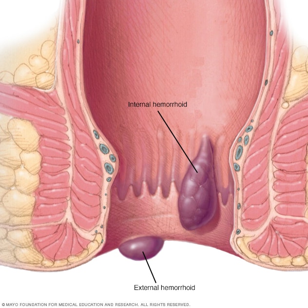
External Hermorrhoids: Causes, Symptoms & Treatments

What causes external hemorrhoids?

The most common cause of external hemorrhoids is repeated straining while having a bowel movement. Hemorrhoids develop when the veins of the rectum or anus become dilated or enlarged and can be either “internal” or “external.” External hemorrhoids are usually found beneath the skin that surrounds the anus.

SYMPTOMS
There is a range of symptoms that can affect a person with hemorrhoids. Symptoms tend to vary depending on the severity of your hemorrhoids. Some of the symptoms that you may have include the following:
  • itching around the anus or rectal area
  • pain around the anus
  • lumps near or around the anus
  • blood in the stool

You may notice bleeding when using the bathroom. This includes seeing blood on toilet paper or in the toilet. Lumps around the anus may feel as if they are swollen.

CAUSES:
What are the causes of external hemorrhoids?

The most common cause of hemorrhoids is repeated straining while having a bowel movement. This is often caused by severe cases of constipation or diarrhea. Straining gets in the way of blood flow into and out of the area. This results in the pooling of blood and enlargement of the vessels in that area.

Pregnant women may also be at an increased risk of hemorrhoids because of the pressure that the uterus places on these veins.

Treating external hemorrhoids:

Hemorrhoids can be treated a few ways depending on severity. Your doctor may ask if you have any preference for certain types of medications or treatments.

Some general treatments that your doctor might suggest include ice packs to reduce swelling, suppositories, or hemorrhoid creams.

These options can offer relief to individuals who have a milder case of hemorrhoids. If you have a more severe case, your doctor may suggest treatment with a surgical procedure.

Surgical treatments include:

  • removal of hemorrhoids, known as hemorrhoidectomy
  • burning of hemorrhoid tissue with infrared photo, laser, or electrical coagulation
  • sclerotherapy or rubber band ligation to reduce the hemorrhoids
PREVENTION:

The main factor for preventing external hemorrhoids is to avoid straining during bowel movements. If you have severe constipation, you may want to try using aids such as laxativesor including more fiber in your diet.

Stool softeners are another popular over-the-counter option that can help you with temporary constipation due to pregnancy or other factors.  If these options don’t help, you may want to talk with your doctor about other options to reduce strain.

Fibroid in Uterus: Symptoms & Treatments

If you have uterine fibroids, you may or may not need treatment. It depends on whether they cause you any problems.

Not all fibroids grow. Even large ones may not cause any symptoms, and most shrink after menopause.

Still, you and your doctor should check on their growth, especially if you develop symptoms like bleeding or pain. So you should at least get pelvic exams every year.

Home Remedies?

You can’t cure fibroids on your own. But you can do things that help you feel better. When fibroids grow on the outside of the uterus, you may become aware of a mass on your abdomen. You can lie down and put a hot pack or hot water bottle on your lower belly to ease the pain. You’d need to do this several times a day.

What Medicines Help?

You can take pain relievers, such as ibuprofen. But be sure to follow the instructions on the label so you don’t accidentally take too much. If you and your doctor decide you need to take something for your fibroids, you may want to consider these other options:

  • Hormone therapy. To help prevent more growth of the fibroid, your doctor may recommend that you stop taking birth control pills or hormone replacement therapy. But in some cases, your doctor may prescribe birth control pills to help control the bleeding and anemiafrom fibroids, even though the hormones may cause fibroids to grow.
  • GnRH agonists. GnRH is a hormone your body naturally makes. An “agonist” medicine counters that hormone, and your doctor may prescribe one to shrink fibroids and reduce anemia. These drugs are expensive. You shouldn’t take them for more than 6 months because they can make you more likely to get osteoporosis, which makes your bones too frail. Your doctor may also prescribe a low dose of progestin, another hormone, to make osteoporosis less likely. (When you stop taking a GnRH agonist, your fibroids can grow back).
  • SERMs. SERMs are a type of medicine that works on your estrogen levels. (SERMs stands for selective estrogen receptor modulators.) They may be able to shrink fibroids without causing menopause symptoms. But researchers aren’t yet sure how well they work for this purpose.

What Procedures Might Work?

There are several possibilities that you and your doctor can consider.

  • Fibroid embolization can shrink a fibroid. Your doctor will inject polyvinyl alcohol (PVA) into the arteries that feed the fibroid. The PVA blocks the blood supply to the fibroid, which makes it shrink. It’s not surgery, but you may need to spend several nights in the hospital because you may have nausea, vomiting, and pain in the first few days afterward.
  • Endometrial ablation is a procedure in which doctors destroy the lining of uterus to cut down on the bleeding linked to small fibroids.
  • Myomectomy is a surgery to remove fibroids. If you plan to become pregnant, your doctor may recommend this over other procedures. But it may cause scarring that can lead to infertility. You’ll need to wait 4 to 6 months after surgery before you try to conceive. In most women, symptoms go away following a myomectomy. But in others, the fibroids come back. Whether it works will partly depend on how many fibroids you have and whether the surgeon could remove them all. A myomectomy may be abdominal surgery, or your surgeon may use a hysteroscope or laparoscope to remove the fibroids without having to make a large cut on your abdomen. There is also an experimental method that uses MRI-guided intense ultrasound energy to pinpoint the fibroids and shrink or destroy them.
  • Hysterectomy is surgery to remove the uterus. Many women don’t need treatment that’s this drastic. You won’t be able to get pregnantafter this operation.
Pages:12