Global Treatment Services Pvt. Ltd.

Global Treatment Services

VP SHUNT Surgery

Ventricular-operational shunting

Ventricular-operational shunting is surgery to treat excess cerebro spinal fluid (CSF) in the cavities (ventricles) of the brain (hydrocephalus).

Description

This procedure is done in the operating room under general anesthesia. It takes about 1 1/2 hours. A tube (catheter) is passed from the cavities of the head to the abdomen to get rid of the excess cerebrospinal fluid (CSF). A pressure valve and an anti-syphon device ensure that just the right amount of fluid is drained.

The procedure is done as follows:

  • An area of hair on the head is shaved. This may be behind the ear or on the top or back of the head.
  • The surgeon makes a skin incision behind the ear. Another small surgical cut is made in the belly.
  • A small hole is drilled in the skull. One end of the catheter is passed into a ventricle of the brain. This can be done with or without a computer as a guide. It can also be done with an endoscope that allows the surgeon to see inside the ventricle.
  • A second catheter is placed under the skin behind the ear. It is sent down the neck and chest, and usually into the belly area. Sometimes, it stops at the chest area. In the belly, the catheter is often placed using an endoscope. The doctor may also make a few more small cuts, for instance in the neck or near the collarbone, to help pass the catheter under the skin.
  • A valve is placed underneath the skin, usually behind the ear. The valve is connected to both catheters. When extra pressure builds up around the brain, the valve opens, and excess fluid drains through the catheter into the belly or chest area. This helps lower intracranial pressure. A reservoir on the valve allows for priming (pumping) of the valve and for collecting the CSF if needed.
  • The person is taken to a recovery area and then moved to a hospital room.

Down’s Syndrome in Children

People with Down syndrome tend to have certain physical features in common. For example, they often have flat noses and small ears.

Their mental abilities will vary, but most have mild to moderate issues with thinking, reasoning, and understanding. They’ll learn and pick up new skills their whole lives, but may take longer to reach important goals like walking, talking, and developing social skills.

Many people with Down syndrome don’t have any other health issues, but some do. Common conditions include heart problems and trouble hearing and seeing.

Causes

Normally, each cell in your body has 23 pairs of chromosomes. One chromosome in each pair comes from your mother. The other comes from your father.

But with Down syndrome, something goes wrong and you get an extra copy of chromosome 21. That means you have three copies instead of two, which leads to the signs and symptoms of Down syndrome. Doctors aren’t sure why this happens.

It varies, but people with Down syndrome often share certain physical traits.

For facial features, they may have:

  • Eyes shaped like almonds (may be shaped in a way that’s not typical for their ethnic group)
  • Flatter faces, especially the nose
  • Small ears, which may fold over a bit at the top
  • Tiny white spots in the coloured part of their eyes
  • A tongue that sticks out of the mouth

Proton treatment

Proton therapy, also called proton beam therapy, is a type of radiation therapy. It uses protons rather than x-rays to treat cancer.

A proton is a positively charged particle. At high energy, protons can destroy cancer cells. Doctors may use proton therapy alone. They may also combine it with x-ray radiation therapy, surgery, chemotherapy, and/or immunotherapy.

Like x-ray radiation, proton therapy is a type of external-beam radiation therapy. It painlessly delivers radiation through the skin from a machine outside the body.

How proton therapy works

A machine called a synchrotron or cyclotron speeds up protons. The high speed of the protons creates high energy. This energy makes the protons travel to the desired depth in the body. The protons then give the targeted radiation dose in the tumor.

With proton therapy, there is less radiation dose outside of the tumor. In regular radiation therapy, x-rays continue to give radiation doses as they leave the person’s body. This means that radiation damages nearby healthy tissues, possibly causing side effects.

What to expect

People usually receive proton therapy in an outpatient setting. This means they do not need to have treatment in the hospital. The number of treatment sessions depends on the type and stage of the cancer.

Sometimes, doctors deliver proton therapy in 1 to 5 proton beam treatments. They generally use larger daily radiation doses for a fewer number of treatments. This is typically called stereotactic body radiotherapy. If a person receives a single, large dose of radiation, it is often called radiosurgery.

Treatment planning

Proton therapy requires planning. Before treatment, you will have a specialised computed tomography (CT) or magnetic resonance imaging (MRI) scan. During this scan, you will be in the exact same position as during treatment.

Movement should be limited while having the scan. So you may be fitted with a device that helps you stay still. The type of device depends on where the tumor is in the body. For example, a person may need to wear a custom-made mask for a tumor in the eye, brain, or head. He or she would also need to wear this device later for the radiation planning scan.

During a radiation planning scan, you will lie on a table and the doctor will figure out the exact places where the radiation therapy will be given on your body or the device. This helps make sure your position is accurate during each proton treatment.

The devices are designed to fit snugly so there is no motion during the radiation treatment. But the health care team wants each person to be as comfortable as possible during treatment. It is important for you to talk with the team to find a comfortable position for treatment.

Some people, particularly with tumours around the head and neck region, feel anxious when they need to lie still in one position with the device. It is important to talk with your medical team if this causes you anxiety. Your doctor can give medication to help you relax for the scans.

The health care team will use the radiation treatment scan to mark where the tumours are on the body. They will also mark where the normal tissues are so they can avoid that area. This process is similar to the radiation planning process with x-rays.

Receiving treatment

People receive proton therapy in a special treatment room. For each treatment, a member of the health care team will place the person into the device on the treatment table in the room. For some areas around the head and neck such as the eye, the person is positioned in a special chair, instead of on a table.

The treatment team will make sure the person is in the correct position before starting treatment. This involves using a laser to center on the marks that were placed on the body or the device during the treatment planning scan. The team takes x-rays or CT scan pictures before every treatment. This helps them position the person in the exact same position for every treatment. This is so that the protons hit the tumour and not the tissues near the tumour.

Some proton treatment rooms have a machine called a gantry. It rotates around the person. This way, the treatment is delivered to the tumor from the best angles. During treatment, the gantry will also rotate around the person so that the machine’s nozzle is in its proper position. The nozzle is where the protons come out of the machine.,

Once the person is positioned, the team will leave the treatment room and go to the delivery controls outside the room. They will use these controls to deliver the proton treatment. The team will be able to see and hear you through a video placed inside the treatment room.


The protons travel through the machine and then magnets direct them to the tumour. Sometimes the gantry will also be used. During the treatment, the person must stay still to avoid moving the tumour out of the focused proton beam.

Time needed for each treatment

In general, a proton radiation treatment lasts about 15 to 30 minutes, starting from the time you enter the treatment room. The time will depend on the part of the body being treated and the number of treatments. It will also depend on how easily the team can see the tumour site with x-rays or CT scans during the positioning process.

Ask your health care team how long each treatment will take. Sometimes, the doctor will need to give treatment from different gantry angles. Ask your team if this will happen for your treatment. Find out if they will come back into the room during treatment to move the gantry or if the gantry will be rotated around you.

It is also important to know that total time in the treatment room may vary from day to day. This is because the doctor may target different areas that require other radiation “fields.” This may require using various kinds of proton beam segments. For example, one treatment may deliver a part of the total radiation dose to lymph nodes and healthy tissues around the tumour that may contain tiny amounts of tumour. Another treatment may deliver a radiation dose to the main tumour.

Other factors can also affect the total time needed, such as waiting for the proton beam to be moved after another person’s treatment is finished. Most proton treatment centres have only one proton machine.

In centers that have more than one treatment room, the protons are magnetically steered from one room to the next. On some days, 2 rooms may be ready at nearly the same time to deliver the proton treatment to the person in each room. This means that one person may have to wait a couple of minutes until the other person’s treatment has been delivered.

Side effects

The treatment itself is painless. Afterwards, you may experience fatigue. You may also have skin problems, including redness, irritation, swelling, dryness, or blistering and peeling.

You may have other side effects, especially if you are also receiving chemotherapy. The side effects of proton therapy depend on the part of the body being treated, the size of the tumour, and the types of healthy tissue near the tumour. Ask your health care team which side effects are most likely to affect you.

Cancers treated with proton therapy

Proton therapy is useful for treating tumours that have not spread and are near important parts of the body. For instance, cancers near the brain and spinal cord. It is also used for treating children because it lessens the chance of harming healthy, growing tissue. Children may receive proton therapy for cancers of the brain and spinal cord. It is also used for cancer of the eye, such as retinoblastoma and orbital rhabdomyosarcoma.

Proton therapy also may be used to treat these cancers:

  • Central nervous system cancers, including chordoma, chondrosarcoma, and malignant meningioma
  • Eye cancer, including uveal melanoma or choroidal melanoma
  • Head and neck cancers, including nasal cavity and paranasal sinus cancer and some nasopharyngeal cancers
  • Lung cancer
  • Liver cancer
  • Prostate cancer
  • Spinal and pelvic sarcomas, which are cancers that occur in the soft-tissue and bone
  • Noncancerous brain tumours

Risks and benefits

Compared with x-ray radiation therapy, proton therapy has several benefits:

  • Usually, up to 60% less radiation can be delivered to the healthy tissues around the tumour. This lowers the risk of radiation damage to these tissues.
  • It may allow for a higher radiation dose to the tumour. This increases the chances that all of the tumour cells targeted by the proton therapy will be destroyed.
  • It may cause fewer and less severe side effects such as low blood counts, fatigue, and nausea during and after treatment.

But there are also some drawbacks to proton therapy:

  • Because proton therapy requires highly specialised and costly equipment, it is available at just a few medical centres in the United States. Find a list of centres that currently offer proton therapy.
  • It may cost more than x-ray radiation therapy. Insurance provider rules differ about which cancers are covered and how much a person needs to pay. Talk with your insurance provider to learn more.
  • Not all cancers can be treated with proton therapy.

Endovascular embolization

In endovascular embolization, your doctor inserts a long, thin tube (catheter) into a leg artery and threads it through blood vessels to your brain using X-ray imaging.

Endovascular embolization (EE) is an invasive surgical procedure. It’s used to treat abnormal blood vessels found in your brain, as well as other areas of your body. This procedure is an alternative to open surgery. It blocks blood vessels to cut off blood flow to an affected area.

Your doctor may recommend EE if you experience one of the following conditions:

  • brain aneurysms, which are bulging weak spots in the walls of blood vessels in your brain
  • tumors such as uterine fibroids, which can be shrunk by blocking their blood flow
  • abnormal growths in your circulatory system
  • arteriovenous malformations (AVMs) of your brain and spine, which are knots of blood vessels that are susceptible to bleeding
  • excessive nosebleeds

EE can be used as the sole form of treatment, or it can be done before another surgery. Blocking off the blood flow to a damaged area can make surgery safer.

Vesicoureteral reflux in Children

Overview

Vesicoureteral  reflux is the abnormal flow of urine from your bladder back up the tubes (ureters) that connect your kidneys to your bladder. Normally, urine flows only down from your kidneys to your bladder.

Vesicoureteral reflux is usually diagnosed in infants and children. The disorder increases the risk of urinary tract infections, which, if left untreated, can lead to kidney damage.

Vesicoureteral reflux can be primary or secondary. Children with primary vesicoureteral reflux are born with a defect in the valve that normally prevents urine from flowing backward from the bladder into the ureters. Secondary vesicoureteral reflux occurs due to a urinary tract malfunction, often caused by abnormally high pressure inside the bladder.

Children may outgrow primary vesicoureteral reflux. Treatment, which includes medication or surgery, aims at preventing kidney damage.

Symptoms

Urinary tract infections commonly occur in people with vesicoureteral reflux. A urinary tract infection (UTI) doesn’t always cause noticeable signs and symptoms, though most people have some.

These signs and symptoms includes:

  • A strong, persistent urge to urinate
  • A burning sensation when urinating
  • Passing frequent, small amounts of urine
  • Blood in the urine (hematuria) or cloudy, strong-smelling urine
  • Fever
  • Pain in your side (flank) or abdomen
  • Hesitancy to urinate or holding urine to avoid the burning sensation

A UTI may be difficult to diagnose in children, who may have only nonspecific signs and symptoms.

  • An unexplained fever
  • Diarrhea
  • Lack of appetite
  • Irritability

As your child gets older, untreated vesicoureteral reflux can lead to:

  • Bed-wetting
  • Constipation or loss of control over bowel movements
  • High blood pressure
  • Protein in urine
  • Kidney failure

Another indication of vesicoureteral reflux, which may be detected before birth by sonogram, is swelling of the kidneys or the urine-collecting structures of one or both kidneys (hydronephrosis) in the fetus, caused by the backup of urine into the kidneys.

When to see a doctor

Call your doctor about fever if your child:

  • Is younger than 3 months old and has a rectal temperature of 100.4 F (38 C) or higher
  • Is 3 months or older and has a fever of 102 F (38.9 C) or higher without any other explainable factors, such as a recent vaccination

In addition, call your doctor immediately if your infant has the following signs or symptoms:

  • Changes in appetite. If your baby refuses several feedings in a row or eats poorly, contact the doctor.
  • Changes in mood. If your baby is lethargic or unusually difficult to rouse, tell the doctor right away. Also let the doctor know if your baby is persistently irritable or has periods of inconsolable crying.
  • Diarrhea. Contact the doctor if several of your baby’s stools are especially loose or watery.
  • Vomiting. Occasional spitting up is normal. Contact the doctor if your baby spits up large portions of multiple feedings or vomits forcefully after feedings.

Causes

Your urinary system includes your kidneys, ureters, bladder and urethra. All play a role in removing waste products from your body.

The kidneys, a pair of bean-shaped organs at the back of your upper abdomen, filter waste, water and electrolytes — minerals, such as sodium, calcium and potassium, that help maintain the balance of fluids in your body — from your blood.

Tubes called ureters carry urine from your kidneys down to your bladder, where it is stored until it exits the body through another tube (the urethra) during urination.

Risk factors

Risk factors for vesicoureteral reflux include:

    • Bladder and bowel dysfunction (BBD). Children with BBD hold their urine and stool and experience recurrent urinary tract infections, which can contribute to vesicoureteral reflux.
    • Race. White children appear to have a higher risk of vesicoureteral reflux.
    • Sex. Generally, girls have about double the risk of having this condition as boys do. The exception is for vesicoureteral reflux that’s present at birth, which is more common in boys.
    • Age. Infants and children up to age 2 are more likely to have vesicoureteral reflux than older children are.
    • Family history. Primary vesicoureteral reflux tends to run in families. Children whose parents had the condition are at higher risk of developing it.

Siblings of children who have the condition also are at higher risk, so your doctor may recommend screening for siblings of a child with primary vesicoureteral reflux.

Complications

Kidney damage is the primary concern with vesicoureteral reflux. The more severe the reflux, the more serious the complications are likely to be.

Complications may include:

  • Kidney (renal) scarring. Untreated UTIs can lead to scarring, also known as reflux nephropathy, which is permanent damage to kidney tissue. Extensive scarring may lead to high blood pressure and kidney failure.
  • High blood pressure (hypertension). Because the kidneys remove waste from the bloodstream, damage to your kidneys and the resultant buildup of wastes can raise your blood pressure.
  • Kidney failure. Scarring can cause a loss of function in the filtering part of the kidney. This may lead to kidney failure, which can occur quickly (acute kidney failure) or may develop over time (chronic kidney disease).

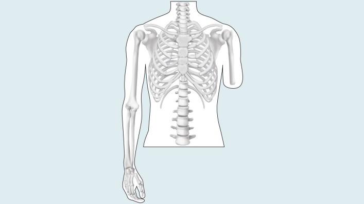
Amputation

Amputation is the surgical removal of all or part of a limb or extremity such as an arm, leg, foot, hand, toe, or finger.Reasons for Amputation

There are many reasons an amputation may be necessary. The most common is poor circulation because of damage or narrowing of the arteries, called peripheral arterial disease. Adequate blood flow is not supplied, the body’s cells cannot get oxygen and nutrients they need from the bloodstream. As a result, the affected cells begins to die and infection may set in.Image result for amputation

Other causes for amputation may include:

  • Severe injury (from a vehicle accident or serious burn, for example)
  • Cancerous tumour in the bone or muscle of the limb
  • Serious infection that does not get better with antibiotic other treatment
  • Thickening of nerve tissue, called a neuron

The Amputation Procedure

An amputation usually requires a hospital stay of five to 14 days or more, depending on the surgery and complications. The procedure itself may vary, depending on the limb or extremity being amputated and the patient’s general health.

Amputation may be done under general anaesthesia (meaning the patient is asleep) or with spinal anaesthesia, which numbs the body from the waist down.

When performing an amputation, the surgeon removes all damaged tissue while leaving as much healthy tissue as possible.

Sleeve Gastrectomy

Sleeve gastrectomy, also called a vertical sleeve gastrectomy, is a surgical weight-loss procedure. This procedure is typically performed laparoscopically, which involves inserting small instruments through multiple small incisions in the upper abdomen. During sleeve gastrectomy, about 80 percent of the stomach is removed, a tube-shaped stomach about the size and shape of a banana.

Limiting the size of your stomach restricts the amount of food you are able to consume. In addition, the procedure triggers the hormonal changes that assist with weight loss. The same hormonal changes also help relieve conditions  being overweight, such as high blood pressure or heart disease.

Why it’s done

Sleeve gastrectomy is done to help you lose excess weight and reduce your risk of potentially life-threatening weight-related health problems, including:

  • Gastroesophageal reflux disease
  • Heart disease
  • High blood pressure
  • High cholesterol
  • Obstructive sleep apnea
  • Type 2 diabetes
  • Stroke
  • Infertility

Sleeve gastrectomy is typically done only after you’ve tried to lose weight by improving your diet and exercise habits.

In general, sleeve gastrectomy surgery could be an option for you if:

  • Your body mass index (BMI) is 40 or higher (extreme obesity).
  • Your BMI is 35 to 39.9 (obesity), and you have a serious weight-related health problem, such as type 2 diabetes, high blood pressure or severe sleep apnea. In some cases, you may qualify for certain types of weight-loss surgery if your BMI is 30 to 34 and you have serious weight-related health problems.

You must also be willing to make permanent changes to lead a healthier lifestyle. You may be required to participate in long-term follow-up plans that include monitoring your nutrition, your lifestyle and behaviour, and your medical conditions.

As with any major surgery, sleeve gastrectomy poses potential health risks, both in the short term and long term. Risks associated with the sleeve gastrectomy can include:

  • Excessive bleeding
  • Infection
  • Adverse reactions to anaesthesia
  • Blood clots
  • Lung or breathing problems
  • Leaks from the cut edge of the stomach

Longer term risks and complications of sleeve gastrectomy surgery can include:

  • Gastrointestinal obstruction
  • Hernias
  • Gastrointestinal reflux
  • Low blood sugar (hypoglycemia)
  • Malnutrition
  • Vomiting

Very rarely, complications of sleeve gastrectomy can be fatal.


Food and medications

Before your surgery, give your doctor a list of all medicines, vitamins, minerals, and herbal or dietary supplements you take. You may have restrictions on eating and drinking and which medications you can take.

If you take blood-thinning medications, talk with your doctor before your surgery. Because these medications affect clotting and bleeding, your blood-thinning medication routine may need to be changed.

If you have diabetes, talk with the doctor who manages your insulin or other diabetes medications for specific instructions on taking or adjusting them after surgery.

Other precautions

You’ll be required to start a physical activity program.

You’ll be required to stop any tobacco use 12 weeks before surgery and may be tested for nicotine prior to your surgery.

You may also need to prepare by planning ahead for your recovery after surgery. For instance, arrange for help at home if you think you’ll need it. People who have a sleeve gastrectomy are typically off work for four weeks.

Results

Sleeve gastrectomy can provide long-term weight loss. The amount of weight you lose depends on your change in lifestyle habits. It is possible to lose approximately 60 percent, or even more, of your excess weight within two years.

In addition to weight loss, sleeve gastrectomy may improve or resolve conditions related to being overweight, including:

  • Heart disease
  • High blood pressure
  • High cholesterol
  • Obstructive sleep apnea
  • Type 2 diabetes
  • Stroke
  • Infertility

Sleeve gastrectomy surgery can also improve your ability to perform routine daily activities, and can help improve your quality of life.

Anemia

Anemia is a very common blood disorder wherein the body lacks healthy red blood cells or hemoglobin. This condition leads to insufficient supply of oxygen to body tissues. It affects all age groups  and pre school children are at a higher risk of this disorder. It can be temporary or long term. The treatments for anemia vary depending on the severity of the condition. There are different types of anemia described. Though symptoms of all types of anemia are mostly the same, some types of anemia have specific symptoms as well.

IMG-20190417-WA0006

SYMPTOMS

  • Lack of energy, feeling f tired and weak
  • feeling of fatigue
  • headache and chest pain
  • paleness of skin
  • fast and irregular heart beat.
  • Dizziness
  • Cold hands and feet
  • Shortness of breath

 CAUSES

A person can be anemic due to various reasons. It can occur due to decreased production of red blood cells, blood loss, destruction of red blood cells etc. The cases of anemia are specific to the types of anemia.

TYPES AND TREATMENTS

  • Iron deficiency anemia:  Iron molecules are associated with hemoglobin that carries oxygen to body tissues through blood. Lack of iron caused due to blood loss , heavy menstrual bleeding, ulcer, cancer etc leads to lack of hemoglobin production. This type is  very common in pregnant women without iron supplementation. The treatment includes iron supplementation.
  • Vitamin deficiency anemia:  Apart from iron body requires folate and vit B-12 for the production of healthy red blood cells..Therefore lack of this vitamin can lead to vitamin deficiency anemia.It is also called pernicious anemia. The symptoms include irritability, diarrhea, and a smooth tongue. Treatments include dietary supplements and B-12 shots
  • Aplastic anemia: It occurs when the body doesn’t produce enough red blood cells. It is caused by infections, certain medicines, autoimmune diseases and exposure to toxic chemicals.  The specific symptoms include fever, frequent infections, and skin rashes. The treatment includes blood transfusions or bone marrow transplantation.
  • Anemia of chronic disease: Diseases  such as cancer, HIV/AIDS, rheumatoid arthritis, kidney disease, Crohn’s disease and other chronic inflammatory diseases  can decrease the production of red blood cells.
  • Anemia associated with bone marrow disease: Blood production in the bone marrow is affected by diseases such as leukemia and myelofibrosis. It can cause anemia.
  • Hemolytic anemias: It occurs when red blood cells are destroyed faster than bone marrow can replace them.  The symptoms include jaundice, dark colored urine, fever, and abdominal pains.
  • Sickle cell anemia: This is an inherited hemolytic anemia. It’s caused by a defective form of hemoglobin that forces red blood cells to assume an abnormal sickle shape. These irregular blood cells die prematurely, resulting in a chronic shortage of red blood cells.  Thesymptoms include painful swelling of the feet and hands, fatigue, and jaundice. Treatment includes oxygen therapy, pain relief, and intravenous fluids. There may also be antibiotics, folic acid supplements, and blood transfusions.


Kidney stone

Kidneys are the excretory organs in humans which remove fluid waste from the blood  as urine. Lack of fluid amount added to the accumulation of certain mineral crystals leads to sticking together of clumps of wastes to form the kidney stone. It is also called renal calculi.Usually men are at more risk than women. It is usually accompanied by severe pain while urinating.

IMG-20190417-WA0003


The major risk factors for  the occurrence of  kidney stone are

  • Reduced intake of water
  • Following a diet rich in protein, sodium and/or sugar.
  • Obese nature of the body.
  • Weak and unhealthy kidney.
  • High levels of cystine, oxalate, uric acid or calcium in urine.
  • Swelling or irritation in bowel or joints.
  • Certain medications
  • A family history of kidney stone.

In severe cases kidney stones are accompanied with complications like severe pain while urinating, pain in back or lower abdomen, blood in the urine and nausea and vomiting.Other symptoms of kidney stones include  pus in the urine, reduced amount of urine excreted, burning sensation while urinating, frequent urge to urinate,  fever and chills if there is an infection. It can also lead to other kidney complications like Chronic kidney disease.

To prevent kidney stone is to follow a healthy diet with plenty of water and adequate  consumption of calcium. Limiting sodium and animal protein (meat, eggs) in the diet may also help to prevent kidney stones. However any changes in the diet should be made only with the advice of the doctor. Intake of basil, celery, apples, grapes and pomegranates protect the kidneys from kidney stones.

The treatment for kidney stones is carried out by rehydrating the patient via an intravenous (IV) tube, and administration of an anti-inflammatory medication. Since passing of the stone is accompaned by severe pain narcotics are used to relieve the pain. Lithotripsy  is a treatment that breaks the kidney stone into smaller pieces and allow it to pass.


Urinary Tract Infection

Urinary Tract Infections (UTI) is characterised by a burning sensation while urinating. It is caused by microbes that can overcome the body’s defenses in urinary tract.It affects the kidneys, ureters and the bladder. Though it affect both men and women of all ages groups, mostly women have the risk of developing a lifetime risk of this infection.The chances of recurrent experiences are also higher in women. UTIs are caused by

  1. Escherichia coli (E. coli), usually found in the digestive system.
  2. Chlamydia and mycoplasma which can infect the urethra and not the bladder.

IMG-20190417-WA0002

The factors that increase the chances of a UTI are sexual intercourse, diabetes,  poor personal hygiene, having a urinary catheter ,bowel incontinence ,blocked flow of urine ,kidney stones,  contraception, pregnancy, menopause, suppressed immune system, use of spermicides and tampons, heavy use of antibiotics, which can disrupt the natural flora of the bowel and urinary tract.

SYMPTOMS

  1. Pain or a burning sensation when urinating.
  2. Strong and frequent urge to urinate
  3. Cloudy, bloody, or strong-smelling urine
  4.  Nausea and vomiting
  5.  Abdominal pains

PREVENTION

1.Drink lots of water and urinate frequently.
2.Reduce alcohol and caffeine that can irritate the bladder.
3.Urinate shortly after sex.
4.Wipe from front to back after urinating and bowel movement.
4.Keep the genital area clean.
5. Use sanitary pads or menstrual cups instead of tampons.
6.Avoid use of diaphragm or spermicide for birth control.
7. Wear cotton underwear and loose-fitting clothing to keep the area around the urethra dry.

Pages:1...567891011...80