Global Treatment Services Pvt. Ltd.

Global Treatment Services

MVT Business App;finger tip access to medical tourism

Global Treatment services Pvt Ltd has been a front runner in medical value tourism for several years.It has brought about several groundbreaking developments in the industry.Latest addition to its long list of innovation is the MVT Business App.The application facilitates the complete medical value tourism workflow at the ease of accessing your mobile phone.The app is a new concept and first of its kind it in the industry. MVT app enables tourist operator, medical tourism facilitator & doctors to connect and offer medical tourism service through us without any hassles.The winning feature of the app is that it can be used to avail services from any country and in any speciality.MVTapp

Application details

It is a light weight application which does not eat into your phone memory.It comes with a downloadable size of only  a few megabytes.The interface is very user friendly designed to swiftly take you through the complete process.The data input required is also only essential details and the remaining is taken care by the background processes.The application also include features to store and track patient,insurance and process details along with various useful components.
On creation of query which is the primary step,it reaches the respective hospitals.The app supports downloading visa invitation letter and travel itinerary.Also, the app enables the user to upload passport and ticket copy through the upload link.There is also an option of sending ticket copy directly to the hospitals.

The application can be downloaded from Play Store(android ).

Download the MVT Business app for Android here

Process flow

The main tabs in the workflow are -Query Management,Patient and Payment and Insurance module.

Query Management
  • The MVT app enables the first step in the process flow, namely creating a query
  • During query creation, user can choose preferred country, city, speciality desired
  • The app gives an option of choosing multiple specialities and multiple cities too
  • After registering these choices, the user is given the option of choosing one or more internationally accredited hospitals based on the user preferences.
  • The user has to provide minimum details like medical reports of the patient
  • Query automatically reaches the respective hospitals
  • The generated query can be viewed in the query list with a unique reference number
  • The app also provides the user to download treatment proposal, on clicking the query.
Patient 
 Once the itinerary is generated, it can be viewed in patient list and by selecting the patientuser is able to get daily update and medical details of the patient.
Payment
Payment icon gives the complete information about the patient bill and commission details, payment status, payment reference number  and also provides an option to raise invoice to the company.

Insurance module

The application facilitates claiming insurance by involving insurance companies.This module works similar to the general module in query management and patient.But the third tab is ‘Bill details’ instead of payment.Using this tab,insurance  companies can access all bill details of the patient.The application supports complete client settlement requirements including discharge summary using this tab.
MVT app provides smooth and transparent process flow to the medical tourism business partners.Its is the one stop app for all you need to manage the medical tourism process.
For any assistance and queries,kindly email us at query@gtsmeditour.com

Understanding Cancer

The very word ‘Cancer’ is dreaded.A cancer diagnosis makes most of the people feel that life has gone out of control.Surely, it is normal to be afraid of the unknown. Uncertainty can make you feel afraid at first. Anxiety and angriness follows. Some people become irritated and frustrated with themselves.Once people start understanding cancer clearly and in depth,they can take charge.They start accepting that they have cancer and they often feel a sense of hope. Surely,there are many reasons to feel hopeful. Millions of cancer patients have survived. Living with cancer and beyond it have become possible more than before. So, let’s dive into cancer basics.21.March2


Cancer start

Cancer is caused by uncontrolled division of abnormal cells.Usually,we have right number of cells in our body.This is maintained by the cells through control signals produced in them.Cells may start to grow or multiply if these signals go faulty.The cells multiply rigorously to form a tumor where the cancer starts.
The instructions are generated by thousands of genes inside the cell nuclei.Normally genes make sure that cells grow and reproduce in an orderly and controlled way. Sometimes a change happens in the genes when a cell divides. This is a mutation.Mutation means that abnormal proteins may be produced in the cells that work differently to normal triggering cells to divide more and more. And once cells start growing too fast, they are more likely to pick up further mutations and less likely to be able to repair the damaged genes.

 Cancer growth

A tumour may contain millions of cancer cells.Cancer cells can break through basement membrane,the membrane which keeps the cells of every body tissues inside.Once this happens, the cancer is called invasive. As a tumour gets bigger, it takes up more space in the body. The cancer can then cause pressure on surrounding structures. It can also grow into body structures nearby. This is called local invasion. A cancer may grow out in a random direction from the place where it started. Cancer cells can move about more easily than normal cells. So it seems likely that one of the ways that cancers spread through nearby tissues is by the cells directly moving.

Tumours  can be benign or cancerous (malignant).

Benign tumours

Benign tumours are made up of cells that are quite similar to normal cells.They usually grow quite slowly and do not spread to other parts of the body.They will  cause a problem only in some cases such as when theygrow very large and press on other body organs,become painful or release hormones that affect how the body works and take up space inside the skull (such as a brain tumour)

Malignant tumours

They are made up of cancer cells. They:

  • usually grow faster than benign tumours
  • spread into surrounding tissues and cause damage
  • may spread to other parts of the body in the bloodstream or through the lymph system to form secondary tumours. This is called metastasis

Cancer spread

The place where a cancer starts in the body is called the primary cancer or primary site. Cells from the primary site may break away and spread to other parts of the body. These escaped cells can then grow and form other tumours, which are known as secondary cancers or metastases.
Cancer cells can spread to other parts of the body through the bloodstream or lymphatic system. There they can start to grow into new tumours. Cancers are named according to where they first started developing. For example, if you have bowel cancer that has spread to the liver, it’s called bowel cancer with liver metastases or secondaries. It is not called liver cancer. This is because the cancerous cells in the liver are actually bowel cells. They are not liver cells that have become cancerous.

Cancer groups

Cancers are divided into groups according to the type of cell they start from. They include

  • Carcinomas – cancer that begins in the skin or in tissues that line or cover internal organs.
  • Lymphomas – cancers that begin in the cells of the immune system
  • Leukaemias – cancer that starts in blood forming tissue such as the bone marrow
  • Brain tumours and spinal cord cancer – central nervous system cancers
  • Sarcomas – cancer that begins in the connective or supportive tissues such as bone, cartilage, fat, muscle or blood vessels

Cancer staging and grading

Staging is a way of describing the size of a cancer and how far it has grown. When doctors first diagnose a cancer, they carry out tests to check how big the cancer is and whether it has spread into surrounding tissues.Cancer staging may sometimes include the grading of the cancer. Tumour grade describes a tumour in terms of how abnormal the tumour cells are when compared to normal cells.

Common staging systems are TNM system and number system.Number staging systems usually use the TNM system to divide cancers into stageswhich is the most commonly communicated system to the patients

Stage 1 –  usually means that a cancer is relatively small and contained within the organ it started in
Stage 2 – usually means that the tumour is larger than in stage 1, but the cancer has not started to spread into the surrounding tissues.
Stage 3 – usually means the cancer is larger. It may have started to spread into surrounding tissues and there are cancer cells in the lymph nodes in the area
Stage 4 – means the cancer has spread from where it started to another body organ. This is also called secondary or metastatic cancer

Generally the grading is as follows.

Grade 1 – the cancer cells look very similar to normal cells and are growing slowly
Grade 2 – the cells don’t look like normal cells and are growing more quickly than normal
Grade 3 – the cancer cells look very abnormal and are growing quickly

Is Cancer genetic?

Most cancers start due to gene changes that happen over a person’s lifetime. More rarely cancers start due to inherited faulty genes passed down in families.

For more information and treatment facilities,email us at query@gtsmeditour.com 

Read about:Restorartive and cosmetic dentistry

Restorative and cosmetic dentistry

Who would not want a perfect smile in your selfie!Nowadays, there are several options for repairing, worn, decayed, damaged or missing teeth in  restorative dentistry. In addition to fixing these major issues , cosmetic measures can be taken then to put the finishing touches on a new smile.20.feb29dent

Pneumonia in children

Pneumonia will kill nearly 11 million children under five by 2030, experts warn based on an analysis conducted by Johns Hopkins University .Pneumonia is the biggest infectious killer of infants worldwide.More than 880,000 children , mainly aged less than two years old , died from pneumonia in 2016 alone.It is a severe lung infection which can be easily prevented with adequate measures.
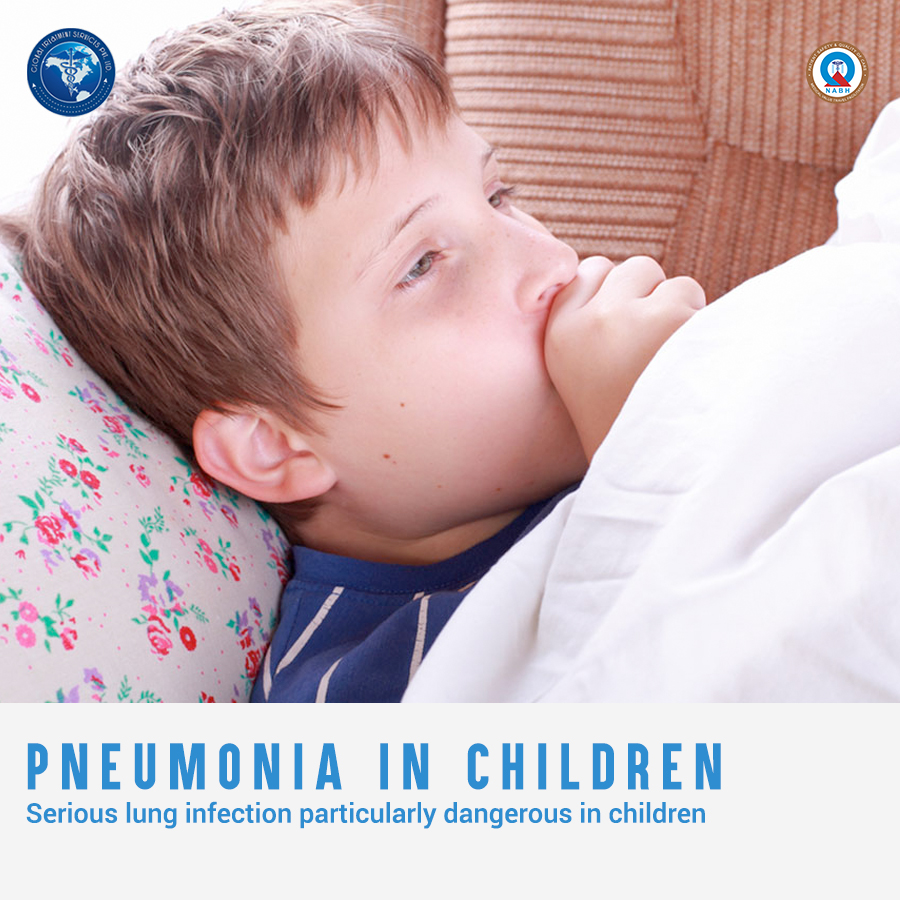
19.feb28Pneumonia


What is pneumonia?

Pneumonia is a serious infection of lungs in which the tiny air sacs, or alveoli , and terminal air spaces gets filled with pus and other fluids making it difficult for oxygen to reach the blood stream.It is more common in children less than  5 years old.The inflamamtion may be caused by bacteria, viruses,fungi or chemical irritants.The infectious agents are introduced into the lungs through blood or inhalation. There are two variants of pneumonia.

Lobar pneumonia:This affects one or more sections (lobes) of the lungs.

Bronchial pneumonia:This affects patches throughout both lungs.

Why children?

  • Unhealthy children with a compromised immune system has weak defenses.
  • Children who suffer from malnutrion, particularly inadequate zinc intake and lack of exclusive breastfeeding have a higher risk of developing pneumonia.
  • Other risk factors include:
    • Being born premature
    • Having asthma or genetic disorder such as sickle-cell disease
    • Having heart defects such as ventricular septal defect (VSD), atrial septal defect (ASD) or patent ductus arteriosus (PDA)
  • Several environmental factors such as overcrowding homes and exposure to parental smoke increases a child’s susceptibility to pneumonia and its complications

Causes and symptoms

Pneumonia begins after an infection of the nose and throat.The sypmtoms start after 2-3 days of a cold/sore throat.It then move downwards to the lungs.Fluid, white blood cells, and debris start to accumulate in the air spaces of the lungs and block gaseous exchange .
Pneumonia is caused by a variety of germs -viruses, bacteria, fungi, and parasites. The length of time between exposure to the germ and when someone starts feeling sick varies, depending on which virus or bacteria is causing the pneumonia.Some symptoms give important clues about which germ is causing the pneumonia.
Viral pneumonia :Most cases, though, are caused by viruses. These include adenoviruses, rhinovirus,influenza virus (flu), respiratory syncytial virus (RSV), and parainfluenza virus.Early symptoms of viral pneumonia are the same as those of bacterial pneumonia.
Bacterial pneumonia :This is caused by various bacteria. The streptococcus pneumoniae is the most common bacterium that causes bacterial pneumonia.Many other bacteria may cause bacterial pneumonia including Group B streptococcus,Staphylococcus aureus,Group A streptococcus.
Mycoplasma pneumonia :This presents slightly different symptoms than other types of pneumonia. They generally cause a mild, widespread pneumonia that affects all age groups.Symptoms usually do not start with a cold, and may include fever and cough are the first to develop,cough that is persistent and may last three to four weeks and a severe cough that may produce some mucus.
Other less common pneumonias may be caused by the inhaling of food, liquid, gases or dust, or by fungi.

Sometimes a child’s only sign may be rapid breathing and often when pneumonia exist in the lower part of the lungs, no breathing problems may be present but rather fever, abdominal pain or vomiting.If pneumonia is caused by bacteria, the infected child becomes sick relative quickly and is prone to developing high fever and rapid breathing.If pneumonia is caused by viruses, symptoms may appear gradually and less severe than the bacterial pneumonia .Parents should be aware of the following signs and symptoms:

  • Nostril flaring
  • Sternal retraction
  • Increased breath rate
    • > 60 breaths/min for newborns up to 2 months
    • > 50 breaths/min for 2 months to 12 months
    • > 40 breaths/min for a child older than 1 years of age

Four Stages

Pneumonia has four stages, namely consolidation, red hepatization, grey hepatization and resolution.
The first stage called Consolidation, which occurs within 24 hours of infection,  is characterized by coughing and deep breathing. Many bacteria and few neutrophils are present.Cellular exudates containing neutrophils, lymphocytes and fibrin replaces the alveolar air.Capillaries in the surrounding alveolar walls become congested.The infections spreads to the lung roots (hilum) and lung membranes(pleura) rapidly.
The stage of Red hepatization , so called because of its similarity to the consistency of liver, is characterized by the presence of many erythrocytes, neutrophils, desquamated epithelial cells, and fibrin within the alveoli.It occurs 2-3 days after consolidation.Alveolar capillaries are engorged with blood.
In the stage of Grey Hepatization (2-3 d fter red hepatization), the lung is gray-brown to yellow because of fibrinopurulent exudate, disintegration of RBCs, and hemosiderin. The final stage of resolution is characterized by resorption and restoration of the pulmonary architecture.

Diagnosis

Sometimes a thorough physical examination is enough for the doctor to make pneumonia diagnosis.Folowing tests may be used to for further confirmation

  • Chest X-ray. A diagnostic test which uses invisible electromagnetic energy beams to produce images of internal tissues, bones, and organs onto film.
  • Blood tests. Blood count for evidence of infection,arterial blood gas to analyze the amount of carbon dioxide and oxygen in the blood.
  • Sputum culture. A diagnostic test performed on the sputum to determine if an infection is present.
  • Pulse oximetry:Used to measure the amount of oxygen in blood
  • Chest CT scan:  To take images of the structures in the chest
  • Bronchoscopy. A procedure used to look inside the airways of the lungs
  • Pleural fluid culture. A culture of fluid sample taken from the pleural space (space between the lungs and chest wall) to identify the bacteria that cause pneumonia

Treatment

Specific treatment for pneumonia will be determined by your child’s doctor based on child’s age, overall health, and medical history,extent of the condition,cause of the condition,your child’s tolerance for specific medications, procedures, or therapies,expectations for the course of the condition.

Treatment may include antibiotics for bacterial and mycoplasma pneumonia. There is no clearly effective treatment for viral pneumonia, which usually resolves on its own.Treatment will vary depending on how bad the symptoms are, and what the cause of the infection is.Other treatment may include appropriate diet,increased fluid intake,cool mist humidifier in the child’s room,acetaminophen and medication for cough.

For severe breathing problems, treatment may include

  • Intravenous (IV) or oral antibiotics,Intravenous (IV) fluids
  • Oxygen therapy
  • Breathing treatments
  • Analgesic administration
  • Cough suppressant medication

Prevention

There is a pneumococcal vaccine to protect from a common form of bacterial pneumonia. Children younger than age 5 and adults ages 65 and older should get this shot.The pneumococcal shot is also recommended for all children and adults who are at increased risk of pneumococcal disease due to other health conditions.Kids should receive  immunisation against Haemophilus Influenzae and Pertussis at  2 months of age.

For any queries regarding  treatment facilities,email us at query@gtsmeditour.com 

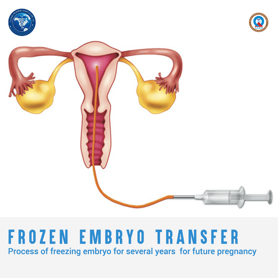
Read about:Frozen embryo transfer


Frozen embryo transfer

Emma Wren Gibson, frozen as an embryo in 1992, was born in 2017, more than 25 years later.Frozen embryo transfer is one of the procedures in invitro fertilisation(IVF).IVF is one assistive reproductive technology(ART) used to establish pregnancy.It involves retrieving eggs from a woman’s ovaries and fertilizing them with sperm in the laboratory. This fertilized egg  known as an embryo can then be frozen for storage or transferred to a woman’s uterus. Emmas’s is the longest an embryo is known to have been frozen before being born as baby.18.feb27FrozenEmbryoTransfer


ART procedures and IVF

IVF is used in women experiencing difficulty to conceive.This may include male or female genetic defects,abnormal sperm production in male,ovulation problems in female among many.Major ARTs are In vitro fertilization (IVF),Intracystoplasmic sperm injection (ICSI),Gamete intrafallopian Transfer (GIFT),Zygote Intrafallopian Transfer (ZIFT) and Tubal Embryo Transfer (TET).Most of these techniques relies on embryo transfer.
In Vitro Fertilisation involves uniting the ovum with the spermatozoon in vitro in order to obtain fertilised embryos for transfer to the mother’s uterus.First step to IVF is ovarian stimulation.Ovarian stimulation consists of the administration of daily injections which cause the ovaries, instead of producing a single ovum which is what they do naturally each month, to produce more oocytes so that a larger number of embryos can be obtained.
Multiple eggs are desired because some eggs will not develop or fertilize after retrieval.Eggs are retrieved through a minor surgical procedure that uses ultrasound imaging to guide a hollow needle through the pelvic cavity to remove the eggs. Medication is provided to reduce and remove potential discomfort.The male is asked to produce a sample of sperm, which is prepared for combining with the eggs.In a process called insemination, the sperm and eggs are mixed together and stored in a laboratory dish to encourage fertilization.The eggs are monitored to confirm that fertilization and cell division are taking place. Once this occurs, the fertilized eggs are considered embryos.
The embryos are usually transferred into the woman’s uterus three to five days following egg retrieval and fertilization. A catheter or small tube is inserted into the uterus to transfer the embryos.If the procedure is successful, implantation typically occurs around six to ten days following egg retrieval.

Frozen Embryo Transfer

A frozen embryo transfer (FET) is a cycle where a frozen embryo from an earlier cycle is thawed and transferred back into a woman’s uterus. This means that woman has an egg or embryo ready  for assistive reproduction procedure.She does not have to undergo a cycle of hormone stimulation and egg collection. Frozen embryo cycles can be undertaken on natural cycles  or ovulation stimulation.
As mentioned earlier, during an IVF cycles more than one embryo may be created.But it is recommended to transfer only one and freeze others. This is due to the serious risks associated with multiple pregnancies if more than one embryo is transferred at a time.Once the embryo transfer has been made,remaining good quality embryos are vitrified so that they can be used in a later IVF cycles.
Embryo freezing gives you more chances for a pregnancy . If pregnancy is not achieved  from the first transfer , we can transfer a frozen embryo during a frozen embryo transfer cycle.
This cuts down on the amount of time that is needed for each ART cycle. The quality of eggs deteriorates with age, leading to the possibility of having a more difficult time conceiving. The more quality of eggs, the better the chance that they will be of high enough quality to result in pregnancy. FET offers the patient the chance to use more quality possible eggs as this is vitrified during earlier ovulation cycles.

Fertility Preservation

The biggest benefit of FET is fertility preservation.There can be cases where some have a serious illness such as cancer that will potentially risk damage to your eggs or sperm from chemotherapy, radiotherapy or other treatments, including surgery.This can affect the fertility in future. FET can also help those people who are not in a position to have a babies right now but would want later and also those who would like the opportunity to start a family beyond the age at which fertility naturally declines.
Fertility preservation options for women include egg freezing, embryo freezing and ovarian tissue freezing.

Embryo freezing is used when there is partner or donor .Frozen embryos can be stored for many years.

Egg freezing is a method of storing a woman’s unfertilised eggs. To obtain eggs for freezing, a woman would usually have hormonal stimulation for 10 – 12 days. Frozen eggs may be stored for many years without significant deterioration. When the woman is ready to use her eggs, they are warmed, and then fertilised with sperm.

Ovarian tissue freezing involves removing a small piece of ovarian tissue from one ovary, cutting it into tiny slices and then freezing it. Later, when you are ready to try to conceive, the ovarian tissue slices are grafted back into your pelvis. Around nine months later, the grafted ovarian tissue can start to produce reproductive hormones and follicular development. Pregnancy may be achieved either with ovarian stimulation and IVF, or perhaps even naturally. Moaza Al Matrooshi was born with a serious blood disorder.By the age of nine ,she needed chemotherapy treatment for the disorder. She is  the first to give birth after having her fertility restored using ovarian tissue frozen before the onset of puberty.She delivered a healthy baby boy.

 For any queries regarding  treatment facilities,email us at query@gtsmeditour.com 

Read about:LASIK or Laser eye surgery


LASIK or Laser eye surgey

As the saying goes “The eyes are the windows to the soul”.This expression conveys that eyes reflect what a person really is inside. However, I think it can be perceived in a different sense.The eyes provide vision which soothes the soul.Feels inappropriate?Anyways,you would agree with me on that ‘one should have clear vision to connect with the world around us’.Here comes the importance of correcting eye treatments. LASIK is a surgical procedure intended to correct a person’s vision and to reduce his dependency on glasses or contact lenses.17.feb26LASIK

Understanding LASIK

The cornea is a part of the eye that helps focus light to create an image on the retina.When the shape of the cornea and the eye are not perfect,the image on the retina is out-of-focus or distorted. These imperfections in the focusing power of the eye are called refractive errors. There are three primary types of refractive errors: myopia, hyperopia and astigmatism. Myopia is nearsightedness, hyperopia is farsightedness and astigmatism is a distortion of the image on the retina caused by irregularities in the cornea or lens of the eye. LASIK is performed  to treat myopia, hyperopia and astigmatism.
Surgical procedures aimed at improving the focusing power of the eye are called refractive surgery. Like other types of refractive surgery, the LASIK procedure reshapes the cornea to enable light entering the eye to be properly focused onto the retina for clearer vision. LASIK stands for Laser-Assisted In Situ Keratomileusis and the  procedure  permanently changes the shape of the cornea.In LASIK surgery, precise and controlled removal of corneal tissue is done by a special laser reshaping the cornea and changing its focusing power.

Procedure

The procedure starts with numbing of the eye with a few drops of topical anesthetic. An eyelid holder is placed between the eyelids to keep them open and prevent the patient from blinking. A suction ring placed on the eye lifts and flattens the cornea and helps keep the eye from moving.
Eye surgeon cuts a flap in the cornea,the clear covering of the front of the eye,using a mechanical surgical tool called microkeratome  or  a laser device called femtosecond laser. A hinge is left at one end of this flap using which the flap is folded back revealing the stroma, the middle section of the cornea. Excimer laser ,preprogrammed with the patient’s unique eye measurements,  is positioned above the patient’s eye .As patient looks at fixation light,excimer laser vaporize a portion of the stroma. This highly specialized laser uses a cool ultraviolet light beam to remove microscopic amounts of tissue from the cornea to reshape it so that it focuses light on the retina more accurately.The flap is then replaced, covering the area where the corneal tissue was removed and edges are smoothed.
For myopia, the cornea is flattened; with farsighted people, a steeper cornea is desired. Excimer lasers correct astigmatism by shaping an irregular cornea into a more normal shape.The flap sticks to the underlying corneal tissues in 2-5 minutes.Laser eye surgery does not require bandages or stitches ,it requires only topical anesthetic drops.The cornea is then allowed to heal naturally.
The ophthalmologist will prescribe eye drops to help the eye heal and relieve dryness.The patient can start doing his normal activities soon after a nap but it may take about six months after the surgery for the improvements in a person’s vision to fully stabilize and any side effects to go away.

Any risks and side effects?

LASIK is not for everyone.Eye doctors carefully test and evaluate to determine if patients are appropriate candidates for the surgery.Some of the reasons you may not qualify for the LASIK may be having changes in power of glasses in the past year,having certain medical conditions like diabetes ,having eye diseases like glaucoma,inflammation etc or having a previous eye surgery.People with dry eyes,large pupils and thin corneas also cannot undergo LASIK.
Benefits outweigh any risks if done on right patients.It can have mild and temporary side effects such as dry eyes,sensitivity to light,blurred vision  and other visual symptoms such as glare,halos etc.A small percentage of people may need a LASIK enhancement procedure.Some people may still need to wear glasses following laser vision correction but the power level typically will be much lower than before.
Laser eye surgery offers numerous benefits and can significantly improves the quality of life. Most people achieve 20/20 vision or better after the surgery.

For any queries regarding  treatment facilities,email us at query@gtsmeditour.com 

Read about:Caesarean section


Cesarean Section

When it comes to delivery,cesarean section is often frowned upon.People shaming c-section says it is not ‘real birth’.They argue that it is an easy way out and you did not experience pain and the baby did not come out of you and on and on.Alas,vaginal birth is not the only thing a mother does for her baby.If  C-section babies are not really born,they would still be in their ‘trimesters’!!Let us see the details of c-section mode of delivery here to accept and appreciate it.16.feb25Csection

What is c-section

Cesarean section delivery is the surgical delivery of the baby by making incisions in the abdomen and uterus of the mother.Emergency c-section is performed when the labour has already begun but complications crop up.Health care team decides immediately that it is the safest option.Emergency c-section can be life saving for both mother and the baby.
When the mode of delivery is already chosen to be c-section before one goes into labour,then it is called planned or elective c-section.In most cases, c-sections are done for the safety of the child.Vaginal delivery poses danger when baby is lying in difficult position for labour or there are problems with the placenta such as a low lying placenta.

Reasons for planned Cesarean Section 

There are several reasons why cesarean sections may be planned prior to the labour or at the beginning of labour. As mentioned earlier,most of the reasons attribute to the safety of the baby and/or the mother.

Placenta Previa
This is where the placenta is near of covering the cervix. This blocks the pathway for a vaginal birth or would present a bleeding risk during labor.

Certain Fetal Positions
Your baby’s position in the uterus may mean that a vaginal birth is not possible, nor safe for them to be born. This can include: Transverse Lie, some breech positions, etc.

Higher Order Multiples
With every baby that you have in the uterus the risk of a cesarean birth increases. While there are births of twins and triplets vaginally, the more babies, the less likely a vaginal birth will be possible. This is many times due to odd positions in the uterus.

Genital herpes
If you have herpes occurring late in pregnancy and you have an active lesion on your genitals, you may be encouraged to have a cesarean rather than delivery vaginally. This is to prevent transmission to your baby.

Pre-eclampsia
You have pregnancy-related high blood pressure

Certain Fetal Conditions
Your baby isn’t getting enough oxygen and nutrients – sometimes this may mean the baby needs to be delivered immediately

Other conditions
Your labour isn’t progressing or there’s excessive vaginal bleeding,diseases that may make vaginal birth difficult like pulmonary or coronary disease of the mother,HIV positive with a high viral load at time of birth.Previous invasive uterine surgery, including a previous classical cesarean incision

Some mothers may choose planned c-section if they feel they cant cope with the pain involved in a vaginal birth especially if they had had a difficult vaginal delivery with their last baby.Also for some mothers,it may have caused severe postpartum depression.You never know what she goes through physically and emotionally.So ,there is no reason in shaming mothers going ahead with elective c-section.

C section Sugery

Most caesareans are carried out under spinal or epidural anaesthetic. Both types of anaesthetic are given as an injection in your spine that numbs the lower part of your body. This mean you’ll be awake.You will either lie on your side or sit leaning forward, curving your back, while the anaesthetist inserts a very fine needle into your spine.Occasionally a general anaesthetic, where you’re asleep, may be used, particularly if the baby needs to be delivered more quickly.This means you will be asleep for the operation

Before the procedure:You will be given

  • fluids through a needle in your arm (a drip), to stop you getting dehydrated and to reduce the risk of low blood pressure during the operation
  • anti-sickness medicine to stop you feeling or being sick
  • a small tube (catheter) into your bladder to drain urine – this will stay in place for at least 12 hours and until you feel able to walk to the toilet.

During the procedure:

  • a screen is placed across your body so that what’s being done is not seen.
  • a cut about 10-20cm long will usually be made across your lower tummy and womb so your baby can be delivered.you may feel some tugging and pulling during the procedure
  •  Baby is then lifted out.

The whole operation normally takes about 40-50 minutes.

After the procedure:

  • regular checks to make sure the anaesthetic is wearing off , your breathing, heart rate, blood pressure, wound dressing and pain relief for the first few hours
  • regular checks on the amount of vaginal bleeding
  • a catheter to drain urine from your bladder
  • compression stockings to reduce your risk of blood clots
  • a needle in your arm (drip) to give you fluids until you’re eating and drinking again

Recovery

C section is a major surgery meaning it risks associated with it like any other surgery.Moms are at greater risk during c section than vaginal birth.Recovery takes more time than normal delivery. Most women experience some discomfort for the first few days after a caesarean, and for some women the pain can last several weeks.The wound in your tummy will eventually form a scar.Regular pain killers are advised to control pain and bleeding.Once the anaesthetic has worn off, you’ll be able to stand up and do short walks.It’s important to move around soon after your c-section to reduce the risk of blood clot.

Benefits far outweigh any disadvanatges.No reasons for shaming mothers!

For any queries regarding  treatment facilities,email us at query@gtsmeditour.com 

Read about:Autism


Autism

Autism spectrum disorder (ASD) is a developmental disability that affects the way an individual interacts with environment and with other people. It is called a ‘developmental disorder’ because symptoms becomes noticeable in the first two years of life.15.feb24autismASD is broad term used to describe a group of developmental disorders;autism has many subtypes.According to Diagnostic and Statistical Manual of Mental Disorders, five different ASD subtypes are

  • with or without accompanying intellectual impairment
  • with or without accompanying language impairment
  • associated with a known medical or genetic condition or environmental factor
  • associated with another neurodevelopmental, mental, or behavioral disorder
  • with catatonia

It merges autistic disorder, childhood disintegrative disorder, pervasive developmental disorder-not otherwise specified (PDD-NOS) and Asperger syndrome under ASD.
Although ASD can be a lifelong disorder, treatments and services can improve a person’s symptoms and ability to function.

Signs and Symptoms 

People with ASD have difficulty with social communication and interaction, restricted interests, and repetitive behaviors.

Social communication / interaction behaviors :

  • issues with communication, including difficulties sharing emotions, sharing interests, or maintaining a back-and-forth conversation
  • issues with nonverbal communication, such as trouble maintaining eye contact or reading body language
  • difficulties developing and maintaining relationships
  • Failing to respond to calling their names
  • Having gestures that do not match what is being said
  • Having an unusual tone of voice that may sound sing-song or flat and robot-like

Restrictive / repetitive behaviors may include:

  • Repetitive movements, motions, or speech patterns
  • Getting upset by slight changes in a routine
  • Being more or less sensitive than other people to sensory input, such as light, noise, clothing, or temperature
  • Fixated interests or preoccupations

People with ASD may also experience sleep problems and irritability.
Although people with ASD experience many challenges, they may also have many strengths, including:

  • Being able to learn things in detail
  • Being able to remember information for long periods of time
  • Being strong visual and auditory learners
  • Excelling in math, science, music, or art

Causes and Risk Factors

Scientists don’t know the exact causes of ASD. Research suggests that both genetics and environment may determine whether a person develops autism. Some risk factors include:
  • Having a immediate family member with ASD.
  • Being born to older parents
  • Having certain genetic conditions like Down syndrome, fragile X syndrome, and Rett syndrome
  • Very low birth weight
  • Metabolic imbalances
  • Exposure to heavy metals and environmental toxins
  • Fetal exposure to the medications valproic acid (Depakene) or thalidomide (Thalomid)

Diagnosis

 ASD can usually be reliably diagnosed by the age of two  by analysing a child’s behavior and development.

Diagnosis in young children is often a two-stage process.

  1. General Developmental Screening during  Checkups
  2. Additional Evaluation
    This second cycle of assessment is carried out by  e a team of doctors and other health professionals who are experienced in diagnosing ASD.The evaluation may assess cognitive level or thinking skills,language abilities and age-appropriate skills needed to complete daily activities independently, such as eating, dressing, and toileting.The comprehensive evaluation may also include blood tests and hearing tests.

Diagnosis in older children and adolescents are often recognized by parents and teachers.Older children and adolescents may have trouble understanding figures of speech, humor, or sarcasm. Parents may also find that their child has trouble forming friendships with peers.
Diagnosing ASD in adults is often more difficult than diagnosing ASD in children. In adults, some ASD symptoms can overlap with symptoms of other mental-health disorders, such as anxiety or attention-deficit/hyperactivity disorder (ADHD).They have issues with social interaction and communication challenges,sensory issues,repetitive behaviors and restricted interests.

Treatments and Therapies

Treatment for ASD should begin as soon as possible after diagnosis. Early treatment for ASD is important as proper care can reduce individuals’ difficulties while helping them learn new skills and make the most of their strengths.

Medication
A doctor may use medication to treat some symptoms that are common with ASD like irritability,aggression,repetitive behavior,hyperactivity,attention problems,anxiety and depression

Behavioral, psychological, and educational therapy
These programs are typically highly structured and intensive and may involve parents, siblings, and other family members. Programs may help people with ASD to learn life-skills necessary to live independently,reduce challenging behaviors,increase or build upon strengths and learn social, communication, and language skills.

With treatments started early,autistic child can lead a normal life.High quality early intervention can improve learning, communication and social skills, as well as underlying brain development.

For any queries regarding  treatment facilities,email us at query@gtsmeditour.com 

Read about:Depression


Depression

Everyone of us have had periods of feeling unmotivated and low moods.But in major depressive disorder,these feelings are intense and prolonged.The persistent feeling of sadness ill affects and impair how you feel, think, and handle daily activities.It troubles normal day to day routines such as sleeping,eating,working etc.You may lose interest even in your favourite activities or hobbies.This eventually culminates in serious physical and emotional complications.The person feels unworthy and becomes suicidal14.feb22depression

Depression is a serious mood disorder and is common.According to WHO,there are 3oo million people affected worldwide.Although it can affect anyone and all ages,more women are affected than men.A person is said to suffer from depression,if he or she experiences the  symptoms for at least two weeks.Depression do not go away on its own.But the good news is that it is treatable.

Signs and Symptoms

There are multiple types of depression but many of them have similar symptoms.Also,not everyone experiencing depression have all symptoms.

Thoughts and Behaviour : Persistent sad, anxious, or “empty” mood,not getting things done at work,withdrawing from close family and friends,unable to concentrate or remember,difficulty making decisions,Thoughts of death or suicide, or suicide attempts
Feelings : Feeling restless or having trouble sitting still,feeling worthless,pessimistic,guilty,irritable,miserable,Loss of interest or pleasure in hobbies and activities
Physical : Decreased energy or fatigue,moving or talking more slowly,difficulty sleeping, early-morning awakening, or oversleeping,appetite and/or weight changes,aches or pains, headaches, cramps, or digestive problems without a clear physical cause and/or that do not ease even with treatment

Causes

Life events
Research suggests that continuing difficulties or recent events  or a combination of events can ‘trigger’ depression if you’re already at risk because of previous bad experiences or personal factors.
Personal factors

  • Family history :Depression can run in families and some people will be at an increased genetic risk. However, having a parent or close relative with depression doesn’t mean you’ll automatically have the same experience.
  • Personality : Some people may be more at risk of depression because of their personality, particularly if they have a tendency to worry a lot, have low self-esteem, are perfectionists, are sensitive to personal criticism, or are self-critical and negative.
  • Serious medical illness :Certain conditions may put you at higher risk, such as chronic illness, insomnia, chronic pain, or attention deficit hyperactivity disorder (ADHD).The stress and worry of coping with a serious illness can lead to depression.
  • Drug and alcohol use :Drug and alcohol use can both lead to and result from depression.
  • Early childhood trauma :Some events impact the way that body reacts to fear and stressful situations.

Changes in the brain
Depression is not simply the result of a ‘chemical imbalance’, for example because you have too much or not enough of a particular brain chemical. There’s a greater risk for depression if the frontal lobe of your brain is less active. However, scientists don’t know if this happens before or after the onset of depressive symptoms.

Types of Depression

  • Persistent depressive disorder (also called dysthymia) is a depressed mood that lasts for at least two years. A person diagnosed with persistent depressive disorder may have episodes of major depression along with periods of less severe symptoms.
  • Postpartum depression : Many women experience depression after giving birth. Women with postpartum depression experience full-blown major depression during pregnancy or after delivery (postpartum depression) making it difficult for new mothers to complete daily care activities for themselves and/or for their babies.
  • Psychotic depression occurs when a person has severe depression plus some form of psychosis, such as having disturbing false fixed beliefs or hearing or seeing upsetting things that others cannot hear or see
  • Seasonal affective disorder is characterized by the onset of depression during the winter months, when there is less natural sunlight. Winter depression, typically accompanied by social withdrawal, increased sleep, and weight gain, predictably returns every year in seasonal affective disorder.
  • Bipolar disorder is different from depression, but it is included in this list is because someone with bipolar disorder experiences episodes of extremely low moods that meet the criteria for major depression called bipolar depression

Examples of other types of depressive disorders newly added to the diagnostic classification of DSM-5 include disruptive mood dysregulation disorder (diagnosed in children and adolescents) and premenstrual dysphoric disorder (PMDD).

Treatment and Therapies

Different types of anxiety or depression require different types of treatment. This may include lifestyle changes such as regular physical activity, healthy eating and adequate sleep, family and peer support, and psychological therapy for mild-moderate anxiety and depression, through to more specialized psychological and medical treatments for severe depression and/or anxiety provided by a team of health and mental health professionals.
Most modern antidepressants have an effect on your brain’s chemical transmitters (serotonin and noradrenaline), which relay messages between brain cells – this is thought to be how medications work for more severe depression. Psychological treatment can also help you to regulate your moods.

Psychotherapies
Several types of psychotherapy can help people with depression like cognitive-behavioral therapy (CBT), interpersonal therapy (IPT), and problem-solving therapy.

Brain Stimulation Therapies
If medications do not reduce the symptoms of depression, electroconvulsive therapy (ECT) may be an option.Based on the latest research ,ECT can provide relief for people with severe depression who have not been able to feel better with other treatments.

Effective treatment can stimulate the growth of new nerve cells in circuits that regulate your mood, which is thought to play a critical part in recovering from the most severe episodes of depression.There is no one proven way that people recover from anxiety or depression, and it’s different for everybody. The important thing is finding the right treatments and the right health professionals and support team that works for you.

For any queries regarding  treatment facilities,email us at query@gtsmeditour.com 

Read about:Nuclear Medicine


Nuclear medicine

Did not expect word ‘nuclear’ in the field of medicine?Well,’Nuclear Medicine’ exists and let us understand it here.
Nuclear medicine is a subspeciality of medical imaging/radiology.But unlike radiology where radiation enter the body from outside,radioactive materials are introduced to the body in Nuclear medicine.These radioactive tracers in combination with imaging instrumentation is used to assess bodily functions and to diagnose , determine the severity of or treat a variety of diseases, including many types of cancers, heart disease, gastrointestinal, endocrine, neurological disorders and other abnormalities within the body.13.feb21nuclearmedicine - Copy

Technique explained

Small amounts of radioactive materials are attached to special compounds to form radiopharmaceuticals. The radioactive part is referred to as a radioactive label or a radioactive tracer.The pharmaceutical part,complex molecules, helps to carry the radioactive part to the area of the body being studied. It is mostly the pharmaceutical part that determines where the radiopharmaceutical will go inside the body.
Radiopharmaceutical is injected,inhaled or swallowed depending on the type of medical examination.This makes the body slightly radioactive for a short time. A special nuclear medicine camera detects the radiation, which is emitted from the body and produces an image of the tracer’s distribution in the body.Because the tracers follow physiological pathways, the result is a functional image.It can show disease processes based on increased or decreased uptake of the tracer compared to normal in various organs. As nuclear medicine procedures are able to pinpoint molecular activity within the body, they offer the potential to identify disease in its earliest stages as well as a patient’s immediate response to therapeutic interventions.

Nuclear medicine Vs Other imaging techniques

  • Nuclear medicine scans detect the radiation coming from a radiopharmaceutical that is inside a patient’s body. In contrast, other imaging procedures (for example, X-ray and CT scan) obtain images by using machines that send radiation through the body.
  • Nuclear medicine is also different from other imaging procedures in that it determines the presence of disease based on biological changes in tissue rather than changes in anatomy.
  • Nuclear medicine procedures are among the safest diagnostic imaging exams available; the amount of radiation received from a nuclear medicine scan is comparable to that of many diagnostic X-ray and CT procedures.

Types of Scans

Single Photon Emission Computed Tomography or SPECT and Positron Emission Tomography or PET scans are the two most common imaging modalities in nuclear medicine.

SPECT imaging instruments provide three-dimensional (tomographic) images of the distribution of radioactive tracer molecules that have been introduced into the patient’s body. The 3D images are computer generated from a large number of projection images of the body recorded at different angles. SPECT imagers have gamma camera detectors that can detect the gamma ray emissions from the tracers that have been injected into the patient. Gamma rays are a form of light that moves at a different wavelength than visible light. The cameras are mounted on a rotating gantry that allows the detectors to be moved in a tight circle around a patient who is lying motionless on a pallet.
SPECT scans are primarily used to diagnose and track the progression of heart disease, such as blocked coronary arteries. There are also radiotracers to detect disorders in bone, gall bladder disease and intestinal bleeding. SPECT agents have recently become available for aiding in the diagnosis of Parkinson’s disease in the brain, and distinguishing this malady from other anatomically-related movement disorders and dementias.

PET scans also use radiopharmaceuticals to create three-dimensional images. The main difference between SPECT and PET scans is the type of radiotracers used. While SPECT scans measure gamma rays, the decay of the radiotracers used with PET scans produce small particles called positrons. A positron is a particle with roughly the same mass as an electron but oppositely charged. These react with electrons in the body and when these two particles combine they annihilate each other. This annihilation produces a small amount of energy in the form of two photons that shoot off in opposite directions. The detectors in the PET scanner measure these photons and use this information to create images of internal organs.
The major purpose of PET scans is to detect cancer and monitor its progression, response to treatment, and to detect metastases. Glucose utilization depends on the intensity of cellular and tissue activity so it is greatly increased in rapidly dividing cancer cells. In fact, the degree of aggressiveness for most cancers is roughly paralleled by their rate of glucose utilization. In the last 15 years, slightly modified radiolabeled glucose molecules (F-18 labeled deoxyglucose or FDG) have been shown to be the best available tracer for detecting cancer and its metastatic spread in the body.A combination instrument that produces both PET and CT scans of the same body regions in one examination (PET/CT scanner) has become the primary imaging tool for the staging of most cancers worldwide.

Other types

  • Bone or Joint Scan: The scan is done to identify abnormal areas within the bones or joints. A small amount of radioactive material is injected into the vein and then images are taken 2 to 3 hours after the injection.
  • Gallium Scan: This is done to detect infection or tumor. A special camera is used to capture images. After the injection of a radioactive material, the images are taken after 24, 48 or 96 hours, depending on the medical history of the patient.
  • Gastric Emptying: This is done to evaluate the function of the stomach.
  • Hepatobiliary Scan: This test is done to evaluate the function of gall bladder and to assess the bile ducts. After the injection of the radioactive material, images are taken immediately with a special camera for a minimum of one hour that can extend up to three hours.
  • Liver or Spleen Scan: This is a type of scan done to find out the size and function of the liver and spleen.
Thyroid Scan, Kidney Scan, MUGA Scan and White Blood Cell Scan are the other types of nuclear medicine scans.

Diagnosis

Nuclear medicine imaging procedures are noninvasive and, with the exception of intravenous injections, are usually painless medical tests that help physicians diagnose and evaluate medical conditions.The most commonly used radiotracer is F-18 fluorodeoxyglucose, or FDG, a molecule similar to glucose. Cancer cells may absorb glucose at a higher rate, being more metabolically active. This higher rate can be seen on PET scans, and that allows your doctor to identify disease before it may be seen on other imaging tests. FDG is just one of many radiotracers in use or in development for a variety of conditions throughout the body.
In many centers, nuclear medicine images can be superimposed with computed tomography (CT) or magnetic resonance imaging(MRI) to produce special views, a practice known as image fusion or co-registration. These views allow the information from two different exams to be correlated and interpreted on one image, leading to more precise information and accurate diagnoses. In addition, manufacturers are now making single photon emission computed tomography/computed tomography (SPECT/CT) and positron emission tomography/computed tomography (PET/CT) units that are able to perform both imaging exams at the same time. An emerging imaging technology, but not readily available at this time is PET/MRI.

Therapy

Nuclear medicine also offers therapeutic procedures, such as radioactive iodine (I-131) therapy that use small amounts of radioactive material to treat cancer and other medical conditions affecting the thyroid gland, as well as treatments for other cancers and medical conditions.

Non-Hodgkin’s lymphoma patients who do not respond to chemotherapy may undergo radioimmunotherapy (RIT).Radioimmunotherapy (RIT) is a personalized cancer treatment that combines radiation therapy with the targeting ability of immunotherapy, a treatment that mimics cellular activity in the body’s immune system

Any risks ?

The total radiation dose conferred to patients by the majority of radiopharmaceuticals used in diagnostic nuclear medicinestudies is no more than what is conferred during routine chest x-rays or CT exams. There are legitimate concerns about possible cancer induction even by low levels of radiation exposure from cumulative medical imaging examinations, but this risk is accepted to be quite small in contrast to the expected benefit derived from a medically needed diagnostic imaging study.
Like radiologists, nuclear medicine physicians are strongly committed to keeping radiation exposure to patients as low as possible, giving the least amount of radiotracer needed to provide a diagnostically useful examination.

Nuclear medicine examinations provide unique information—including details on both function and anatomic structure of the body that is often unattainable using other imaging procedures.For many diseases, nuclear medicine scans yield the most useful information needed to make a diagnosis or to determine appropriate treatment, if any.A nuclear medicine scan is less expensive and may yield more precise information than exploratory surgery.Nuclear medicine offers the potential to identify disease in its earliest stage, often before symptoms occur or abnormalities can be detected with other diagnostic tests.By detecting whether lesions are likely benign or malignant, PET scans may eliminate the need for surgical biopsy or identify the best biopsy location.PET scans may provide additional information that is used for radiation therapy planning.

For any queries regarding  treatment facilities,email us at query@gtsmeditour.com .

Read about:India’s growth as a medical tourism hub

Pages:1...11121314151617...86