Global Treatment Services Pvt. Ltd.

Global Treatment Services

Gullain-Barré Syndrome

Are you or someone you know diagnosed with Gullain-Barré Syndrome? You may want to read up here to find more about it.IMG-GBS
Gullain-Barré Syndrome (GBS) is a serious neurological condition in which the immune system attack peripheral nervous system.As you might have guessed,the name originates from two scientists who first described it,George Guillan and Jean Alexandre Barré. Although this autoimmune disease cause lingering symptoms for a very long time,individuals can achieve spontaneous treatment induced recovery.Also,GBS is neither contagious nor hereditary.

Understanding GBS and variants

The peripheral nervous system -network of nerves outside brain and spinal cord- controls the ability to feel and move by transmitting electrochemical impulses.Neurons in these network have nerve extensions called axons,coated by myelin sheath.In GBS,the myelin sheath is damaged/removed(demyelinating neuropathy) or axon is degenerated (Axonal neuropathy) hindering the messages being delivered to the brain properly and thus muscles become unable to respond to brain commands.

It is not clearly discovered how immune system attacks healthy nerve cells.As GBS is usually preceded by a microbial infection,most established theory is the “molecular mimicry/innocent bystander theory”.According to this theory,some nerve cells resemble molecules of certain microorganisms making it vulnerable to attack when the immune system fights off the infection.
Until last three deacdes,GBS was thought to be only demyelinationg polyneuropathy.But several variants have been identified.

Acute Variants
Acute Inflammatory demyelinationg polyneuropathy (AIDP)
Acute motor axonal neuropathy (AMAN) and Acute motor-sensory axonal neuropathy (AMSAN)
Miller Fischer Syndrome

Chronic Variants
Paraproteinamic demyelinating neuropathy(PDN)
Multi focal motor neuropathy(MMN)
Lewis-Sumner syndrome(MADSAM)
chronic axonal neuropathy
Sub acute inflammatory demyelinating polyradiculoneuropathy(SIDP)

Symptoms

The onset of GBS is generally rapid from muscle weakness,tingling to paralysis.It is also accompanied by loss of bladder control,severe pain in the lower back,inability to breathe and difficulty moving eyes,face,talking or swallowing.
In AIDP,the paralysis is ascending indicating that it travels from limps towards upper part of the body. AMAN is characterized by symmetrical weakness and respiratory failure. ASMAN typically appears in adults as severe motor and sensory dysfunction.Miller-Fischer syndrome presents itself with ataxia-loss of muscle coordination, areflexia-stimuli non responsive muscles and ophthalmoplegia-progressive paralysis of movement muscles in eyes.
In most severe cases,people with GBS can be fully paralysed. Paralysis may eventually affect diaphragm or chest muscles preventing proper breathing becoming fatal.

Diagnosis

Lumbar puncture
Cerebrospinal fluid is tapped to test for levels of protein.
People with GBS have higher levels of specific proteins without any increase in WBCs.

Electromyogram (EMG)
In this effective nerve function test,electrical activity from muscles is recorded to check if the muscle weakness is caused by nerve damage.

Nerve conduction velocity (NCV)
NCV records the speed at which signals travel along the nerves. These signals are surely slowed in GBS.

Treatment

It is critical that patients are immediately moved for treatments.Prognosos is that 70% of affected individualy fully recover in 6 weeks provided the treatment was started in two weeks from the onset of symptoms.Treatment is usually followed by rehabilitative care to regain muscle strength and mobility.

Plasmapherisis(plasma exchange)
Plasma from the patient is extracted using a machine to remove the antibodies attacking the nerve cells and then the blood cells is returned back to the person.

Intravenous immunoglobulin
The immunoglobulins developed from a pool of donors which contains healthy antibodies are administered to the patient.This helps to commission off the harmful antibodies.

It is also very important to combine treatment with rehabilitative care.As the recovery can be non uniform,stronger muscles tend to take over the functions of weaker muscles.This process is called substitution.During the treatment,specific exercises may be needed to prevent substitution,blood stagnation and sludging. After the recovery,rehabilitation therapy facilitates the individual to resume physical strength to carry out daily activities normally.

Narcolepsy 

1.feb1Narcolepsy
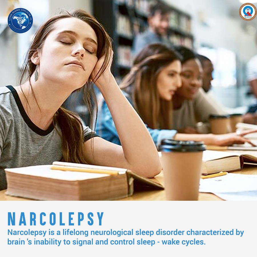
Can you recall a time when you dozed off during work? Probably, it was on the day after you partied whole night long. You were really exhausted and managed only a few hours of sleep. But, can you imagine a situation in which you fall into sleep instantly every now and then? While driving or eating? Isn’t it stressful? This is what happens to people with Narcolepsy.
Narcolepsy is a chronic neurological disorder that affects the brain’s ability to control sleep-wake cycles. It is characterized by sleep attacks, sleep paralysis, hallucination and in some cases episodes of cataplexy (explained in a later section). People with narcolepsy are overwhelmingly drowsy throughout much of the day. They also experience intermittent, uncontrollable episodes of falling asleep during the daytime and uneven interrupted sleep during night. 

Narcolepsy explained

Narcolepsy can affect greatly affect daily activities ,ofcourse adversely. These sudden sleep attacks may occur during any type of activity at any time of the day. Sleep invades even in the middle of an activity like driving, eating, talking or dancing. Yes, you read that right! So, if left undiagnosed or untreated, this neurological condition can interfere with overall psychological, social and cognitive function and development which can in turn inhibit academic, work and social life.
In a normal sleep cycle, we initially enter early stages of sleep followed by deeper sleep stages and ultimately REM (rapid eye movement) sleep. It takes around 60-90 minutes. Dreams happen during REM sleep. Now during this stage, brain keeps muscles “limp” and as a result, people are incapacitated to act out of their dreams.
In contrast, narcoleptic people enter REM sleep rapidly in about 15 minutes of falling asleep without experiencing non-rapid eye movement sleep (NREM), both at night and during the day. The boundary between awake and sleep is blurred. Consequently, aspects of REM sleep intrude on wakefulness, while wakefulness intrudes on their sleep. 

Who is affected?

It can affect irrespective of the gender. Typical onset of narcolepsy is between the ages of 10-25; but can occur anytime in life, even in early childhood. Statistics show that about 1,35,000-2,00000 in US have narcolepsy. In India, it is fewer than a million. But in reality, the numbers may be higher since people with narcolepsy are often misdiagnosed with other conditions such as psychiatric disorders and emotional problems. It is estimated that up to 50% of patients with Narcolepsy may be undiagnosed.  

Causes of Narcolepsy

The cause of narcolepsy is not clearly established. Some experts suggest that narcolepsy may be due to the deficiency of certain chemicals in brain which signals sleep-wake cycles called hypocretin.        Current research points that narcolepsy involves multiple factors, which causes the neurological dysfunction and REM sleep disturbances, working together to cause a lack of hypocretin. 

Symptoms of Narcolepsy 

Narcolepsy is a lifelong condition. But symptoms can partially improve over time, but never disappear completely. All narcoleptics experience excessive daytime sleepiness, but all of the symptoms are seen only in 10-25 % of affected individuals. 

Excessive daytime sleepiness (EDS)
This most obvious symptom is the inability to stay awake and alert during the day regardless of how much sleep an individual gets at night. The real scare is that the sleepiness is not normally drowsiness, but ‘sleep attacks’ where the individual falls off to sleep suddenly.

Sleep disruption and insomnia
Narcoleptic sleep is disrupted by frequent awakening, also called fragmented sleep. Sleep may be also disrupted by insomnia, vivid dreams, sleep apnea, acting out while dreaming and rapid leg movements.

Cataplexy
Cataplexy is a sudden and uncontrollable, brief loss of voluntary muscle control (actually the ‘muscle paralysis of REM sleep) often triggered by experiencing strong and intense emotions such as laughter, stress, anger, fear or excitement. Cataplexy attacks may vary in severity from partial cataplexy, which involve the slurring of speech to full cataplexy where an individual collapse to the ground, awake but unable to move, speak or keep eyes open.
In about 10% of narcoleptics, this is the first symptom to appear and could be misdiagnosed as a seizure disorder. But unlike fainting or seizure disorder, people remain conscious even during the most severe attack.

Sleep Paralysis
While falling asleep or waking up, sleep paralysis causes temporary inability to move or speak. It usually lasts for only a few seconds or minutes. It resembles cataplexy except that it occurs on the edges of sleep.

Hallucinations
Very vivid and frightening images can accompany sleep paralysis and usually occur when people are falling asleep or waking up. Most often, the content is primarily visual, but any of the other senses can be involved too.

Automatic behavior
Sometimes, narcoleptics experience temporary sleep episodes that can be very brief, lasting no more than a few seconds. Person automatically continues any activity they were doing, for example, eating or talking for a few seconds or minutes without consciousness.
They cannot recall their actions and their performance is almost always impaired. If episodes occur during driving, individuals may get lost or have an accident. 

Types of Narcolepsy 

Type 1 narcolepsy (Narcolepsy with cataplexy).
It is diagnosed by low levels of hypocretin or reporting cataplexy/excessive sleepiness on a special nap test. 

Type 2 narcolepsy (Narcolepsy without cataplexy)
These people experience severe day time sleepiness but usually do not have muscle weakness triggered by emotions. 

Diagnosis

On average it takes 10 years from onset of symptoms to an official diagnosis of narcolepsy. Prior to diagnosis, it is not uncommon for people to be misdiagnosed with other sleep disorder, depression, psychiatric conditions, ADHD and even epilepsy.
If doctor suspects narcolepsy, he or she will most likely recommend the following sleep studies: 

Polysomnography (PSG)
During the PSG, a technician places multiple sensor, which are connected to a computer, on the patient’s head, neck, chest and legs to record brain waves, oxygen levels, eye movements, muscle tone and heat and breathing rates. Also, a video is recorded for monitoring the body movement. 

Multiple Sleep Latency Test (MSLT)
MSLT is a day time sleep study conducted after a PSG to assess the quality of night time sleep and the degree of sleepiness during the day. The MSLT involves several naps scheduled at 2-hour intervals beginning a couple of hours after you wake up in the morning. It documents how readily a person can fall asleep, REM patterns, eye movements, heart rate, chin movements and certain brain activities. 

Lumbar Puncture
The lumbar puncture is used to test the level of Hypocretin-I. It involves a spinal tap to collect the fluid. This test is specific to narcolepsy, as there are no other conditions known that can cause a decrease in hypo cretin levels. 

Treatment 

While is no cure for narcolepsy, individuals with narcolepsy can lead full and productive lives with a combination of pharmacological treatment and lifestyle adjustments.
Every individual with narcolepsy is different and medications that work best for one individual may not work for best for another. Therefore, it is necessary to talk to your doctor or health care professional about the option for specific specialized treatments.
Medications for narcolepsy can include, sodium oxybate (Xyrem), stimulants, antidepressants, and selective serotonin reuptake inhibitors (SSRIs) and serotonin/norepinephrine reuptake inhibitors (SNRIs). 

Life style changes
Improve sleep hygiene: 
Sleep hygiene is the practice of healthy sleep habits that optimize nighttime sleep quality.
Exercise: Daily exercise performed more than 3 hours before bedtime promotes better quality sleep.
Rise: When it’s time to wake up, let in as much natural light as possible. Natural light assists internal regulation of our sleep-wake cycle so getting outdoors during the day is important too.
Listen to your body: Scheduled daytime naps are effective ways recharging your energy level.
Avoid smoking: especially at night 

Due to narcolepsy being poorly understood and largely invisible, many people experience feelings of isolation, loss of self-confidence, depression and anxiety. Additionally, the diagnosis of narcolepsy may affect education and career choice, as not all activities and routines fit into the type of schedule an individual with narcolepsy may require. Children and adolescents with narcolepsy may struggle with emotional regulation and display aggressive behaviors that lead to social exclusion and feelings of self-loathing and shame.  

With adequate medications and life style changes, narcolepsy can be brought under control. It is believed, Thomas Alwa Edison was narcoleptic. Late night show host Jimmy Kmmel is narcoleptic. What more assurance is required to prove that narcoleptics can be successful in any direction they choose! But make sure to use proper medications. 


Keratoconus: Causes, Symptoms & Treatments

Symptoms and signs:

As the cornea becomes more irregular in shape, it causes progressive nearsightedness and irregular astigmatism to develop, creating additional problems with distorted and blurred vision. Glare and light sensitivity also may occur.


Normal eye vs. eye with keratoconus

Causes:

New research suggests the weakening of the corneal tissue that leads to keratoconus may be due to an imbalance of enzymes within the cornea. This imbalance makes the cornea more susceptible to oxidative damage from compounds called free radicals, causing it to weaken and bulge forward.

Risk factors for oxidative damage and weakening of the cornea include a genetic predisposition, explaining why keratoconus often affects more than one member of the same family.

Keratoconus also is associated with overexposure to ultraviolet rays from the sun, excessive eye rubbing, a history of poorly fitted contact lenses and chronic eye irritation.

What Warnings Should I Know About Corneal Cross-Linking?

Ulcerative keratitis, a potentially serious eye infection, can occur. Your doctor should monitor defects in the outermost corneal layer of the eye for resolution.

Who Should Not Receive CXL?

The safety and effectiveness of CXL has not been studied in pregnant women, women who are breastfeeding, patients who are less than 14 years of age and patients 65 years of age or older.

What Are The Side Effects Of CXL?

In progressive keratoconus patients, the most common side effects in any CXL treated eye were haze, inflammation, fine white lines, disruption of surface cells, eye pain, reduced sharpness of vision, and blurred vision. In patients with corneal ectasia following refractive surgery, the most common side effects in any CXL treated eye were haze, disruption of surface cells, fine white lines, dry eye, eye pain, inflammation, light sensitivity, reduced sharpness of vision, and blurred vision.

These are not all of the side effects of the CXL treatment. Tell your healthcare provider if you have any side effect that bothers you or that does not go away.

Keratoconus treatment:

In the mildest form of keratoconus, eyeglasses or soft contact lenses may help. But as the disease progresses and the cornea thins and becomes increasingly more irregular in shape, glasses and regular soft contact lens designs no longer provide adequate vision correction.

Corneal crosslinking diagram that shows anchors between collagen fibers in the cornea.
The aim of corneal cross-linking is to strengthen the cornea by increasing the number of “anchors” that bond collagen fibers together. 
Normal eye vs. eye with keratoconus. The keratoconic cornea bulges slightly, for a more cone-shaped surface.

Treatments for progressive keratoconus include:

1. Corneal crosslinking

Treatment by providing contact lenses

  1.  Custom soft contact lenses
  2.  Gas permeable contact lenses
  3.  Silicone hydrogel contact lenses
  4.  Hybrid contact lenses
  5.  Scleral and semi-scleral lenses
  6.  Prosthetic lenses

Surgery Option:

  1.  Intacs (Addition Technology)
  2. Topography-guided conductive keratoplasty
  3. Corneal transplant


Nephrogenic Systemic Fibrosis: Causes, Symptoms & Treatments

Nephrogenic systemic fibrosis is a rare disease that occurs mainly in people with advanced kidney failure with or without dialysis. Nephrogenic systemic fibrosis may resemble skin diseases, such as scleroderma and scleromyxedema, with thickening and darkening developing on large areas of the skin.

Nephrogenic systemic fibrosis can also affect internal organs, such as the heart, kidneys and lungs, and it can cause a disabling shortening of muscles and tendons in the joints (joint contracture).

For some people with advanced kidney disease, being exposed to certain gadolinium-containing contrast agents during magnetic resonance imaging (MRI) and other imaging studies has been identified as a trigger for development of this disease.

Symptoms

Nephrogenic systemic fibrosis can begin days to months after exposure to gadolinium-containing contrast, but progresses quickly. Some signs and symptoms of nephrogenic systemic fibrosis may include:

  • Swelling and tightening of the skin
  • Thickening and hardening of the skin, typically on the arms and legs and sometimes on the body, but almost never on the face or head
  • Skin that may feel “woody” and develop an orange-peel appearance and darkening (excess pigmentation)
  • Burning, itching or severe sharp pains in areas of involvement
  • Skin thickening that inhibits movement, resulting in loss of joint flexibility
  • Rarely, blisters or ulcers

In some people, involvement of muscles and body organs may cause:

  • Muscle weakness
  • Limitation of joint motion caused by muscle tightening (contractures) in arms, hands, legs and feet
  • Bone pain
  • Reduced internal organ function, including heart, lung, diaphragm, gastrointestinal tract, or liver, but direct evidence is often lacking
  • Yellow plaques on the white surface (sclera) of the eyes
  • Blood clots

The condition is generally long term (chronic), but some people may improve. In a few people, it can cause severe disability, even death.

Causes

The exact cause of nephrogenic systemic fibrosis isn’t fully understood. Exposure to gadolinium-containing contrast agents during magnetic resonance imaging (MRI) has been identified as a trigger for development of this disease. The Food and Drug Administration (FDA) recommends avoiding gadolinium-containing contrast agents in people with acute kidney injury or chronic kidney disease.

Other conditions that may lead to or promote the disease when severe kidney disease and exposure to gadolinium-containing contrast are present include:

  • Use of high-dose erythropoietin (EPO), a hormone that promotes the production of red blood cells, often used to treat anemia
  • Recent vascular surgery
  • Blood-clotting problems
  • Severe infection

Treatment

There is no cure for nephrogenic systemic fibrosis, and no treatment is consistently successful in halting or reversing the progression of the disease. Nephrogenic systemic fibrosis only occurs rarely, making it difficult to conduct large studies.

  • Hemodialysis.
  • Physical therapy.
  • Kidney transplant.
  • Ultraviolet A phototherapy.
  • Extracorporeal photopheresis.
  • Plasmapheresis.

Osteoarthritis: Causes, Symptoms & Treatments

Osteoarthritis is the most common form of arthritis, affecting millions of people worldwide. It occurs when the protective cartilage on the ends of your bones wears down over time. Although osteoarthritis can damage any joint in your body, the disorder most commonly affects joints in your hands, knees, hips and spine.

Symptoms

Osteoarthritis symptoms often develop slowly and worsen over time. Signs and symptoms of osteoarthritis include:

  • Pain. Your joint may hurt during or after movement.
  • Tenderness. Your joint may feel tender when you apply light pressure to it.
  • Stiffness. Joint stiffness may be most noticeable when you wake up in the morning or after a period of inactivity.
  • Loss of flexibility. You may not be able to move your joint through its full range of motion.
  • Grating sensation. You may hear or feel a grating sensation when you use the joint.
  • Bone spurs. These extra bits of bone, which feel like hard lumps, may form around the affected joint.

Causes

Osteoarthritis occurs when the cartilage that cushions the ends of bones in your joints gradually deteriorates. Cartilage is a firm, slippery tissue that permits nearly frictionless joint motion.

In osteoarthritis, the slick surface of the cartilage becomes rough. Eventually, if the cartilage wears down completely, you may be left with bone rubbing on bone.

Treatment

Currently, the process underlying osteoarthritis cannot be reversed, but symptoms can usually be effectively managed with lifestyle changes, physical and other therapies, medications, and surgery. Exercising and achieving a healthy weight are generally the most important ways to treat osteoarthritis. Your doctor may also suggest:

Medications:

  • Acetaminophen.
  • Nonsteroidal anti-inflammatory drugs (NSAIDs).
  • Duloxetine (Cymbalta).

Therapy

  • Physical therapy.
  • Occupational therapy.
  • Tai chi and yoga.

Surgical and other procedures:

If conservative treatments don’t help, you may want to consider procedures such as:

  • Cortisone injections. Injections of corticosteroid medications may relieve pain in your joint. During this procedure your doctor numbs the area around your joint, then places a needle into the space within your joint and injects medication. The number of cortisone injections you can receive each year is generally limited to three or four injections, because the medication can worsen joint damage over time.
  • Lubrication injections. Injections of hyaluronic acid may offer pain relief by providing some cushioning in your knee, though some research suggests these injections offer no more relief than a placebo. Hyaluronic acid is similar to a component normally found in your joint fluid.
  • Realigning bones. If osteoarthritis has damaged one side of your knee more than the other, an osteotomy might be helpful. In a knee osteotomy, a surgeon cuts across the bone either above or below the knee, and then removes or adds a wedge of bone. This shifts your body weight away from the worn-out part of your knee.
  • Joint replacement. In joint replacement surgery (arthroplasty), your surgeon removes your damaged joint surfaces and replaces them with plastic and metal parts. Surgical risks include infections and blood clots. Artificial joints can wear out or come loose and may need to eventually be replaced.

Hemodynamic instability: Causes, Symptoms & Treatments

Hemodynamic stability can be simply explained as the stable blood flow. If a person is hemodynamically stable, it means that he/she has a stable heart pump and good circulation of blood. Hemodynamic instability is defined as any instability in blood pressure which can lead to inadequate arterial blood flow to organs. It is also a state where there is a requirement for physiological and mechanical support to ensure there is adequate cardiac input and output or blood pressure.

Abnormal hemodynamic parameters include heart rate, blood pressure, cardiac output, central venous pressure, and pulmonary artery pressure.

Signs and symptoms

Following are some signs and symptoms of hemodynamic instability:

1. Hypotension

2. Abnormal heart rate

3. Shortness of breath

4. Pulmonary congestion

5. Cold extremities

6. Peripheral cyanosis

7. Decreased urine output

8. Alternative consciousness (restlessness, loss of consciousness, confusion)

9. Chest pain

Hypotension can produce insufficient perfusion in the brain and heart due to autoregulation of these organs. The individual is mostly affected, when the particular threshold of the blood pressure become drop down and may cause cerebral and coronary ischemia.

Who is at risk?

Hemodynamic instability is common during surgery conducted under general anesthesia. An anesthesiologist is continuously monitoring the patient for maintaining the normal hemodynamic. Usually anesthesiologist plan a clinical management before commencing the surgery, depending upon the pre-existing condition of the patient.

For made an effective plan for hemodynamic management, it is necessary to have thorough knowledge of medical history. The risk of hemodynamic instability is more with patient having cardiovascular diseases and major surgery need relatively more complex planning for  hemodynamic management than healthy patient.

Recognizing hemodynamic instability

For recognizing the critically ill patient and avoidance of hemodynamic instability, the following diagnostic tests should pursue:

Clinical assessment in the seriously ailing is mainly utilized for the initial three rationales: for example to decide whether the individual patient has hemodynamic instability, to decide whether the individual patient is countering to treatment and to stratify threat of instability.

When a healthcare staff checking the systolic BP (blood pressure) or vessels fills up time the measuring of the result should be exact, precise, and offer further direction to specify the presence of any cardiac syndrome. If the obtained result provides any information which can direct the clinician about the presence of hemodynamic instability or shock, then it is easier to make some decision about the management of the hemodynamic instability, before onset of the condition.

What Does a High Red Blood Cell Count Mean for a Cancer Patient?

Blood pressure recording

Method of clinical assessment of Hemodynamic Instability

The assessment of  vital sign and surrogate bio markers for assessing the organ perfusion, including urine output and capillary refill time have frequently used clinical assessment method to estimate hemodynamic instability. The below mentioned techniques are used as diagnostic tests for hemodynamic instability.

Vital signs

The monitoring of the vital signs is the primary clinical assessment for evaluating hemodynamic instability of a patient. Vital signs measurement is very important in judging the severity of the patient’s condition and to decide the any emergency medical intervention need to support for life saving of the patient or not. The following are the included elements of the vital signs.

Pulse

Variation of pulse rate may provide initial symptom of the development of the hemodynamic instability. The factors which mainly affect the pulse rate are fever, physical exhaustion or exercise, disorder of the thyroid gland and certain medications.

The increase level of catecholamine hormone during stress, haemorrage or dehydrations are different causes which influence pulse rate.  The postural alteration can affect pulse rate and provide the indication of  hypovolemia. Increase or decrease rate of pulse is a marker for arranging of a medical emergency team.

Respiratory rate

Respiratory rate is an important element to assessing the hemodynamic instability, as it is a marker of severity of illness. Respiratory rate become drop down for acute respiratory failure and immediate emergency medical assistance is required  to manage the patient ill health. Respiratory rate also provides guidance of patient reaction after giving any therapy.

Mean Arterial Pressure / Blood Pressure

Proper maintenance of the blood pressure is required to maintain the autoregulation of the heart and brain by keeping sustain perfusion. Mean Arterial Pressure / Blood Pressure is a good marker of hemodynamic instability. Less than 90 mm Hg in systolic blood pressure or less than 65 mm Hg of  Mean Arterial Pressure indicates severe sepsis and septic shock.

Temperature

High body temperature is also used as an indicator of clinical condition of the patient, though it is not sensitive marker of hemodynamic instability.

Toe- Temperature Gradient

Most of the clinicians suggest Toe- Temperature or rectal temperature gradient is useful diagnostic measure for adequacy of circulation. Toe- Temperature Gradient is a better indicator than  Mean Arterial Pressure, cardiac index or lactate to discriminate between survivors or non survivors for patient admitted to a ICU.

Organ perfusion

Organ perfusion is very important marker for diagnosing hemodynamic instability. hypoperfusion in specific organ indicative of insufficient perfusion in kidney or cardiac output.

Urine Output

The small quantity of urine production (oliguria) is another way to diagnose hemodynamic instability, but may associate with other clinical conditions, so it is an important but not specific clinical marker for hemodynamic instability.  insufficient renal perfusion is one of the cause of oliguria. 0.5ml/kg/hour urine output is a sarogate marker for severe sepsis.

Capillary refill time (CRT)

This clinical element is used to diagnosis the severity of instability of a patient treating under emergency care unit or ICU. The CRT is usually measure by preseeing the finger nail bed to occlute the circulation and then release the pressure and time is noted for how long normal coloration is achieved. CRT more than 6 seconds indicates emergency condition. The abnormal level of CRT along with cardiac index and urine output may indicate acute lung injury.


Cellulitis: Causes, Symptoms & Treatments

Cellulitis is a common infection of the skin and the soft tissues underneath. It happens when bacteria enter a break in the skin and spread. The result is infection, which may cause swelling, redness, pain, or warmth.

You’re at risk if you have:

  • Trauma to the skin
  • Diabetes
  • Circulatory problems, such as not enough blood flow to your arms and legs, poor drainage of your veins or lymphatic system, or varicose veins — twisted, enlarged veins near the surface of the skin
  • Liver disease such as chronic hepatitis or cirrhosis
  • Skin disorders such as eczema, psoriasis, or infectious diseases that cause sores, such as chickenpox

Causes:

  • Injuries that tear the skin
  • Infections after surgery
  • Long-term skin conditions such as eczema or psoriasis
  • Foreign objects in the skin
  • Bone infections underneath the skin. (An example is a long-standing, open wound that is deep enough to expose the bone to bacteria.)

Symptoms:

Cellulitis can appear on almost any part of the body. It usually shows up on damaged skin such as inflamed wounds, dirty cuts, and areas with poor circulation. It needs to be treated by a doctor. Common symptoms include:

  • Redness
  • Red streaking
  • Swelling
  • Warmth
  • Pain or tenderness
  • Leaking of yellow, clear fluid or pus

When to see doctor:

Go to the emergency room if you have any of the following:

  • High fever or chills
  • Nausea and vomiting
  • Enlarging or hardening of the reddened area
  • Increased pain
  • Numbness of the area when touched
  • Other medical problems that may be affected by even a minor infection

Exams and Tests for Cellulitis

Your doctor will do a medical history and physical exam. Additional procedures include:

  • A blood test if the infection is suspected to have spread to your blood
  • An X-ray if there’s a foreign object in the skin or the bone underneath is possibly infected
  • A culture. Your doctor will use a needle to draw fluid from the affected area and send it to the lab.

Treatment for Cellulitis

  • Rest the area.
  • Elevate the area to help reduce swelling and relieve discomfort.
  • Use over-the-counter pain relievers such as acetaminophen (Tylenol) or ibuprofen (Motrin) to ease the pain, as well as keep your fever down
If the infection isn’t too bad, you can take antibiotics by mouth for a week to 14 days. Your doctor will schedule a follow-up appointment. Your doctor may use IV or intramuscular antibiotics if:

  • The infection is severe.
  • You have other medical problems.
  • You are very young or very old.
  • The cellulitis covers large areas, is on your hands, or is close to body parts like your eyes.
  • The infection worsens even after taking antibiotics for 2 to 3 days.

In serious cases, you may need to stay in the hospital. You’ll get IV antibiotics until the infection is under control (2 to 3 days), and then go home with oral medicines.

Surgery for Cellulitis

Rarely, severe cases may need surgery. For example, doctors may need to open and drain an abscess or pus that has collected in the tissue. They may also need to cut away dead tissue to allow healing.

Prevention of Cellulitis

  • Practice good personal hygiene and keep your skin clean.
  • Wear sturdy, well-fitting shoes or slippers with loose-fitting cotton socks. Avoid walking barefoot outdoors.
  • Wash injured skin with soap and water. Make sure it heals over the next few days.

Some injuries are at greater risk for cellulitis than others. Be sure to contact your doctor if you have:

  • Animal or human bites
  • Puncture wounds deeper than a half-inch, such as from stepping on a nail
  • Crushed tissue that bleeds
  • Burns that blister
  • Frostbite
  • Deep injuries with dirt in them
  • Injuries that touch sea water (making them more prone to infection), especially if you have liver disease
  • Diabetes or other significant medical conditions, such as liver or kidney disease
  • Swelling in your arms and legs that does not go away

Edema (brain swelling) : Causes, Symptoms & Treatments

Brain swelling, also known as cerebral edema in medical terminology is a serious neurological condition. Swelling can occur in any organ of the body. It is body’s response to any type of injury, infection or an overuse of that particular organ or part of the body. Brain edema can develop as a result of an injury to brain or infection in the brain. It may involve certain part of brain or the whole brain.

Brain swelling produces increase in intracranial pressure. Intracranial pressure prevents proper blood flow to the brain as a result the brain cells are deprived of vital food that is oxygen and glucose.

Raised pressure in brain also blocks the cerebrospinal fluid which makes the matter worse. Edema in brain can cause damage to brain cells which may lead to cognitive and behavioral defect. Brain swelling needs to be treated immediately as the condition has fatal outcome if left untreated.

Causes:


  • Brain Injury: An injury on the head can damage the brain. Usually a severe injury to the skull such as in vehicular accidents, falling from a height, etc is responsible for swelling in brain.

    The injury can cause swelling of the brain tissue. The broken pieces of skull can injure the blood vessels which can lead to swelling of brain.

  • Stroke and high blood pressure: Brain stroke caused due to ischemia of the blood vessel in the brain or hemorrhage inside the brain caused due to trauma or high blood pressure can cause cerebral edema.
  • Infections: Bacterial infection or viral infection such as meningitis, encephalitis can cause swelling of brain tissues.
  • Brain tumor: Edema of brain can be caused due to brain tumor. The growing brain tumor can compress the adjacent area of brain and block the circulating spinal fluid. These may cause rise in intracranial pressure and brain swelling. The growing blood vessels in and around the tumor can also cause brain edema.
  • High altitude: Brain swelling can be one of the symptoms of high altitude sickness although not very common. The swelling is more likely to develop at an altitude 5000 feet and above. The condition is often accompanied with acute mountain sickness.

 Symptoms:

  • Headache
  • Neck pain or stiffness
  • Nausea or vomiting
  • Dizziness
  • Irregular breathing
  • Vision loss or changes
  • Memory loss
  • Inability to walk
  • Difficulty speaking
  • Stupor
  • Seizures
  • Loss of consciousness

Treatment for Edema:

Minor cases of brain swelling due to causes such as moderate altitude sickness or a slight concussion often resolve within a few days. In most cases, however, more treatment is needed quickly.

The goal is to assure that the brain receives enough blood and oxygen to remain healthy while the swelling is relieved and any underlying causes are treated. This may require a combination of medical and surgical treatments. Prompt treatment usually results in quicker and more complete recovery. Without it, some damage may remain.

Treatment for brain edema may include any combination of the following:

  • Oxygen therapy: Providing oxygen through a respirator or other means helps make sure that the blood has enough oxygen in it. The doctor can adjust the respirator to help reduce the amount of swelling.
  • IV fluids: Giving fluids and medicine through an IV can keep blood pressure from dropping too low. This helps to make sure that the body — including the brain — is receiving enough blood. However, some fluids can make swelling worse. Doctors attempt to use the right amounts of the right fluids in someone with brain swelling.
  • Lowering body temperature (hypothermia): Lowering the temperature of the body and brain helps relieve swelling and allows the brain to heal. Hypothermia as a treatment for brain swelling is not widely used because it is difficult to perform correctly.
  • Medication: In some cases of brain edema, your doctor may start a drug to help relieve the swelling. Medication may also be given for other reasons, such as to slow your body’s response to the swelling or to dissolve any clots. The drugs your doctor gives you depend on the cause and symptoms of brain swelling.
  • Ventriculostomy: In this procedure, a surgeon cuts a small hole in the skull and inserts a plastic drain tube. Cerebrospinal fluid is drained from inside the brain, helping to relieve the pressure.
  • Surgery: Surgery may have one or more of these goals:
    • Removing part of the skull to relieve intracranial pressure; this procedure is called decompressive craniectomy.
    • Removing or repairing the source of the swelling, such as repairing a damaged artery or vein or removing a growth

Hydrocephalus: Causes, Symptoms & Treatments

Hydrocephalus is the buildup of fluid in the cavities (ventricles) deep within the brain. The excess fluid increases the size of the ventricles and puts pressure on the brain. Cerebrospinal fluid normally flows through the ventricles and bathes the brain and spinal column. But the pressure of too much cerebrospinal fluid associated with hydrocephalus can damage brain tissues and cause a range of impairments in brain function. Hydrocephalus can happen at any age, but it occurs more frequently among infants and adults 60 and over. Surgical treatment for hydrocephalus can restore and maintain normal cerebrospinal fluid levels in the brain. Many different therapies are often required to manage symptoms or functional impairments resulting from hydrocephalus.

Symptoms

The signs and symptoms of hydrocephalus vary somewhat by age of onset.

Infants

Common signs and symptoms of hydrocephalus in infants include:

Changes in the head

  • An unusually large head
  • A rapid increase in the size of the head
  • A bulging or tense soft spot (fontanel) on the top of the head

Physical signs and symptoms

  • Vomiting
  • Sleepiness
  • Irritability
  • Poor feeding
  • Seizures
  • Eyes fixed downward (sunsetting of the eyes)
  • Deficits in muscle tone and strength
  • Poor responsiveness to touch
  • Poor growth

Toddlers and older children

Among toddlers and older children, signs and symptoms may include:

Physical signs and symptoms

  • Headache
  • Blurred or double vision
  • Eyes fixed downward (sunsetting of eyes)
  • Abnormal enlargement of a toddler’s head
  • Sleepiness or lethargy
  • Nausea or vomiting
  • Unstable balance
  • Poor coordination
  • Poor appetite
  • Seizures
  • Urinary incontinence

Behavioral and cognitive changes

  • Irritability
  • Change in personality
  • Decline in school performance
  • Delays or problems with previously acquired skills, such as walking or talking

Young and middle-aged adults

Common signs and symptoms in this age group include:

  • Headache
  • Lethargy
  • Loss of coordination or balance
  • Loss of bladder control or a frequent urge to urinate
  • Impaired vision
  • Decline in memory, concentration and other thinking skills that may affect job performance

Older adults

Among adults 60 years of age and older, the more common signs and symptoms of hydrocephalus are:

  • Loss of bladder control or a frequent urge to urinate
  • Memory loss
  • Progressive loss of other thinking or reasoning skills
  • Difficulty walking, often described as a shuffling gait or the feeling of the feet being stuck
  • Poor coordination or balance

When to see a doctor

Seek emergency medical care for infants and toddlers experiencing these signs and symptoms:

  • A high-pitched cry
  • Problems with sucking or feeding
  • Unexplained, recurrent vomiting
  • An unwillingness to move the head or lay down
  • Breathing difficulties
  • Seizures

Diagnosis

A diagnosis of hydrocephalus is usually based on:

  • Your answers to the doctor’s questions about signs and symptoms
  • A general physical
  • A neurological exam
  • Brain imaging tests

Neurological exam

The type of neurological exam will depend on a person’s age. The neurologist may ask questions and conduct relatively simple tests in the office to judge muscle condition, movement, well-being and how well the senses are functioning.

Brain imaging

Brain imaging tests can show enlarged ventricles caused by excess cerebrospinal fluid. They may also be used to identify underlying causes of hydrocephalus or other conditions contributing to the symptoms. Imaging tests may include:

  • Ultrasound. Ultrasound imaging, which uses high-frequency sound waves to produce images, is often used for an initial assessment for infants because it’s a relatively simple, low-risk procedure. The ultrasound device is placed over the soft spot (fontanel) on the top of a baby’s head. Ultrasound may also detect hydrocephalus prior to birth when the procedure is used during routine prenatal examinations.
  • Magnetic resonance imaging (MRI) uses radio waves and a magnetic field to produce detailed 3-D or cross-sectional images of the brain. This test is painless, but it is noisy and requires lying still.Children may need mild sedation for some MRI scans. However, some hospitals use a very fast version of MRI that generally doesn’t require sedation.
  • Computerized tomography (CT) scan is a specialized X-ray technology that can produce cross-sectional views of the brain. Scanning is painless and quick. But this test also requires lying still, so a child usually receives a mild sedative.Drawbacks to CT scanning include less detailed images than an MRI, and exposure to a small amount of radiation. CT scans for hydrocephalus are usually used only for emergency exams.

Treatment

One of two surgical treatments may be used to treat hydrocephalus.

1. Shunt

The most common treatment for hydrocephalus is the surgical insertion of a drainage system, called a shunt. It consists of a long, flexible tube with a valve that keeps fluid from the brain flowing in the right direction and at the proper rate.

One end of the tubing is usually placed in one of the brain’s ventricles. The tubing is then tunneled under the skin to another part of the body where the excess cerebrospinal fluid can be more easily absorbed — such as the abdomen or a chamber in the heart.

People who have hydrocephalus usually need a shunt system for the rest of their lives, and regular monitoring is required.

2. Endoscopic third ventriculostomy

Endoscopic third ventriculostomy is a surgical procedure that can be used for some people. In the procedure, your surgeon uses a small video camera to have direct vision inside the brain. Your surgeon makes a hole in the bottom of one of the ventricles or between the ventricles to enable cerebrospinal fluid to flow out of the brain.

Complications of surgery

Both surgical procedures can result in complications. Shunt systems can stop draining cerebrospinal fluid or poorly regulate drainage because of mechanical malfunctions, blockage or infections. Complications of ventriculostomy include bleeding and infections.

Any failure requires prompt attention, surgical revisions or other interventions. Signs and symptoms of problems may include:

  • Fever
  • Irritability
  • Drowsiness
  • Nausea or vomiting
  • Headache
  • Vision problems
  • Redness, pain or tenderness of the skin along the path of the shunt tube
  • Abdominal pain when the shunt valve is in the abdomen
  • Recurrence of any of the initial hydrocephalus symptoms

Other treatments

Some people with hydrocephalus, particularly children, may need additional treatment, depending on the severity of long-term complications of hydrocephalus.

A care team for children may include a:

  • Pediatrician or psychiatrist, who oversees the treatment plan and medical care
  • Pediatric neurologist, who specializes in the diagnosis and treatment of neurological disorders in children
  • Occupational therapist, who specializes in therapy to develop everyday skills
  • Developmental therapist, who specializes in therapy to help your child develop age-appropriate behaviors, social skills and interpersonal skills
  • Mental health provider, such as a psychologist or psychiatrist
  • Social worker, who assists the family with accessing services and planning for transitions in care
  • Special education teacher, who addresses learning disabilities, determines educational needs and identifies appropriate educational resources

Retinal Detachment: Causes, Symptoms & Treatments

Retinal detachment describes an emergency situation in which a thin layer of tissue (the retina) at the back of the eye pulls away from its normal position. Retinal detachment separates the retinal cells from the layer of blood vessels that provides oxygen and nourishment. The longer retinal detachment goes untreated, the greater your risk of permanent vision loss in the affected eye.

Warning signs of retinal detachment may include one or all of the following: the sudden appearance of floaters and flashes and reduced vision. Contacting an eye specialist (ophthalmologist) right away can help save your vision.

Symptoms

Retinal detachment itself is painless. But warning signs almost always appear before it occurs or has advanced, such as:

  • The sudden appearance of many floaters — tiny specks that seem to drift through your field of vision
  • Flashes of light in one or both eyes (photopsia)
  • Blurred vision
  • Gradually reduced side (peripheral) vision
  • A curtain-like shadow over your visual field

Causes

There are three different types of retinal detachment:

  • Rhegmatogenous 
  • Tractional
  • Exudative

Treatment

Surgery is almost always used to repair a retinal tear, hole or detachment. Various techniques are available. Ask your ophthalmologist about the risks and benefits of your treatment options. Together you can determine what procedure or combination of procedures is best for you.

Retinal tears

When a retinal tear or hole hasn’t yet progressed to detachment, your eye surgeon may suggest one of the following procedures to prevent retinal detachment and preserve vision.

  • Laser surgery (photocoagulation). The surgeon directs a laser beam into the eye through the pupil. The laser makes burns around the retinal tear, creating scarring that usually “welds” the retina to underlying tissue.
  • Freezing (cryopexy). After giving you a local anesthetic to numb your eye, the surgeon applies a freezing probe to the outer surface of the eye directly over the tear. The freezing causes a scar that helps secure the retina to the eye wall.

Both of these procedures are done on an outpatient basis. After your procedure, you’ll likely be advised to avoid activities that might jar the eyes — such as running — for a couple of weeks or so.

Retinal detachment

Illustration depicting pneumatic retinopexy

Pneumatic retinopexy

If your retina has detached, you’ll need surgery to repair it, preferably within days of a diagnosis. The type of surgery your surgeon recommends will depend on several factors, including how severe the detachment is.

  • Injecting air or gas into your eye.
  • Indenting the surface of your eye.
  • Draining and replacing the fluid in the eye.Vitrectomy may be combined with a scleral buckling procedure.

After surgery your vision may take several months to improve. You may need a second surgery for successful treatment. Some people never recover all of their lost vision.

Pages:1...13141516171819...86