Retinal detachment describes an emergency situation in which a thin layer of tissue (the retina) at the back of the eye pulls away from its normal position. Retinal detachment separates the retinal cells from the layer of blood vessels that provides oxygen and nourishment. The longer retinal detachment goes untreated, the greater your risk of permanent vision loss in the affected eye.
Warning signs of retinal detachment may include one or all of the following: the sudden appearance of floaters and flashes and reduced vision. Contacting an eye specialist (ophthalmologist) right away can help save your vision.
Symptoms
Retinal detachment itself is painless. But warning signs almost always appear before it occurs or has advanced, such as:
- The sudden appearance of many floaters — tiny specks that seem to drift through your field of vision
- Flashes of light in one or both eyes (photopsia)
- Blurred vision
- Gradually reduced side (peripheral) vision
- A curtain-like shadow over your visual field
Causes
There are three different types of retinal detachment:
- Rhegmatogenous
- Tractional
- Exudative
Treatment
Surgery is almost always used to repair a retinal tear, hole or detachment. Various techniques are available. Ask your ophthalmologist about the risks and benefits of your treatment options. Together you can determine what procedure or combination of procedures is best for you.
Retinal tears
When a retinal tear or hole hasn’t yet progressed to detachment, your eye surgeon may suggest one of the following procedures to prevent retinal detachment and preserve vision.
- Laser surgery (photocoagulation). The surgeon directs a laser beam into the eye through the pupil. The laser makes burns around the retinal tear, creating scarring that usually “welds” the retina to underlying tissue.
- Freezing (cryopexy). After giving you a local anesthetic to numb your eye, the surgeon applies a freezing probe to the outer surface of the eye directly over the tear. The freezing causes a scar that helps secure the retina to the eye wall.
Both of these procedures are done on an outpatient basis. After your procedure, you’ll likely be advised to avoid activities that might jar the eyes — such as running — for a couple of weeks or so.
Retinal detachment
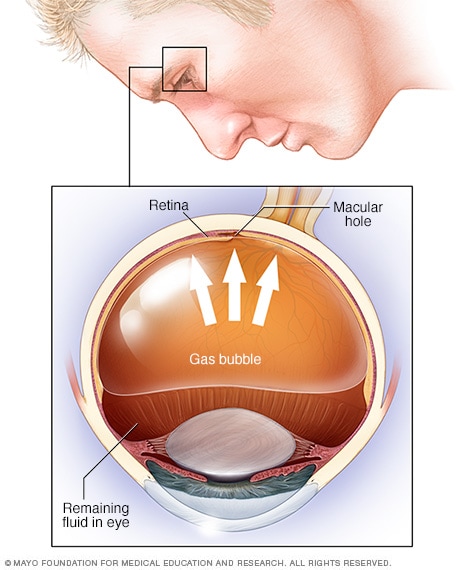
Pneumatic retinopexy
If your retina has detached, you’ll need surgery to repair it, preferably within days of a diagnosis. The type of surgery your surgeon recommends will depend on several factors, including how severe the detachment is.
- Injecting air or gas into your eye.
- Indenting the surface of your eye.
- Draining and replacing the fluid in the eye.Vitrectomy may be combined with a scleral buckling procedure.
After surgery your vision may take several months to improve. You may need a second surgery for successful treatment. Some people never recover all of their lost vision.
Post a comment