Did not expect word ‘nuclear’ in the field of medicine?Well,’Nuclear Medicine’ exists and let us understand it here.
Nuclear medicine is a subspeciality of medical imaging/radiology.But unlike radiology where radiation enter the body from outside,radioactive materials are introduced to the body in Nuclear medicine.These radioactive tracers in combination with imaging instrumentation is used to assess bodily functions and to diagnose , determine the severity of or treat a variety of diseases, including many types of cancers, heart disease, gastrointestinal, endocrine, neurological disorders and other abnormalities within the body.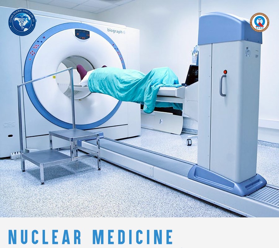
Technique explained
Small amounts of radioactive materials are attached to special compounds to form radiopharmaceuticals. The radioactive part is referred to as a radioactive label or a radioactive tracer.The pharmaceutical part,complex molecules, helps to carry the radioactive part to the area of the body being studied. It is mostly the pharmaceutical part that determines where the radiopharmaceutical will go inside the body.
Radiopharmaceutical is injected,inhaled or swallowed depending on the type of medical examination.This makes the body slightly radioactive for a short time. A special nuclear medicine camera detects the radiation, which is emitted from the body and produces an image of the tracer’s distribution in the body.Because the tracers follow physiological pathways, the result is a functional image.It can show disease processes based on increased or decreased uptake of the tracer compared to normal in various organs. As nuclear medicine procedures are able to pinpoint molecular activity within the body, they offer the potential to identify disease in its earliest stages as well as a patient’s immediate response to therapeutic interventions.
Nuclear medicine Vs Other imaging techniques
- Nuclear medicine scans detect the radiation coming from a radiopharmaceutical that is inside a patient’s body. In contrast, other imaging procedures (for example, X-ray and CT scan) obtain images by using machines that send radiation through the body.
- Nuclear medicine is also different from other imaging procedures in that it determines the presence of disease based on biological changes in tissue rather than changes in anatomy.
- Nuclear medicine procedures are among the safest diagnostic imaging exams available; the amount of radiation received from a nuclear medicine scan is comparable to that of many diagnostic X-ray and CT procedures.
Types of Scans
Single Photon Emission Computed Tomography or SPECT and Positron Emission Tomography or PET scans are the two most common imaging modalities in nuclear medicine.
SPECT imaging instruments provide three-dimensional (tomographic) images of the distribution of radioactive tracer molecules that have been introduced into the patient’s body. The 3D images are computer generated from a large number of projection images of the body recorded at different angles. SPECT imagers have gamma camera detectors that can detect the gamma ray emissions from the tracers that have been injected into the patient. Gamma rays are a form of light that moves at a different wavelength than visible light. The cameras are mounted on a rotating gantry that allows the detectors to be moved in a tight circle around a patient who is lying motionless on a pallet.
SPECT scans are primarily used to diagnose and track the progression of heart disease, such as blocked coronary arteries. There are also radiotracers to detect disorders in bone, gall bladder disease and intestinal bleeding. SPECT agents have recently become available for aiding in the diagnosis of Parkinson’s disease in the brain, and distinguishing this malady from other anatomically-related movement disorders and dementias.
PET scans also use radiopharmaceuticals to create three-dimensional images. The main difference between SPECT and PET scans is the type of radiotracers used. While SPECT scans measure gamma rays, the decay of the radiotracers used with PET scans produce small particles called positrons. A positron is a particle with roughly the same mass as an electron but oppositely charged. These react with electrons in the body and when these two particles combine they annihilate each other. This annihilation produces a small amount of energy in the form of two photons that shoot off in opposite directions. The detectors in the PET scanner measure these photons and use this information to create images of internal organs.
The major purpose of PET scans is to detect cancer and monitor its progression, response to treatment, and to detect metastases. Glucose utilization depends on the intensity of cellular and tissue activity so it is greatly increased in rapidly dividing cancer cells. In fact, the degree of aggressiveness for most cancers is roughly paralleled by their rate of glucose utilization. In the last 15 years, slightly modified radiolabeled glucose molecules (F-18 labeled deoxyglucose or FDG) have been shown to be the best available tracer for detecting cancer and its metastatic spread in the body.A combination instrument that produces both PET and CT scans of the same body regions in one examination (PET/CT scanner) has become the primary imaging tool for the staging of most cancers worldwide.
Other types
- Bone or Joint Scan: The scan is done to identify abnormal areas within the bones or joints. A small amount of radioactive material is injected into the vein and then images are taken 2 to 3 hours after the injection.
- Gallium Scan: This is done to detect infection or tumor. A special camera is used to capture images. After the injection of a radioactive material, the images are taken after 24, 48 or 96 hours, depending on the medical history of the patient.
- Gastric Emptying: This is done to evaluate the function of the stomach.
- Hepatobiliary Scan: This test is done to evaluate the function of gall bladder and to assess the bile ducts. After the injection of the radioactive material, images are taken immediately with a special camera for a minimum of one hour that can extend up to three hours.
- Liver or Spleen Scan: This is a type of scan done to find out the size and function of the liver and spleen.
Diagnosis
Nuclear medicine imaging procedures are noninvasive and, with the exception of intravenous injections, are usually painless medical tests that help physicians diagnose and evaluate medical conditions.The most commonly used radiotracer is F-18 fluorodeoxyglucose, or FDG, a molecule similar to glucose. Cancer cells may absorb glucose at a higher rate, being more metabolically active. This higher rate can be seen on PET scans, and that allows your doctor to identify disease before it may be seen on other imaging tests. FDG is just one of many radiotracers in use or in development for a variety of conditions throughout the body.
In many centers, nuclear medicine images can be superimposed with computed tomography (CT) or magnetic resonance imaging(MRI) to produce special views, a practice known as image fusion or co-registration. These views allow the information from two different exams to be correlated and interpreted on one image, leading to more precise information and accurate diagnoses. In addition, manufacturers are now making single photon emission computed tomography/computed tomography (SPECT/CT) and positron emission tomography/computed tomography (PET/CT) units that are able to perform both imaging exams at the same time. An emerging imaging technology, but not readily available at this time is PET/MRI.
Therapy
Nuclear medicine also offers therapeutic procedures, such as radioactive iodine (I-131) therapy that use small amounts of radioactive material to treat cancer and other medical conditions affecting the thyroid gland, as well as treatments for other cancers and medical conditions.
Non-Hodgkin’s lymphoma patients who do not respond to chemotherapy may undergo radioimmunotherapy (RIT).Radioimmunotherapy (RIT) is a personalized cancer treatment that combines radiation therapy with the targeting ability of immunotherapy, a treatment that mimics cellular activity in the body’s immune system
Any risks ?
The total radiation dose conferred to patients by the majority of radiopharmaceuticals used in diagnostic nuclear medicinestudies is no more than what is conferred during routine chest x-rays or CT exams. There are legitimate concerns about possible cancer induction even by low levels of radiation exposure from cumulative medical imaging examinations, but this risk is accepted to be quite small in contrast to the expected benefit derived from a medically needed diagnostic imaging study.
Like radiologists, nuclear medicine physicians are strongly committed to keeping radiation exposure to patients as low as possible, giving the least amount of radiotracer needed to provide a diagnostically useful examination.
Nuclear medicine examinations provide unique information—including details on both function and anatomic structure of the body that is often unattainable using other imaging procedures.For many diseases, nuclear medicine scans yield the most useful information needed to make a diagnosis or to determine appropriate treatment, if any.A nuclear medicine scan is less expensive and may yield more precise information than exploratory surgery.Nuclear medicine offers the potential to identify disease in its earliest stage, often before symptoms occur or abnormalities can be detected with other diagnostic tests.By detecting whether lesions are likely benign or malignant, PET scans may eliminate the need for surgical biopsy or identify the best biopsy location.PET scans may provide additional information that is used for radiation therapy planning.
For any queries regarding treatment facilities,email us at query@gtsmeditour.com .
Post a comment