Global Treatment Services Pvt. Ltd.

Global Treatment Services

Varicose vein

Varicose veins are swollen, twisted veins that can be visible just under the surface of the skin. Although it usually occurs in the legs, they also can form in other parts of the body also.Varicose vein is quite a common condition.29.varicosevein

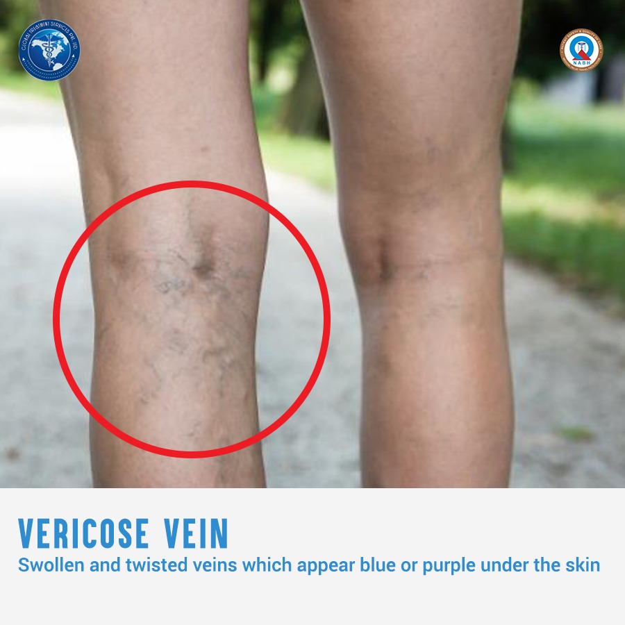
Veins have one-way valves that help keep blood flowing toward your heart. If the valves are weak or damaged, blood can back up and pool in your veins. This causes the veins to swell, which can lead to varicose veins.
Many factors can raise your risk for varicose veins. Examples of these factors include family history, older age, gender, pregnancy, overweight or obesity, lack of movement, and leg trauma.Varicose veins are treated with lifestyle changes and medical procedures. The goals of treatment are to relieve symptoms, prevent complications, and improve appearance.

Causes

Weak or damaged valves in the veins can cause varicose veins. After your arteries and capillaries deliver oxygen-rich blood to your body, your veins return the blood to your heart. The veins in your legs must work against gravity to do this.One-way valves inside the veins open to let blood flow through, and then they shut to keep blood from flowing backward. If the valves are weak or damaged, blood can back up and pool in your veins. This causes the veins to swell.
Weak vein walls may cause weak valves.If these walls become weak, they lose their normal elasticity. They become like an overstretched rubber band. This makes the walls of the veins longer and wider, and it causes the flaps of the valves to separate.When the valve flaps separate, blood can flow backward through the valves. The backflow of blood fills the veins and stretches the walls even more. As a result, the veins get bigger, swell, and often twist as they try to squeeze into their normal space. These are varicose veins.
Many factors can raise your risk for varicose veins. Examples of these factors include family history, older age, gender, pregnancy, overweight or obesity, lack of movement, and leg trauma.

Associated vein problems

Telangiectasias
Telangiectasias are small clusters of blood vessels usually found on the upper body, including the face.These blood vessels appear red. They may form during pregnancy, and often they develop in people who have certain genetic disorders, viral infections, or other conditions, such as liver disease.Telangiectasias can be a sign of a more serious condition.

Spider Veins
Spider veins are a smaller version of varicose veins and a less serious type of telangiectasias. Spider veins involve the capillaries, the smallest blood vessels in the body.Spider veins often appear on the legs and face. They’re red or blue and usually look like a spider web or tree branch. These veins usually aren’t a medical concern.

Varicoceles
Varicoceles are varicose veins in the scrotum and may be linked to male infertility.

Other types of varicose veins include venous lakes, reticular veins, and hemorrhoids. Venous lakes are varicose veins that appear on the face and neck. Reticular veins are flat blue veins often seen behind the knees. Hemorrhoids are varicose veins in and around the anus.

Diagnosis

Duplex Ultrasound
Duplex ultrasound combines traditional with Doppler ultrasound. Traditional ultrasound uses sound waves to create a picture of the structures in your body, in this case the blood vessels and anything that may be blocking the flow of blood. Doppler ultrasound uses sound waves to create pictures of the flow or movement of the blood through the veins. Duplex ultrasound combined with traditional together gives a picture of the blood flow in your arteries and veins.

Angiogram
Although it is not very common, an angiogram givest a more detailed look at the blood flow through your veins.An angiogram helps to confirm whether you have varicose veins or another condition.

Treatment

Varicose veins are treated with lifestyle changes and medical procedures. The goals of treatment are to relieve symptoms, prevent complications, and improve appearance.For more severe symptoms like pain, blood clots, or skin disorders ,one or more medical procedures may be needed.

Medical procedures

Sclerotherapy
Sclerotherapy uses a liquid chemical which is injected into the vein to cause irritation and scarring inside the vein. It causes the vein to close off, and it fades away.This procedure often is used to treat smaller varicose veins and spider veins.

Microsclerotherapy
Microsclerotherapy  is used to treat spider veins and other very small varicose veins.A small amount of liquid chemical is injected into a vein which scars the inner lining of the vein, causing it to close off.

Laser Surgery
The laser light makes the vein fade away.Laser surgery mostly is used to treat smaller varicose veins. The main advantage is that no cutting or injection of chemicals is involved.

Endovenous Ablation Therapy
Endovenous ablation therapy uses lasers or radiowaves to create heat to close off a varicose vein.A device at the tip of the catheter inserted to the vein heats up the inside of the vein and closes it off.

Endoscopic Vein Surgery
For endoscopic vein surgery, a small cut is made in your skin near a varicose vein. A tiny camera is used at the end of a thin tube to move through the vein and a surgical device at the end of the camera is used to close the vein.Endoscopic vein surgery usually is used only in severe cases when varicose veins are causing skin ulcers.

Ambulatory Phlebectomy

For ambulatory phlebectomy,  doctor will make small cuts in your skin to remove small varicose veins. This procedure usually is done to remove the varicose veins closest to the surface of your skin.

Vein Stripping and Ligation
Vein stripping and ligation typically is done only for severe cases of varicose veins. The procedure involves tying shut and removing the veins through small cuts in your skin.


Plastic and cosmetic surgery

We often use plastic and cosmetic surgery interchangeably. But this is technically wrong although both specialities are closely related.Cosmetic surgery is focussed on enhancing appearance whereas plastic surgery is reconstructive in nature.Plastic surgery involves procedures for repairing defects to reconstruct normal functioning an d appearance.28.plasticsurgery

Plastic surgery is defined as a surgical specialty dedicated to reconstruction of facial and body defects due to birth disorders, trauma, burns, and disease. Plastic surgery is intended to correct dysfunctional areas of the body and is reconstructive in nature. Improving aesthetic appeal, symmetry, and proportion are the key goals of cosmetic surgery. Cosmetic surgery can be performed on all areas of the head, neck, and body. Because the treated areas function properly, cosmetic surgery is purely optional.

Types of plastic an cosmetic surgery

 For Head, face, and eyes

  • Facelift
  • Brow/forehead lift
  • Eyelid lift
  • Ear pinning
  • Ear reshaping
  • Hair replacement surgery
  • Nasal surgery
  • Nose reshaping
  • Chin, cheek, or jaw reshaping / facial implants
  • Lip augmentation
  • Cleft lip and cleft palate
  • Craniosynostosis (craniofacial anomaly)
  • Oral and maxillofacial surgery

Breasts

  • Breast augmentation
  • Breast reconstruction
  • Breast reduction in men with gynecomastia
  • Breast lift

Abdomen

  • Tummy tuck (abdominoplasty)
  • Liposuction

Hand and upper limb

  • Chase Hand & Upper Limb Center

Skin

  • Chemical peel
  • Dermabrasion / dermaplaning
  • Collagen/fat injectable fillers
  • Botox/filler injections (restylane, radiesse)
  • Glycolic peels
  • Laser peels
  • Vein removal
  • Scar revision
  • Tattoo removal

Surgical Techniques Used in Plastic Surgery

Foetal medicine

Maternal-fetal medicine (MFM) is a specilaity of Gynecology and Obstetrics which specializes in health of pregnant women foetus.The department provides specialized care of the mother and fetus in complicated, high-risk pregnancies.The speciality is also called perinatology.27.fm

Reasons to visit MFM

Sometimes it is the pregnant women who needs care for unexpected problems that develop during pregnancy such as early labor, bleeding, or high blood pressure.  In other cases, it is the baby who faces the non-routine. If birth defects or growth problems are found in the foetus, treatment can be started  before birth by providing monitoring, blood transfusions, or surgery to support babies with the best possible care until they are ready for delivery.

Reasons why you may see an MFM physician:

  • Pregnant women of advanced maternal age (35 years or older) at the expected time of delivery.
  • Pregnant women who have had an abnormal first trimester screening result (for down syndrome and/or Trisomy 18) or an abnormal second trimester quad screen result (for Down Syndrome, Trisomy 18 and/or spina bifida)
  • Pregnant women who have had a positive carrier test results for genetic conditions such as cystic fibrosis and sickle cell disease
  • Pregnant women experiencing complications such as bleeding, preterm labor, hypertension, diabetes and others
  • Pregnant women with a multifetal gestation (twins, triplets, quadruplets)
  • Pregnant women using medications, alcohol or other drugs which could be harmful to the unborn baby
  • Pregnant women who have an abnormality discovered by ultrasound
  • Couples who are pregnant or considering pregnancy who have a family history of birth defects, mental retardation or genetic conditions
  • Couples with unexplained infertility, recurrent miscarriages or fetal loss.
  • Problems with a previous pregnancy, such as multiple previous miscarriages, premature birth, low birth weight baby, Rh sensitization, prior cesarean delivery or a desire for Trial of Labor after cesarean (TOLAC), prior stillbirth or early neonatal demise
  • Cervical insufficiency, also known as incompetent cervix
  • Intrauterine growth restriction (IUGR)
  • Pre-pregnancy diabetes and gestational diabetes
  • Chronic high blood pressure in addition to gestational hypertension, preeclampsia or eclampsia
  • Maternal heart disease such as repaired congenital heart malformation or coronary artery disease
  • Kidney disease such as chronic renal failure, nephropathy, kidney stones or lupus nephritis
  • Placenta abnormalities such as placenta previa (covering the cervix) or placental abruption (premature separation of the placenta)
  • Premature labor threatening to result in early delivery
  • Hyperemesis gravidarum (excessive nausea and vomiting during pregnancy)
  • Infections that could threaten a pregnancy, such as HIV/AIDS, STDs (sexually transmitted diseases), bacterial vaginosis (BV), cytomegalovirus (CMV), hepatitis B virus (HBV), hepatitis A virus (HAV), listerisosis, parvovirus BI9 infection (also know as Fifth’s disease), toxoplasmosis and urinary tract infections
  • Complication from thyroid diseases (Graves disease, Hashimoto’s disease, hypothyroid)
  • Complication from liver diseases (intrahepatic cholestasis of pregnancy, hepatitis, acute fatty liver of pregnancy
  • Thrombophilias or clotting disorders, such as Factor V Leiden mutation, prothrombin gene mutation, antithrombin III deficiency, protein S and protein C deficiencies
  • Autoimmune diseases, such as systemic lupus erythematosus
  • Reproductive abnormalities, such as double uterus
  • Umbilical cord abnormalities, such as vasa previa, nuchal cord, umbilical cord cysts or knots
  • Rh disease and other cases of alloimmunization
  • Managing pre-existing conditions that require medications, such as seizure disorders, cancer, interstitial cystitis, Crohn’s disease or ulcerative colitis,
  • Management of wide range of obstetrical emergencies, such as maternal hemorrhage

Prenatal Diagnostics

Some fetal problems may be identified during pregnancy through tests.Prenatal diagnosis assists couples in making informed decisions regarding the management of their pregnancy.Genetic counseling assesses the risk of passing an inheritable disease or birth defect to your baby.

Screening tests such as 1st Trimester Screening and Multiple Marker Screening (or Quad Test) provide information regarding the relative risk of having a baby with either a genetic (chromosomal) abnormality such as Down syndrome or a structural anomaly such as spina bifida.

  • 1st trimester screening for fetal chromosomal abnormalities is done between about 11 weeks and 13 weeks 6 days of pregnancy. It combines two modalities, ultrasound and a maternal blood specimen. It provides information about a woman’s risk for having a child with Down syndrome  and Edwards or Patau’s syndrome
  • Multiple Marker Screening or Quad Test assesses the child’s risk for having a chromosomal abnormality or neural tube defect (spina bifida or anencephaly).  It is a screening test, not diagnostic. Performed between the 16th and 20th weeks of pregnancy, a sample of the mother’s blood is drawn to help adjust a pregnant woman’s age-related risk.
  • Diagnostic tests used to identify conditions are amniocentesis, chorionic villus sampling (CVS), targeted sonogram examination and percutaneous umbilical blood sampling (PUBS).
    • Amniocentesis is a technique to obtain amniotic fluid containing fetal cells from the “bag of waters” to analyze the cells for the number of chromosomes. It does require inserting a needle into the “bag of waters” to obtain the fluid containing the cells. It is usually done between 15 and 20 weeks of pregnancy for this purpose.
    • CVS is a procedure to obtain a sample of the placenta to detect fetal chromosomal abnormalities. The test is used when information about the chromosomes of the fetus is desired earlier in pregnancy, since it is done between 10 and 13 weeks of pregnancy. This test involves inserting a needle into the developing placenta to obtain the sample.
    • High resolution ultrasonography, including 3-D ultrasonography when indicated, helps identify fetal abnormalities.
    • Percutaneous umbilical blood sampling (PUBS) detects chromosomal abnormalities and blood abnormalities (fetal hemolytic disease) and may be used to diagnose fetal infection (toxoplasmosis or rubella), abnormal fetal platelet count and Rh incompatibility (alloimmunization). It is rarely needed now but when necessary it typically is done later in pregnancy, at 20 weeks of pregnancy and beyond.
  • Maternal blood tests can determine a woman’s status regarding the Rh factor, immunity to rubella (German measles), Parvo virus, toxoplasmosis and chickenpox.

Treatments

Cervical cerclage is a surgical procedure performed when the cervix cannot hold a pregnancy inside the womb against the forces of gravity.  A stitch is placed to help the developing baby remain inside the uterus as long as possible or until 37-38 weeks of pregnancy. The procedure is often used if the mother has a history of second-trimester miscarriages without labor, a damaged cervix, a previous cone biopsy or LEEP procedure, or an inherited uterine anomaly.There are currently no treatments for fetal chromosomal abnormalities. When the condition is known before birth, full preparations can be made for the arrival of a child who will have special needs.Alloimmunized pregnancy, either from the Rh factor or other red blood cell factors, may require intrauterine transfusion of blood to the fetus to treat or prevent serious fetal low blood count.


First aid

A sudden health illness or injury can occur anytime or anywhere.In these critical situations,it is important to exercise immediate care procedures before emergency services arrive or can be reached.First aid consists of simple life saving techniques.26.Firstaid

Firstly,let us understand some first aid facts.

  • Aims of first aid are 3 P s .It stands for preserve life,prevent harm and promote recovery
  • ABC is how it is performed.It stands for airway,breathing and circulation.
  • CPR is the most important first aid procedure.It stands for cardiopulmonary resuscitation which helps maintain the flow of oxygenated blood.

ABC

  • Airway: Making sure the airway is clear. Choking, which results from the obstruction of airways, can be fatal.
  • Breathing: Once the airways are confirmed to be clear, determine whether the person can breathe, and, if necessary, provide rescue breathing.
  • Circulation: If the person involved in the emergency situation is not breathing, the first aider should go straight for chest compressions and rescue breathing. The chest compressions will promote circulation. This saves valuable time. In emergencies that are not life-threatening, the first aider needs to check the pulse.

Recovery position

Even if the individual is breathing,it needs to be made sure that theere is no risk of airway obstruction. The recover position is critical in keeping the patient alive for longer in emergency situtations. Following are how the recover position should be cleared.

  1. Remove glasses if the individual is wearing them.
  2. Kneel next to the person, and place the arm nearest to you at a right angle to the body.
  3. Bring the other arm across the chest. Hold the back of your hand against their nearest cheek.
  4. With your other hand, hold the thigh furthest from you and pull up the knee. Make sure the foot is flat on the ground.
  5. Slowly pull down on the raised knee, and roll the body over towards you.
  6. Move the upper leg slightly, so that the hip and knee are bent at right angles. This makes sure that they do not roll onto their face.
  7. Gently tilt the head back so that the airway is kept open.

CPR

If a person is not breathing, CPR needs to be done. These chest compressions and rescue breaths can triple the chances of saving the life of the person.If you are not alone, send someone to call for help. While help is on the way, follow these CPR steps:

  1. Position your hand
    Make sure the patient is lying on his back on a firm surface. Kneel beside him and place the heel of your hand on the center of the chest.
  2. Interlock fingers
    Keeping your arms straight, cover the first hand with the heel of your other hand and interlock the fingers of both hands together. Keep your fingers raised so they do not touch the patient’s chest or rib cage.
  3. Give chest compressions
    Lean forward so that your shoulders are directly over the patient’s chest and press down on the chest about two inches. Release the pressure, but not your hands, and let the chest come back up.Repeat to give 30 compressions at a rate of 100 compressions per minute.
  4. Open the airway
    Move to the patient’s head. Tilt his head and lift his chin to open the airway again. Let his mouth fall open slightly.
  5. Give rescue breaths
    Pinch the nostrils closed with the hand that was on the forehead and support the patient’s chin with your other hand. Take a normal breath, put your mouth over the patient’s, and blow until you can see his chest rise.
  6. Watch chest fall
    Remove your mouth from the patient’s and look along the chest, watching the chest fall. Repeat steps five and six once.
  7. Repeat chest compressions and rescue breaths
    Place your hands on the chest again and repeat the cycle of 30 chest compressions, followed by two rescue breaths. Continue the cycle.

For any queries regarding the procedure and treatment facilities,email us at query@gtsmeditour.com .

Skin diseases in sportspersons

The most common disease among sports person in general is skin disease.These dermatological disorders include infections, inflammatory conditions, traumatic entities, environmental encounters, and neoplasms.  Direct and indirect transmission of infectious agents occurs readily by the shear nature of contact competition in the sports environment. So it is important that we recognise  common and uncommon skin disorders of the athlete.

25.March8Skin infections

Skin infection can disrupt both individual and team activities.Below are some of the common skin infections

Herpes gladiatorum
The herpes simplex virus (HSV) causes a recurrent cluster of tender, burning blisters on or around the mouth or lips. In wrestlers, HSV often appears in other places on the body including the neck, torso, and extremities and is commonly referred to as “herpes gladiatorum.” When it affects the fingertip, it is known as “herpetic whitlow.”
Antiviral medications can reduce recurrent infections.

Tinea corporis gladiatorum (Ringworm)
Tinea corporis presents as a circular or ring-shaped, scaly, raised plaque .Tinea corporis, or ringworm, has become a common nuisance in competitive wrestling. Although it is a fairly benign infectious skin disease, it has significant effects on the ability of a wrestler to compete because of infection control issues.
Ringworm of the skin makes the skin itchy and red and creates a round patchy rash that has raised borders and a clear center.Ringworm of the nails may affect one or more nails on the hands or feet. The nails may become thick, white or yellowish, and brittle.
Heaps of sweaty clothes are part of lives of sports persons and so is the risk of getting fungal skin infections.Other fungal infections include is Tinea pedis (athlete’s foot).It  affects the soles and interdigital spaces(between toes and fingers) of the feet. It is commonly associated with peeling, cracking, scaling.Athletes are at increased risk to this condition due to a warm, moist environment inside occlusive footwear, shared pools and treatment tubs, and communal showers.
Treatment for patients with tinea pedis consists of topical antifungal cream unless infection is severe or extensive, in which case oral antifungal therapy is required.

Impetigo
Patients with impetigo develop clusters of red, round, scaly patches with scalloped borders. These patches are often covered with yellow, honey-colored “crust” .There is no burning or tingling sensation in the area before the patches appear. Impetigo typically appears on the lower face, but can quickly and easily spread to the extremities and torso.
It may be transmitted by skin-to-skin contact, by using something infected with the bacteria like an infected towel or sports equipment. Wearing infected clothing is another way to get impetigo.
The treatment of infected athletes requires a dual-pronged approach with both topical and oral antibiotics. Topical mupirocin twice daily in addition to oral dicloxacillin or cephalexin clears impetigo.

Furunculosis
Furunculosis is a deeper infection of the hair follicle generally which presents as  hot, tender inflammatory nodule (boil) from which pus can be expressed.
Treatment of patients with any of these growths includes incision and drainage, use of warm compresses and sterile dressings, and appropriate restriction from sports. Use of antibiotics is optional in treatment unless cellulitis is also present, but antibiotics are required for return to sports participation. 

Other diseases

Anaphylaxis is a serious, life-threatening allergic reaction.Athletes who develop exercise-induced anaphylaxis may prevent outbreaks by avoiding food before exercise and extreme temperatures while they exercise.
Almost all sports enthusiasts are at risk of developing traumatic entities such as nail dystrophies, calluses and blisters.
Other more unusual traumatic skin conditions, such as talon noire, jogger’s nipples and mogul’s palm, occur in specific sports.
Winter sport athletes may develop frostbite and swimmers in both fresh and saltwater may develop swimmer’s itch or seabather’s eruption, respectively. Swimmers with fair skin and light hair may also present with unusual green hair that results from the deposition of copper within the hair.
Finally, athletes are at risk of developing both benign and malignant neoplasms. Hockey players, surfers, boxers and football players can develop athlete’s nodules. Outdoor sports enthusiasts are at greater risk of developing melanoma and non-melanoma skin cancer.
Several techniques and special clothing exist to help prevent traumatic skin conditions in athletes. Almost all athletes, to some degree, interact with the environment.Athletes spend a great deal of time outdoors, typically during peak hours of ultraviolet exposure. The frequent use of sunscreens and protective clothing will decrease the athlete’s sun exposure.

Daiabetic kidney disease

Kidney is the waste disposal system of our body.The main function of the kidneys is to remove waste from the blood and return the cleaned blood back to the body. But once kidney are damaged ,it is very difficult for them to repair themselves.Kidney failure means the kidneys are no longer able to remove waste and maintain the level of fluid and salts that the body needs.One cause of kidney failure is diabetes mellitus. Over time, the high levels of sugar in the blood damage the millions of tiny filtering units within each kidney. This eventually leads to kidney failure.
24.March7DKidney disease

Condition and symptoms

Healthy kidneys filter about a half cup of blood every minute, removing wastes and extra water to make urine. The urine flows from the kidneys to the bladder through two thin tubes of muscle called ureters, one on each side of your bladder. Your bladder stores urine. Urine leaves the bladder via the urethra, the thin tube that connects to the outside of the body.
Kidney damage from diabetes is called diabetic nephropathy.Kidneys affected by diabetic nephropathy no longer work efficiently, and trace amounts of protein appear in the urine (microalbuminuria). The retained water and salts cause the characteristic fluid retention and, frequently, the blood pressure begins to rise.
For people with diabetes, kidney problems are usually picked up during a check-up by their doctor. At first, the only sign is high protein levels in the urine, but this has no symptoms. It may be years before the kidneys are damaged severely enough to cause symptoms. Some of the symptoms may include fluid retention (oedema of the legs or face),fatigue.headache,nausea,vomiting.

Diagnosis

Testing may be the only way to know if you have kidney disease. Get checked if you have diabetes, high blood pressure, heart disease, or a family history of kidney failure. The sooner you know you have kidney disease, the sooner you can get treatment.

Diabetic nephropathy is diagnosed using a number of tests including:

  • Urine tests – to check protein levels. An abnormally high level of protein in the urine is one of the first signs of diabetic nephropathy.Urine test is done to check for albumin. Albumin is a protein that can pass into the urine when the kidneys are damaged.
  • Blood pressure – regular checks for raised blood pressure are necessary. Elevated blood pressure is caused by diabetic nephropathy and also contributes to its progression.
  • Blood tests – blood test that checks degree of kidney function, how well your kidneys are filtering your blood called GFR. GFR stands for glomerular filtration rate.
  • Biopsy – a small tag of tissue is removed from the kidney, via a slender needle, and examined in a laboratory. This is usually only performed when there is doubt about whether kidney damage is due to diabetes or to another cause.
  • Kidney ultrasound – enables the size of the kidneys to be imaged and allows the arteries to the kidneys to be checked for narrowing that can cause decreased kidney function.

Treatment

There is no cure for diabetic nephropathy. Treatment must become ever more aggressive as the kidneys deteriorate towards failure. Medical options include:

  • Prevention – this is the best form of treatment and includes good control of blood glucose levels and blood pressure.Important treatments for kidney disease are tight control of blood glucose and blood pressure. Blood pressure has a dramatic effect on the rate at which the disease progresses. Even a mild rise in blood pressure can quickly make kidney disease worsen. Four ways to lower your blood pressure are losing weight, eating less salt, avoiding alcohol and tobacco, and getting regular exercise.
  • Diet – some doctors use with macroalbuminuria is a low-protein diet. Protein seems to increase how hard the kidneys must work. A low-protein diet can decrease protein loss in the urine and increase protein levels in the blood. Never start a low-protein diet without talking to your health care team.
  • Medications – including medications to reduce high blood pressure, particularly angiotensin converting enzyme (ACE) inhibitors and angiotensin receptor blockers to curb kidney damage.
  • Dialysis – or artificial kidney treatment. End stage kidney failure is the failure of the kidney to function at all. Dialysis involves either shunting the patient’s blood through a special machine (haemodialysis) that helps remove the wastes while preserving water and salts, or removing wastes through fluid introduced into the abdomen (peritoneal dialysis). Dialysis is required several times every week for the rest of the person’s life.
  • Kidney transplant – a healthy donor kidney, obtained either from someone who has died or from a relative or friend, replaces the function of the diseased kidneys.
When diagnosed early, it may be possible to stop diabetic kidney disease and fix the damage. If the disease continues, however, the damage may not be reversible.It is critical to keep blood sugar as well controlled as possible. This not only helps the kidneys, but decreases the risk of other serious problems that can come from diabetes, such as blindness, heart attack and damage to the blood vessels and nerves.Once kidneys fail, dialysis is necessary. The person must choose whether to continue with dialysis or to get a kidney transplant.
For any queries regarding the procedure and treatment facilities,email us at query@gtsmeditour.com .Read about:Bariatric surgery


Bariatric surgery

Bariatrics is a branch of medicine that deals with the control and treatment of obesity and related diseases.Bariatric surgery includes a variety of procedures performed on obese people.The basic principle of bariatric surgery is to restrict food intake and decrease the absorption of food in the stomach and intestines.Evidence suggests that bariatric surgery may lower death rates for patients with severe obesity, especially when coupled with healthy eating and lifestyle changes after surgery.23.March6Bariatric

Principle of Bariatric surgery

Obesity is defined as having a body mass index (BMI) of 30 or more. Classes 2 (BMI 30-40)and 3 BMI(40 and more), also known as severe obesity, are often hard to treat with diet and exercise alone.Bariatric surgery works by alter or interrupt the digestion process so that food is not broken down and absorbed in the usual way. A reduction in the amount of nutrients and calories absorbed enables patients to lose weight and decrease their risk for obesity-related health risks or disorders.Some types of bariatric surgeries make stomach smaller, allowing  to eat and drink less at one time . Other bariatric surgeries also change your small intestine.
Bariatric surgery also may be an option if you have serious health problems, such as type 2 diabetes or sleep apnea, related to obesity. Bariatric surgery can improve many of the medical conditions linked to obesity, especially type 2 diabetes.

Types of Bariatric surgery

Most weight loss surgeries today are performed using minimally invasive techniques or laparoscopic surgery.The most common bariatric surgery procedures are gastric bypass, sleeve gastrectomy, adjustable gastric band, and biliopancreatic diversion with duodenal switch.

Gastric bypass
The Roux-en-Y Gastric Bypass – often called gastric bypass – is considered the ‘gold standard’ of weight loss surgery.It is done by stapling your stomach which creates a small pouch in the upper section. The staples make your stomach much smaller, so you eat less and feel full sooner.The procedure also involves cutting small intestine and atatching the lower part of it directly to the small stomach pouch. Food then bypasses most of the stomach and the upper part of your small intestine so your body absorbs fewer calories.

Sleeve gastrectomy
The Laparoscopic Sleeve Gastrectomy – often called the sleeve – is performed by removing approximately 80 percent of the stomach. The remaining stomach is a tubular pouch (banana shaped section) that is closed with staples. Like gastric band surgery, this surgery reduces the amount of food that can fit in your stomach, making you feel full sooner.

Adjustable gastric band
The Adjustable Gastric Band – often called the band – involves an inflatable band that is placed around the upper portion of the stomach, creating a small stomach pouch above the band, and the rest of the stomach below the band.The band has a circular balloon inside that is filled with salt solution. The surgeon can adjust the size of the opening from the pouch to the rest of your stomach by injecting or removing the solution through a small device called a port placed under your skin.

Biliopancreatic diversion
The Biliopancreatic Diversion with Duodenal Switch – abbreviated as BPD/DS – is a procedure with two components. First, a smaller, tubular stomach pouch is created by removing a portion of the stomach, very similar to the sleeve gastrectomy. Next, a large portion of the small intestine is bypassed.This type of surgery allows you to lose more weight than the other three.

Candidates for bariatric surgery

For adults who have

  • a body mass index (BMI) of 40 or more, OR
  • a BMI of 35 or more with a serious health problem linked to obesity, such as type 2 diabetes, heart disease, or sleep apnea
  • a BMI of 30 or more with a serious health problem linked to obesity, for the gastric band only

For teens who have gone through puberty  and reached their adult height, and have

  • a BMI of 35 or more with serious obesity-related health problems, such as type 2 diabetes or severe sleep apnea, OR
  • a BMI of 40 or more with less severe health problems, such as high blood pressure or high cholesterol

Be it adults or teens,anyone who wants to undergo weight-loss surgery should be prepared for the lifestyle changes they will need to make after the surgery.

For any queries regarding the procedure and treatment facilities,email us at query@gtsmeditour.com 

Read about:MVT Business app

MVT Business App;finger tip access to medical tourism

Global Treatment services Pvt Ltd has been a front runner in medical value tourism for several years.It has brought about several groundbreaking developments in the industry.Latest addition to its long list of innovation is the MVT Business App.The application facilitates the complete medical value tourism workflow at the ease of accessing your mobile phone.The app is a new concept and first of its kind it in the industry. MVT app enables tourist operator, medical tourism facilitator & doctors to connect and offer medical tourism service through us without any hassles.The winning feature of the app is that it can be used to avail services from any country and in any speciality.MVTapp

Application details

It is a light weight application which does not eat into your phone memory.It comes with a downloadable size of only  a few megabytes.The interface is very user friendly designed to swiftly take you through the complete process.The data input required is also only essential details and the remaining is taken care by the background processes.The application also include features to store and track patient,insurance and process details along with various useful components.
On creation of query which is the primary step,it reaches the respective hospitals.The app supports downloading visa invitation letter and travel itinerary.Also, the app enables the user to upload passport and ticket copy through the upload link.There is also an option of sending ticket copy directly to the hospitals.

The application can be downloaded from Play Store(android ).

Download the MVT Business app for Android here

Process flow

The main tabs in the workflow are -Query Management,Patient and Payment and Insurance module.

Query Management
  • The MVT app enables the first step in the process flow, namely creating a query
  • During query creation, user can choose preferred country, city, speciality desired
  • The app gives an option of choosing multiple specialities and multiple cities too
  • After registering these choices, the user is given the option of choosing one or more internationally accredited hospitals based on the user preferences.
  • The user has to provide minimum details like medical reports of the patient
  • Query automatically reaches the respective hospitals
  • The generated query can be viewed in the query list with a unique reference number
  • The app also provides the user to download treatment proposal, on clicking the query.
Patient 
 Once the itinerary is generated, it can be viewed in patient list and by selecting the patientuser is able to get daily update and medical details of the patient.
Payment
Payment icon gives the complete information about the patient bill and commission details, payment status, payment reference number  and also provides an option to raise invoice to the company.

Insurance module

The application facilitates claiming insurance by involving insurance companies.This module works similar to the general module in query management and patient.But the third tab is ‘Bill details’ instead of payment.Using this tab,insurance  companies can access all bill details of the patient.The application supports complete client settlement requirements including discharge summary using this tab.
MVT app provides smooth and transparent process flow to the medical tourism business partners.Its is the one stop app for all you need to manage the medical tourism process.
For any assistance and queries,kindly email us at query@gtsmeditour.com

Understanding Cancer

The very word ‘Cancer’ is dreaded.A cancer diagnosis makes most of the people feel that life has gone out of control.Surely, it is normal to be afraid of the unknown. Uncertainty can make you feel afraid at first. Anxiety and angriness follows. Some people become irritated and frustrated with themselves.Once people start understanding cancer clearly and in depth,they can take charge.They start accepting that they have cancer and they often feel a sense of hope. Surely,there are many reasons to feel hopeful. Millions of cancer patients have survived. Living with cancer and beyond it have become possible more than before. So, let’s dive into cancer basics.21.March2


Cancer start

Cancer is caused by uncontrolled division of abnormal cells.Usually,we have right number of cells in our body.This is maintained by the cells through control signals produced in them.Cells may start to grow or multiply if these signals go faulty.The cells multiply rigorously to form a tumor where the cancer starts.
The instructions are generated by thousands of genes inside the cell nuclei.Normally genes make sure that cells grow and reproduce in an orderly and controlled way. Sometimes a change happens in the genes when a cell divides. This is a mutation.Mutation means that abnormal proteins may be produced in the cells that work differently to normal triggering cells to divide more and more. And once cells start growing too fast, they are more likely to pick up further mutations and less likely to be able to repair the damaged genes.

 Cancer growth

A tumour may contain millions of cancer cells.Cancer cells can break through basement membrane,the membrane which keeps the cells of every body tissues inside.Once this happens, the cancer is called invasive. As a tumour gets bigger, it takes up more space in the body. The cancer can then cause pressure on surrounding structures. It can also grow into body structures nearby. This is called local invasion. A cancer may grow out in a random direction from the place where it started. Cancer cells can move about more easily than normal cells. So it seems likely that one of the ways that cancers spread through nearby tissues is by the cells directly moving.

Tumours  can be benign or cancerous (malignant).

Benign tumours

Benign tumours are made up of cells that are quite similar to normal cells.They usually grow quite slowly and do not spread to other parts of the body.They will  cause a problem only in some cases such as when theygrow very large and press on other body organs,become painful or release hormones that affect how the body works and take up space inside the skull (such as a brain tumour)

Malignant tumours

They are made up of cancer cells. They:

  • usually grow faster than benign tumours
  • spread into surrounding tissues and cause damage
  • may spread to other parts of the body in the bloodstream or through the lymph system to form secondary tumours. This is called metastasis

Cancer spread

The place where a cancer starts in the body is called the primary cancer or primary site. Cells from the primary site may break away and spread to other parts of the body. These escaped cells can then grow and form other tumours, which are known as secondary cancers or metastases.
Cancer cells can spread to other parts of the body through the bloodstream or lymphatic system. There they can start to grow into new tumours. Cancers are named according to where they first started developing. For example, if you have bowel cancer that has spread to the liver, it’s called bowel cancer with liver metastases or secondaries. It is not called liver cancer. This is because the cancerous cells in the liver are actually bowel cells. They are not liver cells that have become cancerous.

Cancer groups

Cancers are divided into groups according to the type of cell they start from. They include

  • Carcinomas – cancer that begins in the skin or in tissues that line or cover internal organs.
  • Lymphomas – cancers that begin in the cells of the immune system
  • Leukaemias – cancer that starts in blood forming tissue such as the bone marrow
  • Brain tumours and spinal cord cancer – central nervous system cancers
  • Sarcomas – cancer that begins in the connective or supportive tissues such as bone, cartilage, fat, muscle or blood vessels

Cancer staging and grading

Staging is a way of describing the size of a cancer and how far it has grown. When doctors first diagnose a cancer, they carry out tests to check how big the cancer is and whether it has spread into surrounding tissues.Cancer staging may sometimes include the grading of the cancer. Tumour grade describes a tumour in terms of how abnormal the tumour cells are when compared to normal cells.

Common staging systems are TNM system and number system.Number staging systems usually use the TNM system to divide cancers into stageswhich is the most commonly communicated system to the patients

Stage 1 –  usually means that a cancer is relatively small and contained within the organ it started in
Stage 2 – usually means that the tumour is larger than in stage 1, but the cancer has not started to spread into the surrounding tissues.
Stage 3 – usually means the cancer is larger. It may have started to spread into surrounding tissues and there are cancer cells in the lymph nodes in the area
Stage 4 – means the cancer has spread from where it started to another body organ. This is also called secondary or metastatic cancer

Generally the grading is as follows.

Grade 1 – the cancer cells look very similar to normal cells and are growing slowly
Grade 2 – the cells don’t look like normal cells and are growing more quickly than normal
Grade 3 – the cancer cells look very abnormal and are growing quickly

Is Cancer genetic?

Most cancers start due to gene changes that happen over a person’s lifetime. More rarely cancers start due to inherited faulty genes passed down in families.

For more information and treatment facilities,email us at query@gtsmeditour.com 

Read about:Restorartive and cosmetic dentistry

Restorative and cosmetic dentistry

Who would not want a perfect smile in your selfie!Nowadays, there are several options for repairing, worn, decayed, damaged or missing teeth in  restorative dentistry. In addition to fixing these major issues , cosmetic measures can be taken then to put the finishing touches on a new smile.20.feb29dent

Pages:12