The back bears a heavy load; it supports the weight of the body, sustains the weight of objects that are lifted or carried, and absorbs the stresses that result when parts of the body move. The back is a complex combination of muscles, ligaments, tendons, and bones-all attached to the backbone. The backbone is a series of interconnected blocks of bone called vertebrae. They form a tube-like “vertebral canal” that contains and protects the spinal cord and its bundles of nerves.
Causes of Low-Back Pain:
Low-back pain may be caused by abnormal development of the backbone, excessive stress on the back, injury, or any one of a number of physical disorders that affect the bones or the discs in the spine. The following are among the most common:
1. Ruptured or Herniated Disc. This is a frequent cause of low-back pain, and is sometimes called a “slipped” disc. Actually, an intervertebral disc cannot “slip” out of position. It can rupture, however, and when it does, some of the disc’s fragments push backward (prolapse posteriorly) into the spinal canal and press on nearby nerves, causing pain, numbness, tingling, and sometimes weakness in the leg or foot. A disc may rupture after a relatively minor stress, such as bending over to pick up an object. Pain may occur immediately after the rupture occurs, or it may grow steadily worse over the next few minutes or hours. Pain from a ruptured disc may involve the center or one side of the back, and it spreads gradually to the leg. This leg pain, which may be accompanied by numbing or tingling sensations, may affect the thigh, the back or outside of the calf, or the edge or top of the foot. Called Sciatica, leg pain or numbness is caused by the pressure that the ruptured disc’s fragments exert on the components of the sciatic nerve, which runs from the spinal cord down the thigh to the calf and foot. Each vertebra has a cylinder-shaped body, a vertebral arch, and several bony protuberances. The body of the vertebra rests on a cushion of tissue, known as an intervertebral disc that can act as a shock absorber. The vertebral arch extends from the body of the vertebra up and over the spinal cord to safeguard the spinal nerves. The bony protuberances are the places at which muscles, ligaments, tendons, and other bones join the backbone; they allow for normal flexibility of spinal movements.
2. Degeneration of the Vertebrae or Discs. Low-back pain occurs when parts of the vertebrae or the intervertebral disc deteriorate. When vertebral joints begin to wear down, the condition is called osteoarthritis. When the intervertebral discs start to degenerate, the spinal canal may become narrow and bone spurs can develop, a condition known as Spondylosis. Osteoarthritis and spondylosis produce intermittent aching or stiffness in the low back. Such low-back pain may spread into the buttocks and the thighs and may be aggravated by exercise or poor posture. People with osteoarthritis or spondylosis often feel stiff when they try to bend forward or stretch backward, because with these diseases, the backbone loses its mobility.
3. Spinal Stenosis. Narrowing of the vertebral canal is known as spinal stenosis. It may be due to overgrowth of vertebral joints associated with backward bulging of the discs or to degenerative diseases such as osteoarthritis or spondylosis (accompanied by thickening of the normal spinal ligaments). Pain from spinal stenosis, which typically occurs during walking or other exercise, develops after a few minutes of activity, accompanied by numbness, tingling, or cramps in the legs, and eases after a few minutes of rest
4. Sprains. Just as a sudden twist of the foot can cause a sprained ankle, an abrupt movement of the spine can sprain the muscles and ligaments of the back. A sprain is a partial tear of a ligament that has been overstretched. The pain from a sprain is located over the damaged ligament.
5. Infection. An infection in one part of the body, such as tuberculosis, can spread to the backbone and produce an inflammation of the bone or, occasionally, an abscess. Back pain from an infection develops slowly and eventually becomes severe. In addition to the back pain, a spinal infection raises the patient’s temperature and brings on an overall feeling of weakness and bouts of chills. The pain is often associated with severe spasms and stiffness of the back.
6. Tumors. Spinal tumors are uncommon. They may arise in the vertebral column or within the spinal cord or nerve roots, or they may spread to the spine from cancer elsewhere in the body. Spinal tumors cause pain in the back and may produce weakness or numbness
7. Ankylosing Spondylitis. Ankylosing Spondylitis is an inflammation of the backbone that causes stiffness. It occurs mainly in men between the ages of 15 and 25. In the most severe form of the disease, the backbone becomes completely rigid. Initially, the low back is stiff and painful, and the pain is aggravated by rest. A person with Ankylosing Spondylitis will often awake with an aching and stiff back and will gain relief only by exercising.
Before an Operation is considered:
Many of the conditions that bring about low-back pain (ankylosing spondylitis, sprains, osteoarthritis, and even a prolapsed disc) can be treated through rest, appropriate medication, and mild exercise. An operation is not considered, in fact, until these and sometimes other conservative measures have proved unsuccessful. If a trial period of conservative therapy produces unsatisfactory results and low-back pain continues to interfere with a person’s day-to-day activities, an operation may be considered.
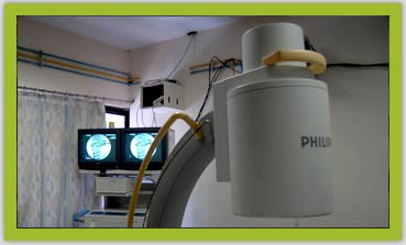
Even when an operation becomes a possibility, it will not be attempted until the spine has been carefully assessed. Before performing a surgical procedure, the surgeon must know the exact nature of the problem in the back. Consequently, he or she will study the back by means of X-rays or other tests, such as myelography, computerized axial tomography (CT), or magnetic resonance imaging (MRI).
In myelography, a radiopaque material is injected into the vertebral canal to outline any disorders that may be found in the vertebrae or discs. Usually, the patient is placed on a special table that makes it possible to change his or her position, thereby distributing the injected material up and down the vertebral canal. Because myelography may cause headaches, which can be aggravated by sitting up or standing, patients may be asked to remain in bed for a day after the test.
During CT, a patient is placed in a large, circular device that projects X rays through a cross-section of the body. The X-rays outline the densities of various tissues, and by analyzing these densities, a physician can detect abnormalities.
Magnetic resonance imaging (MRI) is a relatively new technique for showing the bones and other tissues of the body. MRI scans do not involve the use of X-rays, and they may or may not include the injection of a contrast agent in the vertebral canal to enhance the images seen by the physician. An advantage of this method is that soft tissues (such as ruptured discs) show up much better on an MRI scan than they do on an X-ray or a CT scan. The test takes a longer time to perform than an X-ray or CT, and the patient must lie quietly in a large magnetic tube for the time of the examination. However, this type of examination is proving to be a safe and highly effective way to diagnose spinal disorders.
In addition, electrical studies of the muscles and nerves may be useful in diagnosing and managing spinal disorders.
About Operations on the Back:
The type of operation a surgeon performs depends on the nature of a patient’s back problem. However, most procedures involve a Fenestration, which may require the partial removal of the vertebral arch to gain access to the cause of the patient’s low-back pain. If a disc has ruptured, a surgeon will perform a Microdiscectomy to investigate the vertebral canal, identify the ruptured disc, and remove a good portion of the degenerated disc material, especially those fragments that press on the nerve roots.
The surgeon may consider a second procedure-Spinal Fusion-if he or she feels that stabilization of the spine is necessary. A spinal fusion is performed by fusing the vertebrae together with bone grafts; sometimes, the grafts are combined with metal plates or other types of instruments.
Some types of herniated discs are suitable for treatment by microsurgery or by a technique known as percutaneous discectomy, in which the disc is repaired through the skin without making a surgical incision. For this technique, the surgeon uses an X ray as a guide for inserting a large bore needle into the center of the disc; the central portion of the disc is then removed by using fine instruments that are placed through the needle. You should discuss with your surgeon the various treatment options to determine which is the most appropriate for your specific problems.
To treat spinal stenosis, the surgeon makes an incision that is long enough to allow inspection of all of the vertebrae that have contributed to narrowing of the vertebral canal. After performing a limited laminectomy, the surgeon performs a decompression operation by entering the vertebral canal and removing the material that is pressing on the spinal nerve roots. Occasionally, some form of spinal fusion or other type of stabilization may be indicated.
When a patient has a spinal tumor, the physician may opt to treat the patient with radiation or chemotherapy rather than a surgical operation. If an operation is needed, the surgeon performs a laminectomy, locates the tumor, and removes it from the spine, the spinal cord, and the nerve roots. Some tumors require that the operation be approached from the front of the spine, followed by spinal stabilization. Following the removal of a spinal tumor, the surgeon decides if further radiation therapy and/or chemotherapy should be given.
When a patient has a spinal infection with an abscess in the back part of the spinal canal, the surgeon removes the vertebral arch, locates the abscess, and drains away the pus. If the abscess is toward the front (anterior) in the disc space, the surgeon may make an anterior approach to the vertebral bodies. Appropriate antibiotics will be given to cure the infection.
Recovering from the Operation:
Recovering after back surgery varies with the type of operation that was performed. Following ordinary disc removal, most patients are able to get out of bed and move about on the same day after surgery and get discharged on the very next day. Patients who have undergone a spinal fusion or an operation for stenosis take longer to become mobile (upto 12-24 hours), and these patients may remain in the hospital for longer periods of time (48-72 hours) after the operation. In addition, they may be required to wear a brace or cast for a few weeks to months after surgery.
The length of stay for patients with spinal tumors depends on the type of tumor. Patients who have had an operation to drain an abscess of the spine stay in the hospital until the infection has been controlled.
A common problem after major back surgery is difficulty with urination. This problem usually subsides in three to four days. The insertion of a tube (catheter) into the bladder that will drain the urine may be necessary until the patient is able to void normally. After discharge from the hospital, most back surgery patients will need some time to recuperate before returning to their usual activities. The types of activities the patient can safely resume should be outlined by the operating surgeon and should be followed carefully by the patient. The period of recuperation varies, but it may range from weeks to months, and a back brace or physical therapy program may be recommended