What is an atrial septal defect?
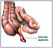
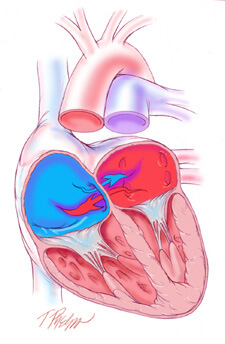
An atrial septal defect or ASD is an opening between the upper two chambers of the heart, known as the right atrium and the left atrium. A congenital heart defect, an ASD permits mixing of deoxygenated blood returning to the heart from the body (right atrium) and freshly oxygenated blood coming from the lungs (left atrium). The degree of mixing is largely related to the size of the defect and the relative pressures in each atrium. Early in life, blood from the left atrium preferentially moves into the right atrium, causing excessive blood to flow through the lungs. If this flow, otherwise known as “shunting” is significant enough, resistance to flow develops in the lung resulting in a gradual reversal of shunting of deoxygenated blood from the right atrium into the left atrium and subsequently through the body. This latter condition can lead to a condition called “cyanosis” whereby inadequately oxygenated blood is delivered to the body causing early fatigue and congestive heart failure.
 Atrial Septal Defect
Causes:
Blood can flow between the two upper heart chambers through the ASD.
When blood flows between the two heart chambers. This is called a shunt. Pressure in the lungs may build up. Over time, there will be less oxygen in the blood that goes to the body.

Small atrial septal defects often cause very few problems, and may be discovered much later in life. Many problems can occur if the opening is large, or if there is more than one opening. ASD is not very common.
Symptoms:
A person with no other heart defect, or a small defect (less than 5 millimeters) may not have symptoms, or the symptoms may not occur until middle age or later.
Symptoms that do occur may begin at any time after birth through childhood. They can include:
- Difficulty breathing (dyspnea)
- Frequent respiratory infections in children
- Feeling the heart beat (palpitations) in adults
- Shortness of breath with activity
How is an atrial septal defect treated?
Small atrial septal defects can often be followed conservatively without surgery, due to minimal shunting. Larger ASDs should be closed to prevent the late irreversible consequences of excessive left-to-right shunting. ASDs can be closed surgically by simply sewing them closed or, in the case of larger ASDs, placing a patch of the patient’s own tissue or synthetic material (e.g., Dacron) over it.
How is an atrial septal defect repaired?
ASD repairs require the use of cardiopulmonary bypass, otherwise known as “the heart-lung machine.” This permits the surgeon to safely open the right atrium and access the ASD in a relatively bloodless field. In some cases, the heart is also stopped for 1 to 2 hours to facilitate the repair. Repairs range from relatively simple operations to more complex procedures depending on the location, size, and characteristics of the ASD. The total duration of the operation ranges from 2 to 3 hours.
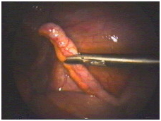
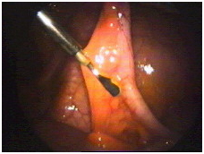
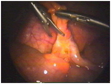
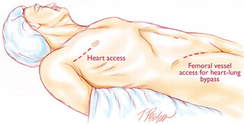
What are minimally invasive approaches to an atrial septal defect?
The most common surgical approach requires the surgeon to saw open the breastbone and spread the edges apart to gain direct access to the heart. Although this approach provides excellent access to the heart, the resulting wound requires several months to heal completely, an extended recovery period with substantial activity restrictions, and can be subject to serious complications including infection, breakdown, and even death.
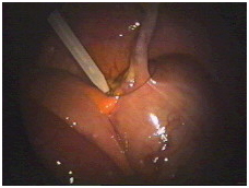
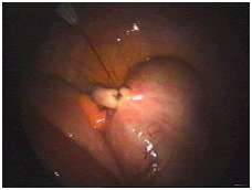
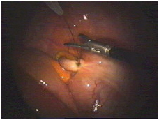
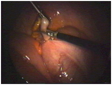
Mini-ASD Repair
 Minimally Invasive Approach to ASD Closure
Currently at The Johns Hopkins Hospital, the most commonly employed minimally invasive approach to ASDs is a “mini-thoracotomy” which consists of a 3 inch incision made through the right side of the chest between the ribs. Heart-lung bypass is instituted with small tubes placed in the main artery and vein of the right leg through a 1 to 2 inch incision placed in the right groin crease. The heart is then stopped and the right atrium is opened to expose the ASD. At this point, specialized hand-held “chopstick” like instruments are inserted through this small incision by the surgeon to repair the defect. After the defect is repaired, the heart is then closed and restarted. Finally, heart-lung bypass is discontinued and the incisions are closed.