Pancreatic Cancer
Pancreatic cancer is a disease in which malignant (cancerous) cells form in the tissues of the pancreas. The pancreas is a gland located behind the stomach and in front of the spine. The pancreas produces digestive juices and hormones that regulate blood sugar. Cells called exocrine pancreas cells produce the digestive juices, while cells called endocrine pancreas cells produce the hormones. The majority of pancreatic cancers start in the exocrine cells.
Symptoms:
Early pancreatic cancers usually cause few symptoms, most of which are vague. Because signs and symptoms of most pancreatic cancer may be mistaken for less-serious digestive problems, the disease is rarely detected before it has spread to nearby tissues or distant organs via the bloodstream or lymphatic system. Symptoms that may arise include:
- Significant weight loss accompanied by abdominal pain — the most likely warning signs.
- Vague but gradually worsening abdominal pain that may decrease when leaning forward and increase when lying down. Pain is often severe at night and may radiate to the lower back.
- Digestive or bowel complaints such as diarrhea, constipation, gas pains, bloating, or belching.
- Nausea, vomiting, and loss of appetite.
- Jaundice, which is usually painless and is indicated by yellowish discoloration of the skin and eye whites, very dark urine, and light-colored stools.
- Sudden onset of glucose tolerance disorder, such as diabetes.
- Black or bloody stool, indicating bleeding from the digestive tract.
- Overall weakness.
- Enlarged liver and gallbladder.
- Itching.
- Blood clots in the legs.
- Mental status changes, such as a new onset of depression.
Causes:
- Episodes of weakness, sweating, rapid heartbeat, irritability, or skin flushing related to low blood sugar
- Severe watery diarrhea
- A new, unusual skin rash
- Severe gastrointestinal symptoms, such as stomach pain and watery diarrhea, which do not respond to antacids or ulcer medications
If you experience any of these symptoms, call your doctor for a full physical exam.
How Is Pancreatic Cancer Diagnosed?
To diagnose pancreatic cancer, a doctor will order certain imaging tests, such as a pancreatic ultrasound or a CT scan of the abdomen. Endoscopic ultrasonography (EUS) uses an ultrasound device connected to the end of a small flexible tube that is inserted into the mouth and is about 85% to 90% accurate in diagnosing pancreatic cancer. If necessary, endoscopic retrograde cholangiopancreatography (ERCP) is used. With ERCP, detailed images are obtained by inserting an endoscope into the mouth to the pancreas, injecting a dye, and then taking X-rays. A tissue sample for biopsy can also be extracted through the scope. If a biopsy confirms cancer, further tests are done to determine how far the disease has advanced. Laparoscopy, may be used. In this technique, a small tube with a small video camera and light source is introduced into the abdominal cavity. The tumor can then be seen. Occasionally, exploratory surgery is needed. The surgeon can then study the tumor directly, determine if nearby lymph nodes are cancerous, and take tissue samples for microscopic exam.
What Are the Treatments for Pancreatic Cancer?
Pancreatic cancer is very hard to control. But if it is caught early and the cancer hasn’t spread beyond the pancreas, it can be treated with surgery. This offers the best outcome for pancreatic cancer. The surgery is called a “Whipple procedure,” or pancreaticoduodenectomy, and is named after Dr. George Hoyt Whipple, the surgeon who pioneered it. If possible, the surgeon removes the malignant tumor, leaving as much of the normal pancreas as possible to allow continued pancreatic function. Less often, the entire pancreas must be removed. If a patient undergoes a total pancreatectomy, a lifelong regimen of replacement enzymes and hormones, including insulin, must be administered.
Unfortunately, pancreatic cancers have subtle, and vague symptoms. Thus the disease is often diagnosed after it has advanced and spread. However, even at later stages, treatment can improve quality of life by controlling uncomfortable symptoms and complications of the disease.
Depending on the type and stage of pancreatic cancer, patients may be given chemotherapy treatments alone or in combination with radiation. Whether or not the tumor is removed surgically, or has spread to lymph nodes, these therapies may be given after surgery in an effort to extend survival time. These treatments may also be given before surgery to shrink the tumor and make it possible for surgery to be performed or used as a means of relieving symptoms, such as pain. Prescription medications, usually narcotics, are given to help manage pain associated with advanced pancreatic cancer.


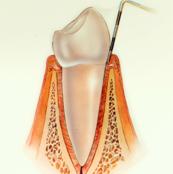 Normal healthy gumsHealthy gums and bone anchor teeth firmly in place.
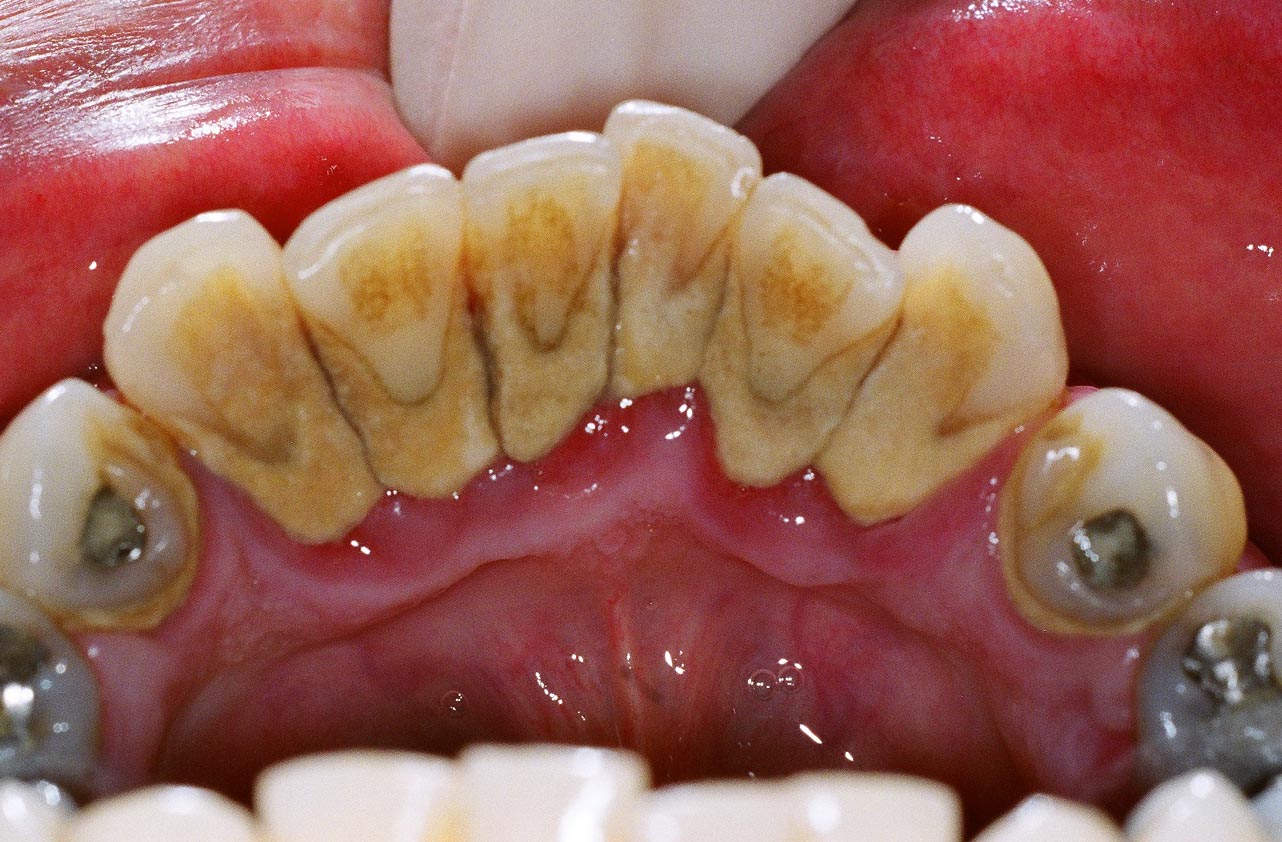
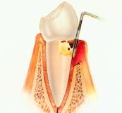 PeriodontitisUnremoved plaque hardens into calculus (tartar), As plaque and calculus continue to build up, the gums begin to recede( pull away) from the teeth, and pockets form between the teeth and gums.
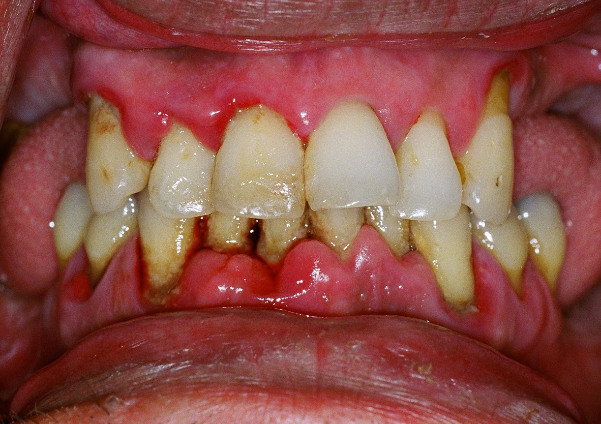
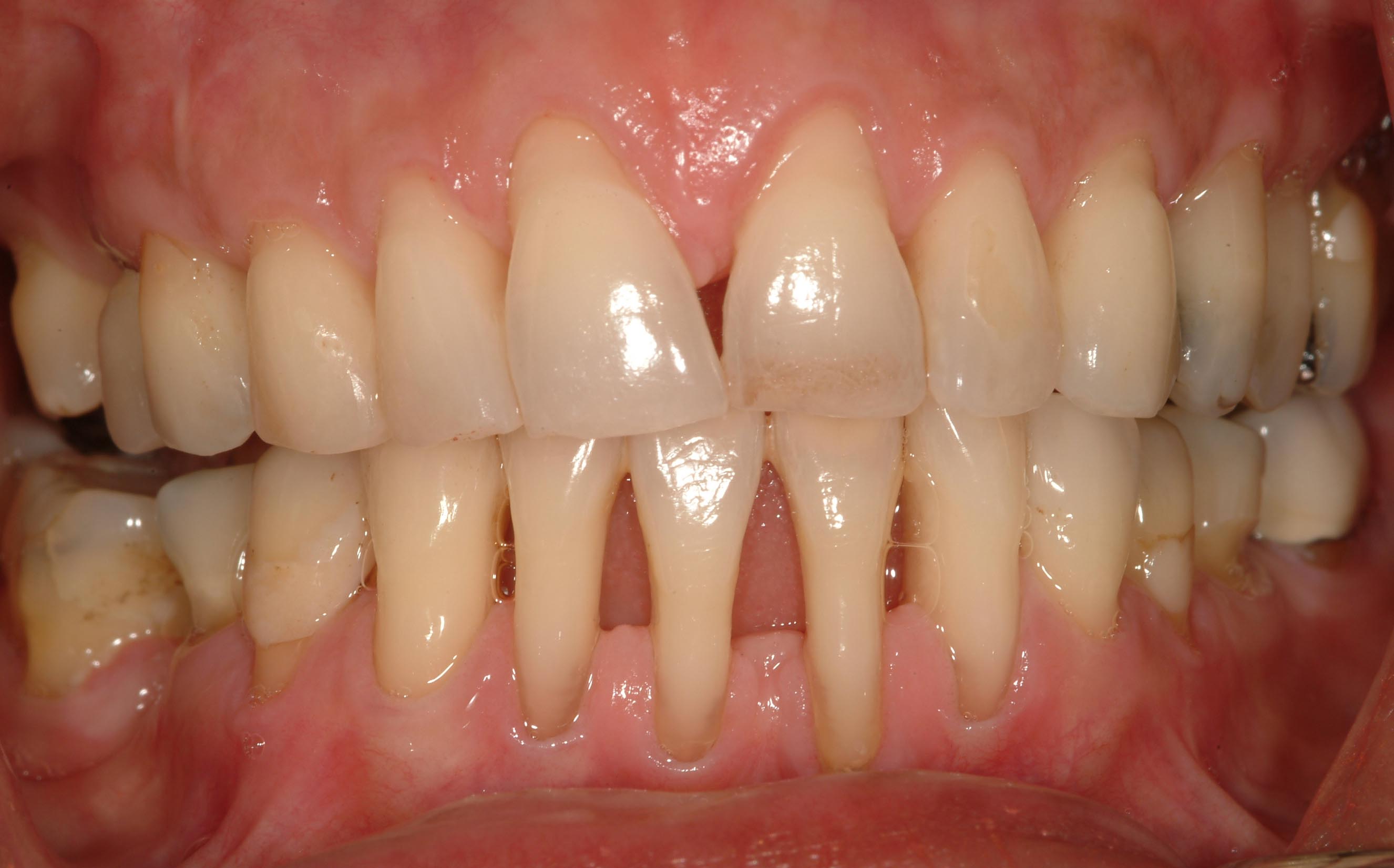
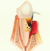 Advanced PeriodontitisThe gums recede further, destroying more bone and the periodontal ligament. Even healthy teeth may become loose and need to be extracted