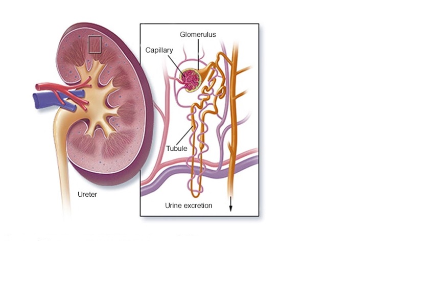
Global Treatment Services Pvt. Ltd.

Global Treatment Services

OCD(Obsessive Compulsive Disorder): Causes, Symptoms & Treatments

Obsessive-compulsive disorder is a type of mental illness. People with OCD can have either obsessive thoughts and urges or compulsive, repetitive behaviors. Some have both obsessions and compulsions.OCD isn’t about habits like biting your nails or always thinking negative thoughts. The disorder can affect your job, school, and relationships and keep you from living a normal life. Your thoughts and actions are beyond your control.An obsessive thought, for example, is to think that your family members might get hurt if they don’t put their clothing on in the exact same order every morning. A compulsive habit, on the other hand, might be to wash your hands 7 times after touching something that might be dirty. Although you may not want to think or do these things, you feel powerless to stop.

Symptoms

Many people with OCD know that their thoughts and habits don’t make sense. They don’t do them because they enjoy them, but because they can’t quit. And if they do stop, they feel so bad that they start again.

Obsessions and compulsions can involve many different things, like a need for order or cleanness, hoarding, and intrusive thoughts about sex, religion, violence, and body parts.

Obsessive thoughts can include:

  • Fear of germs or getting dirty
  • Worries about getting hurt or others being hurt
  • Need for things to be placed in an exact order
  • Belief that certain numbers or colors are “good” or “bad”
  • Constant awareness of blinking, breathing, or other body sensations
  • Unfounded suspicion that a partner is unfaithful

Compulsive habits can include:

  • Washing hands many times in a row
  • Doing tasks in a specific order every time, or a certain “good” number of times
  • Repetitive checking on a locked door, light switch, and other things
  • Need to count things, like steps or bottles
  • Putting items in an exact order, like cans with labels facing front
  • Fear of touching doorknobs, using public toilets, or shaking hands

Causes

Doctors aren’t sure why some people have OCD. Certain areas in the brain may not look normal in people with OCD, but more research is needed. OCD is slightly more common in women than in men. Symptoms often appear in teens or young adults. Stress can make symptoms worse.

There may be a gene or genes linked to OCD, but this is still unknown.

You are more likely to get the disorder if you have:

  • A parent, sibling, or child with OCD
  • Depression, anxiety or tics
  • Experience with trauma
  • A history of physical or sexual abuse as a child

Diagnosis

Your doctor may do a physical exam and bloodwork to make sure that your symptoms aren’t caused by something else. She will also talk with you about your feelings, thoughts, and habits. If your thoughts and habits keep you from doing what you want to do for at least an hour a day, you may have OCD.

Treatment:

There’s no cure for OCD. But with treatments, you may be able to lessen how much your symptoms interfere with your life. Your doctor may prescribe medicine, send you to talk therapy, called psychotherapy, or both.

Benign fibrous histiocytoma: Causes, Symptoms & Treatments

Benign Fibrous Histiocytoma or Dermatofibromas  is a rare tumor of mesenchymal origin. First described in 1958 by Dahlin. They are composed of two cell types, namely atypical fibroblasts and histiocytes, arranged in a storiform pattern. Lipid-filled cells may be conspicuous and occasionally are the major component. These types of benign fibrous histiocytomas are known as xanthomas or fibroxanthomas. Benign Fibrous Histiocytomas are microscopically very similar to non-ossifying fibroma. It is essentially a non ossifying fibroma that grows in and unusual location or in an unusual age group.

The definition of a dermatofibroma is more simple than the complex name for this condition implies. In short, a dermatofibroma is a benign skin bump that occurs most commonly on the legs. It is firm and slightly elevated. Dermatofibroma are larger under the skin than can be seen just by looking.

Dermatofibromas are normally dome-shaped and often darker-colored papules. The growths range from brownish to purplish-red in color. They can begin as red and later change to brown. While common on the legs, they can be found throughout the body but especially on exposed parts.

Causes

Dermatofibromas may itch because they’re often caused by bug bites. Splinters and minor injuries are common culprits for the appearance of dermatofibromas as well.

Is it a Dermatofibroma or a Mole?

Sometimes a dermatofibroma is confused with a mole. The way to tell the difference between the two is to pinch the bump. If you pinch a dermatofibroma, it creates a dimple because it is attached to the underlying subcutaneous tissue. On the other hand, if you pinch a mole, it projects up away from the skin. Moles appear when skin cells grow in clusters.

While dermatofibromas are usually red, brown or purplish, moles can be tan, black, blue or pink in addition to the typical dermatofibroma’s color. Moles can appear in both exposed and unexposed areas of the body, including the armpits or even under nails.

Because dermatofibromas are benign (they do not cause cancer) doctors usually do not excise them. In fact, excising the skin growth may produce a scar that’s more severe in appearance than the original dermatofibroma. If your physician is unclear about whether you have a dermatofibroma or another type of skin growth, you may have to undergo a biopsy.

Treating a dermatofibroma involves everything from surgical removal of the top of the growth to freezing the top with liquid nitrogen or removing the center. Because these treatments don’t completely remove the dermatofibroma, the growths will likely reach their original size again. If that happens, you could have the top removed once more or seek out a procedure to excise the entire growth.

Occipital lobe damage: Causes,Symptoms & Treatments

The cerebral cortex of the brain has 4 lobes on each side: the frontal lobe, the temporal lobe, the parietal lobe and the occipital lobe. Each of these regions carries out specific functions, and damage to any of these areas results in corresponding impairment. The frontal lobes are relatively large compared to other regions of the brain, and the extensive functions controlled by the frontal lobe reflects that large size.

Occipital Lobe Strokes:

The occipital lobes are vital for visual processing. A stroke of the occipital lobe can cause vision loss or partial vision loss of one eye or both eyes. 

Because of the way the blood vessels are arranged in the brain, occipital lobe strokes are less common than strokes affecting the frontal lobes, temporal lobes, and parietal lobes.


Fig: branching point of the right middle cerebral artery (MCA) trunk (red circle), suspicious for thrombus or embolus.

Of the 4 major lobes of the brain, the occipital lobe is the smallest—but in our daily lives, we perhaps lean on it the most. The lobe is located at the back of the skull, thus the name (“occipital” comes from the Latin for “back of the head”).

The occipital lobe’s purpose is to receive visual stimuli from the eyes, process the information, and forward the information to the frontal lobe (which will formulate a response). Put simply, the occipital lobe controls our ability to respond to things we see. However, that role is far more complex than we may understand at first.

While sight is probably our most valued senses, it’s also the one that’s most taken for granted. Because we use our sight so instinctively, we often forget about how incredible our sight truly is. Take a second and look at an object in the distance. A simple task, yes?

Except in that split second it took you to pick an object, your brain not only focused your center of vision on it, but it processed its contour, distinguished it from the objects around it, located its distance away from you, identified its size relative to the objects around it, and probably understood its relative size to you.

That’s a great deal of information to receive and process correctly in a split-second, but it’s what your occipital lobe does thousands of times every day. All told, your “ability to see” is actually a series of abilities that is made possible by the different parts of the occipital lobe.

CONSCIOUS & UNCONSCIOUS PROCESSING

Because our understanding of the world around us leans so heavily on sight, we associate our ability to see with our conscious decisions. For example, there are laws nationwide that address the problem of distracted driving. Lawmakers are facing a whole new generation of people behind the wheel who do not place their complete, conscious focus on the road.

Now, Plasticity Brain Centers is certainly not recommending texting while driving or anything similar, but sight depends on far more than conscious attention. In addition to the things you are focusing on, the occipital lobe is constantly processing visual stimuli that you may not be consciously aware of. This can result in something truly remarkable known as “blindsight.”

SEEING /WITHOUT SEEING: DAMAGE TO THE OCCIPITAL LOBE:

The condition we call “blindness” has two possible, general causes. The common one we know of is caused by damage to the eyes themselves. Because the receptors are damaged, the functioning occipital lobe receives no stimuli, so the brain cannot formulate a response to the outside world. In some of these cases, the occipital lobe becomes an additional site of processing for aural stimuli—which is why blind people seem to have incredible hearing or can respond to obstacles through sound.

However, the less common cause of blindness is a result of damage to the occipital lobe. For example, lesions on the occipital lobe can result in a “hole” in your vision known as a scotoma. Extensive damage can result in total blindness. Unlike other lobes of the brain, damage to one side of the occipital lobe can result in damage to both eyes’ fields of vision.

However, in these cases, the eyes are still completely functionalThey continue to receive information, except the occipital lobe is not processing it correctly. While this results in a kind of blindness, some processing can still occur that isn’t consciously understood. For example, studies have been conducted on patients who are blind in one eye due to occipital lobe damage. Despite not being consciously aware of what they are seeing, these patients were able to navigate obstacles or detect movement without perceiving what they were seeing.

Treatments 

Plasticity Brain Centers leverages the complex relationship between receptors (eyes, skin, tongue, nose, ears), stimuli, and their corresponding lobes (occipital, parietal, temporal, frontal) through clinical neuroscience. In other words, we use receptors of different kinds to create tests and targeted therapies for the damaged parts of our patients’ brains.

For damaged or underperforming occipital lobes, we employ visual stimuli such as eye movement training, visual search exercises, hand-eye coordination exercises, or light therapy to rebuild or create pathways between neurons. These pathways can bypass damaged tissue or rebuild old pathways in order to restore visual processing power.

Our therapies can improve a patient’s ability to perceive depth, distinguish objects from their surroundings, identify objects or people more quickly, and track objects with their sight more accurately. By understanding the state of your occipital lobe, we can more precisely identify why your sight is suffering and how we can restore function to the visual processing part of your brain.

Hiatal Hernia: Causes, Types & Treatments

Any time an internal body part pushes into an area where it doesn’t belong, it’s called a hernia.

The hiatus is an opening in the diaphragm — the muscular wall separating the chest cavity from the abdomen. Normally, the esophagus(food pipe) goes through the hiatus and attaches to the stomach. In a hiatal hernia (also called hiatus hernia) the stomach bulges up into the chest through that opening.

There are two main types of hiatal hernias: sliding and paraesophageal (next to the esophagus).

In a sliding hiatal hernia, the stomach and the section of the esophagus that joins the stomach slide up into the chest through the hiatus. This is the more common type of hernia.

Causes:

Most of the time, the cause is not known. A person may be born with a larger hiatal opening. Increased pressure in the abdomen such as from pregnancy, obesity, coughing, or straining during bowel movements may also play a role.

Risks:

Hiatal hernias occur more often in women, people who are overweight, and people older than 50.

Treatments:

Most people do not experience any symptoms of their hiatal hernia so no treatment is necessary. However, the paraesophageal hernia (when part of the stomach squeezes through the hiatus) can sometimes cause the stomach to be strangled, so surgery is sometimes recommended. Other symptoms that may occur along with the hernia such as chest pain should be properly evaluated. Symptoms of GERD, such as heartburn, should be treated.


When Is Hiatal Hernia Surgery Necessary?

If the hiatal hernia is in danger of becoming constricted or strangulated (so that the blood supply is cut off), surgery may be needed to reduce the hernia, meaning put it back where it belongs.

Hiatal hernia surgery can often be performed as a laparoscopic, or “minimally invasive,” procedure. During this type of surgery, a few small (5 to 10 millimeter) incisions are made in the abdomen. The laparoscope that allows the surgeon to see inside the abdomen and surgical instruments are inserted through these incisions. The surgeon is guided by the laparoscope, which transmits a picture of the internal organs to a monitor. The advantages of laparoscopic surgery include smaller incisions, less risk of infection, less pain and scarring, and a more rapid recovery.

Gastrointestinal Perforation(GP): Causes, Symptoms & Treatments

Gastrointestinal perforation (GP) occurs when a hole forms all the way through the stomach, large bowel, or small intestine. It can be due to a number of different diseases, including appendicitis and diverticulitis. It can also be the result of trauma, such as a knife wound or gunshot wound. A perforation may also occur in the gallbladder. This can have symptoms that are similar to the symptoms of a gastrointestinal perforation.

A hole in your gastrointestinal system or gallbladder can lead to peritonitis. Peritonitis is inflammation of the membrane that lines the abdominal cavity.

It occurs when any of the following enters the abdominal cavity:

  • bacteria
  • bile
  • stomach acid
  • partially digested food
  • stool

GP is a medical emergency that requires immediate medical care. The condition is life-threatening. Chances of recovery improve with early diagnosis and treatment.

This condition is also known as intestinal perforation or perforation of the intestines.

Symptoms:

  • severe stomach pain
  • chills
  • fever
  • nausea
  • vomiting

When you’ve had a gastrointestinal perforation and peritonitis occurs, the abdomen feels very tender. Pain often worsens when someone touches or palpates the area or when the patient moves. Pain is generally better when lying still. The abdomen may stick outward farther than normal and feel hard.

In addition to the general symptoms of perforation, symptoms of peritonitis may include:

  • fatigue
  • passing less urine, stools, or gas
  • shortness of breath
  • a fast heartbeat
  • dizziness
Causes:
  • appendicitis, which is more common among older persons
  • diverticulitis, which is a digestive disease
  • a stomach ulcer
  • gallstones
  • gallbladder infection
  • inflammatory bowel diseases such as Crohn’s disease or ulcerative colitis, which is less common
  • inflamed diverticulum, which is a congenital abnormality of the small intestine that’s similar to the appendix
  • cancer in the gastrointestinal tract

The condition may also be due to:

  • blunt trauma to the abdomen
  • a knife or gunshot wound to the abdomen
  • abdominal surgery
  • stomach ulcers due to taking aspirin, nonsteroidal anti-inflammatory drugs, and steroids (more common in older adults)
  • ingestion of foreign objects or caustic substances

Smoking and excessive use of alcohol increase your risk of GP.

Rarely, the condition may occur due to bowel injuries from an endoscopy or colonoscopy.

In most cases, surgery is necessary to close the hole and treat the condition. The goals of the surgery are to:

  • fix the anatomical problem
  • fix the cause of peritonitis
  • remove any foreign material in the abdominal cavity that might cause problems, such as feces, bile, and food

In rare cases, your doctor may forgo surgery and prescribe antibiotics alone if the hole closed on its own.

Sometimes, a piece of the intestine will need removal. The removal of a portion of either the small or large intestine may result in a colostomy or ileostomy, which allows intestinal contents to drain or empty into a bag attached to your abdominal wall.

Complications:
  • bleeding
  • sepsis, which is a life-threatening bacterial infection
  • abscesses in the belly
  • a wound infection
  • a bowel infarction, which is the death of part of the bowel
  • a permanent ileostomy or colostomy


Leprosy: Causes, Symptoms & Treatment

Leprosy is an infectious disease that causes severe, disfiguring skin sores and nerve damage in the arms, legs, and skin areas around the body. You can catch it only if you come into close and repeated contact with nose and mouth droplets from someone with untreated leprosy. Children are more likely to get leprosy than adults.

Related image


Trophic ulcer on the ball of great toe in left foot.


Fig: Leprosy on hand and leg

Causes: Leprosy is caused by a slow-growing type of bacteria called Mycobacterium leprae .

Symptoms:

Leprosy primarily affects the skin and the nerves outside the brain and spinal cord, called the peripheral nerves. It may also strike the eyes and the thin tissue lining the inside of the nose. The main symptom of leprosy is disfiguring skin sores, lumps, or bumps that do not go away after several weeks or months. The skin sores are pale-colored.

Nerve damage can lead to:

  • Loss of feeling in the arms and legs
  • Muscle weakness

It usually takes about 3 to 5 years for symptoms to appear after coming into contact with the leprosy-causing bacteria. Some people do not develop symptoms until 20 years later. The time between contact with the bacteria and the appearance of symptoms is called the incubation period. Leprosy’s long incubation period makes it very difficult for doctors to determine when and where a person with leprosy got infected.

Types of Leprosy:

  • A mild, less severe form of leprosy. People with this type have only one or a few patches of flat, pale-colored skin (paucibacillary leprosy). The affected area of skin may feel numb because of nerve damageunderneath. Tuberculoid leprosy is less contagious than other forms.
  • Lepromatous. A more severe form of the disease. It has widespread skin bumps nd rashes(multibacillary leprosy), numbness, and muscle weakness. The nose, kidneys, and male reproductive organs may also be affected. It is more contagious than tuberculoid leprosy.
  • People with this type of leprosy have symptoms of both the tuberculoid and lepromatous forms.

Diagnosis:

Affected skin send it to lab for biopsy. A skin smear test may also be done. With paucibacillary leprosy, no bacteria will be detected. In contrast, bacteria are expected to be found on a skin smear test from a person with multibacillary leprosy. 

Treatments: 

Treatment depends on the type of leprosy that you have. Antibiotics are used to treat the infection. Long-term treatment with two or more antibiotics is recommended, usually from six months to a year. People with severe leprosy may need to take antibiotics longer. Antibiotics cannot treat the nerve damage.

Anti-inflammatory drugs are used to control nerve pain and damage related to leprosy. This may include steroids, such as prednisone. Patients with leprosy may also be given thalidomide, a potent medication that suppresses the body’s immune system. It helps treat leprosy skin nodules. Thalidomide is known to cause severe, life-threatening birth defects and should never be taken by women who are pregnant or women who may become pregnant. 

Leprosy Complications

Without treatment, leprosy can permanently damage your skin, nerves, arms, legs, feet, and eyes. Complications of leprosy can include:

  • Blindness or glaucoma.
  • Disfiguration of the face (including permanent swelling, bumps, and lumps).
  • Erectile dysfunctionand infertility in men.
  • Kidney failure.
  • Muscle weakness that leads to claw-like hands or an inability to flex the feet.
  • Permanent damage to the inside of the nose, which can lead to nosebleedsand a chronic, stuffy nose.
  • Permanent damage to the nerves outside the brainand spinal cord, including those in the arms, legs, and feet.

Nerve damage can lead to a dangerous loss of feeling. A person with leprosy-related nerve damage may not feel pain when the hands, legs, or feet are cut, burned, or otherwise injured.

Spinal Muscular Atrophy(SMA) : Causes, Symptoms & Treatments

Spinal muscular atrophy (SMA) most often affects babies and children and makes it hard for them to use their muscles. When your child has SMA, there’s a breakdown of the nerve cells in the brain and spinal cord. The brain stops sending messages that control muscle movement.

When that happens, your child’s muscles get weak and shrink, and he can have trouble controlling head movement, sitting without help, and even walking. In some cases, he can have trouble swallowing and breathing as the disease progresses.


Woman with muscular atrophy

Diagnosis

SMA can sometimes be hard to diagnose because the symptoms may be similar to other conditions. To help figure out what’s going on, your doctor may ask you:

  • Has your baby missed any developmental milestones, such as holding his head up or rolling over?
  • Does your child have trouble sitting or standing on his own?
  • Have you seen your child have trouble breathing?
  • When did you first notice the symptoms?
  • Has anyone in your family had similar symptoms?

Treatment

The FDA has approved one medication to treat SMA: nusinersen (Spinraza). Studies show it helps about 40% of people who use it by slowing down the advance of the disease and making people stronger.

Doctors inject nusinersen into the fluid around your child’s spinal cord. The procedure for the shot-including preparation and recovery time — can take at least 2 hours. Your child will need multiple shots.

Nusinersen is a type of gene therapy. It belongs to a group of drugs called antisense oligonucleotides that have small bits of man-made genetic material. They work by “modulating,” or adjusting, the SMN2 gene.

Everyone who has spinal muscular atrophy has at least one copy of the SMN2 gene, and some people have more. The SMN2 gene isn’t doing a good job of making a key protein that allows the cells that control muscles to work. Adjusting this gene can let it make more protein or a better-quality protein.

Besides adjusting the SMN2 gene, researchers are studying another type of gene therapy that involves replacing or correcting the SMN1 gene. Since spinal muscular atrophy is caused by a mutation — or change — in the SMN1 gene, doctors are checking to see if replacing or fixing it will treat the disease.


Black Fever (leishmaniasis): Causes, Symptoms & Treatments

Black fever or Kala-Azar is a slow progressing indigenous disease caused by infection with leishmania parasites. The parasite primarily infects the immune system and may be found in abundance in bone marrow, spleen and liver.

Medication can cure the disease. However , the treatment is most effective when started before extensive damage to your immune system occurs. Visceral leishmaniasis is often fatal within two years if it’s not treated properly.


Symptoms

  • Recurrent fever intermittent or remittent with often double rise of temperature.
  • Loss of appetite,
  • pallor and weight loss with progressive emaciation
  • Weakness
  • Skin – Dry,
  • thin and scaly and hair may be lost.
  • Light colored person show grayish discoloration of the skin of hands, feet, abdomen and face which gives the Indian name Kala-Azar meaning “Black fever”.
  • Anemia – develops rapidly
  • Splenomegaly – spleen enlarges rapidly to massive enlargement, usually soft and non-tender.
  • Liver – enlargement not to the extent of spleen, soft, smooth surface, sharp edge.

Causes: 

Black fever is caused by bites from female Phlebotomine sandflies. Sand flies are small insects, about one fourth of the size of a mosquito. The length of a sand fly body ranges from 1.5 to 3.5 mm. Sand flies breed in high relative humidity, warm temperature, high subsoil water and abundance of vegetation.

Diagnosis

Symptomatic:  A case of fever of more than two weeks duration not responding to anti-malarials and antibiotics. Clinical laboratory findings may include anemia, progressive leucopenia thrombocytopenia and hypergammaglobulinemia.


Medical Management

Diagnosis and treatment of Kala-Azar are problematic because of a variety of reasons. While treatment is lengthy and relatively costly, definitive diagnosis of Kala-Azar requires tissue specimens, which are conventionally obtained by organ needle aspiration for microscopic demonstration of amastigote forms in stained smears. Bone marrow the spleen, and lymph-node (in some regions) are the tissues most often sampled in patients with suspected infection. The diagnostic sensitivity of splenic aspiration is high (95% – 98%), but the procedure carries a risk of bleeding; the sensitivity of examination of bone marrow specimens is considered to be lower (53% – 95%). Organ aspiration and accurate examination of smears also require technical skills that are not uniformly available in rural areas. Culture or PCR testing of aspirate material improves parasitologic yield, but these methods are seldom undertaken outside of research laboratories.


Prevention

There are no vaccines or preventive drugs for visceral leishmaniasis. The most effective method to prevent infection is to protect from sand fly bites. To decrease the risk of being bitten following precautions are suggested:

Outdoors:
Avoid outdoor activities, especially from dusk to dawn, when sand flies generally are the most active. When outdoors (or in unprotected quarters): Minimize the amount of exposed (uncovered) skin. To the extent that is tolerable in the climate, wear long-sleeved shirts, long pants, and socks; and tuck your shirt into your pants.

Apply insect repellent to exposed skin and under the ends of sleeves and pant legs. Follow the instructions on the label of the repellent. The most effective repellents generally are those that contain the chemical DEET (N,N–diethylmetatoluamide)

Indoors:

  • Stay in well-screened or air-conditioned areas.
  • Keep in mind that sand flies are much smaller than mosquitoes and therefore can get through smaller holes.
  • Spray living/sleeping areas with an insecticide to kill insects. If you are not sleeping in a well-screened or air-conditioned area, use a bed net and tuck it under your mattress.
  • If possible, use a bed net that has been soaked in or sprayed with a pyrethroid-containing insecticide. The same treatment can be applied to screens, curtains, sheets, and clothing (clothing should be retreated after five washings).”


Chronic obstructive pulmonary disease (COPD)

Image result for chronic obstructive pulmonary disease (copd)

Chronic obstructive pulmonary disease (COPD) is a progressive, irreversible inflammatory disease in your lungs that makes it hard to breathe. Common symptoms include a chronic cough, wheezing, production of phlegm, shortness of breath, and a feeling of tightness in your chest, though these symptoms may not be noticeable until you’re in the later stages of the disease. COPD is not curable, but it’s a preventable and treatable illness.

COPD is characterized by the limitation of airflow that is not fully reversible. Airflow limitation in COPD is progressive. It’s associated with an abnormal inflammatory response of your lungs to noxious stimuli, like cigarette smoke, air pollution, or harsh chemicals.

Some causes of COPD include exposure to cigarette smoking, whether you smoke yourself or you’ve been exposed to secondhand smoke; occupational exposure to chemicals; indoor and outdoor air pollution; and, far less often, a genetic disorder called alpha-1-antitrypsin deficiency. People with asthma sometimes develop COPD as well.

COPD is not a death sentence, with proper treatment, it can be controlled. That said, there are factors that influence COPD life expectancy, especially your body mass index, the degree of airway obstruction, dyspnea levels, and your exercise tolerance.The best treatment for COPD if you’re a smoker is to quit as soon as possible. While this won’t reverse the damage you already have, it can help slow the progression of your COPD.

If you have risk factors for COPD or think you may have it, see your doctor as soon as possible. The earlier you begin treatment, the better your prognosis will likely be. There are things you can do to slow some of the progressive symptoms of COPD, too. The biggest and most effective is to quit smoking. Proper nutrition and daily exercise are also key to living well. With treatment and management of your symptoms, you can live the best life possible.

to know more details:- send in your queries to query@gtsmeditour.com or WhatsApp your latest medical reports to +91-9880149003.

ACL Reconstruction

Image result for ACL reconstruction may be used for these knee problems:

ACL reconstruction is surgery to reconstruct the ligament in the center of your knee. The anterior cruciate ligament (ACL) connects your shin bone to your thigh bone. A tear of this ligament can cause your knee to give way during physical activity. Most people have general anesthesia right before surgery. The tissue to replace your damaged ACL will come from your own body or from a donor. A donor is a person who has died and chose to give all or part of his or her body to help others.

The procedure is usually performed with the help of knee arthroscopy. With arthroscopy, a tiny camera is inserted into the knee through a small surgical cut. The camera is connected to a video monitor in the operating room. Your surgeon will use the camera to check the ligaments and other tissues of your knee.

If you do not have your ACL reconstructed, your knee may continue to be unstable. This increases the chance you may have a meniscus tear. ACL reconstruction may be used for these knee problems:

  • Knee that  feels unstable during daily activities
  • Knee pain
  • Inability to return to sports or other activities
  • When other ligaments are also injured
  • When your meniscus is torn

The recovery process for the ACL is usually broken down into different phases of rehabilitation. Each phase has its own objectives, however is intertwined with other phases since the goals are as progressive as the recovery itself. The rehabilitation process is at the pace of the patient. Timelines are sometimes given to help give an idea of where one can be during rehabilitation.

Pages:1...11121314151617...80