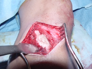
Benign Fibrous Histiocytoma or Dermatofibromas is a rare tumor of mesenchymal origin. First described in 1958 by Dahlin. They are composed of two cell types, namely atypical fibroblasts and histiocytes, arranged in a storiform pattern. Lipid-filled cells may be conspicuous and occasionally are the major component. These types of benign fibrous histiocytomas are known as xanthomas or fibroxanthomas. Benign Fibrous Histiocytomas are microscopically very similar to non-ossifying fibroma. It is essentially a non ossifying fibroma that grows in and unusual location or in an unusual age group.
The definition of a dermatofibroma is more simple than the complex name for this condition implies. In short, a dermatofibroma is a benign skin bump that occurs most commonly on the legs. It is firm and slightly elevated. Dermatofibroma are larger under the skin than can be seen just by looking.
Dermatofibromas are normally dome-shaped and often darker-colored papules. The growths range from brownish to purplish-red in color. They can begin as red and later change to brown. While common on the legs, they can be found throughout the body but especially on exposed parts.
Causes
Dermatofibromas may itch because they’re often caused by bug bites. Splinters and minor injuries are common culprits for the appearance of dermatofibromas as well.
Is it a Dermatofibroma or a Mole?
Sometimes a dermatofibroma is confused with a mole. The way to tell the difference between the two is to pinch the bump. If you pinch a dermatofibroma, it creates a dimple because it is attached to the underlying subcutaneous tissue. On the other hand, if you pinch a mole, it projects up away from the skin. Moles appear when skin cells grow in clusters.
While dermatofibromas are usually red, brown or purplish, moles can be tan, black, blue or pink in addition to the typical dermatofibroma’s color. Moles can appear in both exposed and unexposed areas of the body, including the armpits or even under nails.
Because dermatofibromas are benign (they do not cause cancer) doctors usually do not excise them. In fact, excising the skin growth may produce a scar that’s more severe in appearance than the original dermatofibroma. If your physician is unclear about whether you have a dermatofibroma or another type of skin growth, you may have to undergo a biopsy.
Treating a dermatofibroma involves everything from surgical removal of the top of the growth to freezing the top with liquid nitrogen or removing the center. Because these treatments don’t completely remove the dermatofibroma, the growths will likely reach their original size again. If that happens, you could have the top removed once more or seek out a procedure to excise the entire growth.
Post a comment