Global Treatment Services Pvt. Ltd.

Global Treatment Services

Blepharoplasty

Overview

ble

The eye is an important component of facial aesthetics, blepharoplasty often means removing excess skin, muscle and underlying fatty tissue. Blepharoplasty play a vital positive role in facial harmony and the perception of aging. Blepharoplasty is one of the most commonly performed facial cosmetic procedures. Symptoms such as tired-looking eyes, excess skin, droopy eyelids, or circles around the eyes may benefit from blepharoplasty. You might be a good candidate for this procedure if your vision is affected by sagging skin. Some people may complain that their vision when looking upwards is blocked by the hanging skin.

people who have blepharoplasty say they feel more self-confident and feel they look younger and more rested. For some people, surgery results may last a lifetime. For others, droopy eyelids can recur. Bruising and swelling generally lessen slowly in about 10 to 14 days. Scars from the surgical cuts may take months to fade.

Causes

Blepharoplasty might be an  ideal option for:

  • Baggy or droopy upper eyelids
  • Excess skin of the upper eyelids that partially blocks peripheral vision
  • Excess skin on the lower eyelids
  • Bags under the eyes

Blepharoplasty can be done at the same time as another procedure, such as a brow lift, face-lift or skin resurfacing.

Symptoms

Patient with below symptoms may require blepharoplasty

  • Tired-looking eyes
  • Excess skin
  • Droopy eyelids
  • Circles around the eyes

Risks involved

All surgery has risks, including reaction to anesthesia and blood clots. Besides those, rare risks of eyelid surgery include:

  • Infection and bleeding
  • Dry, irritated eyes
  • Difficulty closing the eyes or other eyelid problems
  • Noticeable scarring
  • Injury to eye muscles
  • Skin discoloration
  • Temporarily blurred vision or, rarely, loss of eyesight
  • The need for follow-up surgery.

Treatment

eye

Depending on your goals and the recommendation of your surgeon, blepharoplasty can involve your upper eyelids, lower eyelids or both.

Upper blepharoplasty

During an upper blepharoplasty, your surgeon will make cuts (incisions) in the natural crease of your upper eyelid. These incisions will be hidden when your eyes are open. Your surgeon will remove excess skin and protruding fat, and then they’ll close the incisions.

Lower blepharoplasty

During a lower blepharoplasty, your surgeon will make an incision just below your lower eyelash line. They’ll remove excess skin in your lower eyelid through this incision. They may also use an incision hidden inside your lower eyelid (transconjunctival incision) to correct lower eyelid issues and redistribute or remove excess fat.

There will be a stitch in each of your upper lids that’ll remain for about a week. The lower lids don’t require stitches if the incisions are made on the inside of your lids. It’s common for swelling and bruising to occur in your upper and lower lids. Expect to stay home from work and limit your activities for several days after surgery to allow your eyelids to heal.

Although the surgery is typically painless, you may have some swelling and bruising. Most people feel comfortable going out in public after 10 to 14 days. But it can take a few months to heal completely.

Recovery

During your blepharoplasty recovery, you can use cold compresses and antibiotic ointment to ease any swelling. Your surgeon will give you specific instructions that may include:

  • How to care for your eyes.
  • Medications to aid healing and reduce the potential for infection.
  • Specific concerns to look for at the surgical site.
  • When to follow up with your surgeon.

Some people experience dry eyes after surgery, but the condition rarely lasts more than two weeks. If you have dry eyes for more than two weeks, contact your healthcare provider.

Further for any kind of medical assistance or free medical opinion please share us your reports/complaints on our email : query@gtsmeditour.com or you can whatsapp us on +91 9880149003 

Happy to help always…:)

Thank you..!


Pressure sores

Overview

You may know pressure sores by their more common name i.e, bedsores, pressure ulcers and decubitus (‘lying down’) ulcers. Bedsores can arise over hours or days. Pressure sores are areas of damage to the skin and the underlying tissue caused by constant pressure or friction. This type of skin damage can develop quickly to anyone with reduced mobility, such as older people or those confined to a bed or chair.

It affects the skin over bony areas such as the heels, elbows, the back of the head and the tailbone (coccyx) is particularly at risk. The lack of enough blood flow can cause the affected tissue to die if left untreated. Pressure sores can be difficult to treat and can lead to serious complications. Pressure sores are graded to four levels, If found early, there’s a good chance these sores will heal in a few days, with little fuss or pain. Without treatment, they can get worse. Most sores heal with treatment, but some never heal completely. You can take steps to put a stop to bedsores and help them heal.

Warning signs of pressure sores

Daily checks are needed to look for early warning signs including:

  • red, purple or blue torn or swollen skin, especially over bony areas
  • signs of infection, such as skin warmth, swelling, cracks, calluses, and wrinkles.

 Pressure Sores Diagnosis   

  • When did the sore appear?
  • Does it hurt?
  • How often do you change positions?
  • Have you ever had a pressure sore before?


Pressure Sores Stages/Grades and their  Symptoms

pressure sore

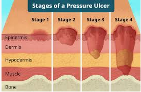
There are four stages of pressure sores from mild to severe:

Stage I

This is the mildest stage. These pressure sores only affect the upper layer of your skin.

Symptoms: Pain, burning, or itching are common symptoms. The spot may also feel different from the surrounding skin: firmer or softer, warmer or cooler.

You may notice a red area on your skin. If you have darker skin, the discolored area may be harder to see. The spot doesn’t get lighter when you press on it or even 10-30 minutes after you stop pressing. This means less blood is getting to the area.

Stage II

This happens when the sore digs deeper below the surface of your skin.

Symptoms: Your skin is broken, has an open wound, or looks like a pus-filled blister. The skin around it may be discolored.

The area is swollen, warm, and/or red. The sore may ooze clear fluid or pus. And it’s painful.

 Stage III

These sores have gone through the second layer of skin into the fat tissue.

Symptoms: The sore looks like a crater and may have a bad odor. It may show signs of infection: red edges, pus, odor, heat, and/or drainage. The tissue in or around the sore is black if it has died.

Stage IV

These sores are the most serious. Some may even go so deep they affect your muscles, ligaments, and bones.

Symptoms: The sore is deep and big. Skin has turned black and shows signs of infection—red edges, pus, odor, heat, and/or drainage. You may be able to see tendons, muscles, and bone.

Other stages

In addition to the four main stages for bed sores, there are two others:

Unstageable is when you can’t see the bottom of the sore because it’s covered in a layer of dead skin. Your doctor can only stage it once it’s cleaned out.

Suspected deep tissue injury is when the surface of the skin looks like a stage I or II sore, but underneath the surface, it’s a stage III or IV sore.

Risk factors for pressure sores

A pressure sore is caused by constant pressure applied to the skin over a period of time. The skin of older people tends to be thinner and more delicate, which means an older person has an increased risk of developing a pressure sore during a prolonged stay in bed.

Other risk factors for pressure sores include:

  • immobility and paralysis – for example due to a stroke or a severe head injury
  • being restricted to either sitting or lying down
  • impaired sensation or impaired ability to respond to pain or discomfort. For example, people with diabetes who experience nerve damage are at increased risk of pressure sores
  • urinary and faecal incontinence – skin exposed to urine or faeces is more susceptible to irritation and damage
  • malnutrition – can lead to skin thinning and poor blood supply, meaning that skin is more fragile
  • obesity – being overweight in combination with, for example, immobility or being restricted to sitting or lying down can place extra pressure on capillaries. This then reduces blood flow to the skin
  • circulation disorders – leading to reduced blood flow to the skin in some areas
  • smoking – reduces blood flow to the skin and, in combination with reduced mobility, can lead to pressure sores. The healing of pressure sores is also a slower process for people who smoke.

If you use a wheelchair you’re most likely to develop a pressure sore on the parts of the body where they rest against the chair. These may include the tailbone or buttocks, shoulder blades, spine and the backs of arms or legs.

If you’re bedridden, pressure sores can occur in a number of areas, including:

  • back or sides of the head
  • rims of the ears
  • shoulders or shoulder blades
  • hipbones
  • lower back or tailbone
  • backs or sides of the knees
  • heels, ankles and toes.

Treatment for pressure sores

There are a variety of treatments available to manage pressure sores and promote healing, depending on the severity of the pressure sore. These include:

  • regular position changes
  • special mattresses and beds that reduce pressure
  • being aware of the importance of maintaining healthy diet and nutrition
  • dressings to keep the sore moist and the surrounding skin dry. There is no advantage of one type of dressing over another.
  • saline gauze dressing may be used if ointments or other dressings (for example foam dressings) are unavailable.
  • light packing of any empty skin spaces with dressings to help prevent infection
  • regular cleaning with appropriate solutions, depending on the stage of the sore
  • there is no advantage of one particular type of antiseptic (e.g. iodine) or antibiotic treatment over another
  • specific drugs and chemicals applied to the area, if an infection persists
  • surgery to remove the damaged tissue that involves thorough debridement of the wound, the removal of underlying or exposed bone, and filling the empty space
  • operations to close the wound, using skin grafts if necessary
  • continuing supportive lifestyle habits such as eating a healthy and nutritious diet, as suggested by the nutritional staff.

Preventing pressure sores

If you are confined to a bed or chair for any period of time, it’s important to be aware of the risk of pressure sores. To prevent skin damage, you or your carer need to relieve the pressure, reduce the time that pressure is applied and improve skin quality. Pressure offloading surfaces such as mattresses and wheelchair cushions may help in providing pressure relief by evenly distributing the pressure.

Pressure injury monitoring devices that measure the skin moisture content, body motion and the pressure in-between may be used to prevent pressure sores and injuries. An example of a devices is pressure-sensing mats placed on beds or wheelchairs.

Develop a plan that your, your carer and any other caregivers can follow. This plan will include position changes, supportive devices, daily skin care, a nutritious diet and lifestyle changes.

A routine nursing assessment may be required if you’re at high risk of pressure sores. As visual skin assessment may sometimes be unreliable, early detection of pressure sores using some bedside technologies may help facilitate preventive interventions.

Pressure from medical devices such as oxygen tubing, catheters, cervical collars, casts and restraints should be minimised or removed.

Position changes to prevent pressure sores

If you use a wheelchair shift position within your chair about every 15 minutes. If you spend most of their time in bed change position at least once every two hours, even during the night and avoid lying directly on your hipbones.

Pillows may be used as soft buffers between your skin and the bed or chair. The head-of-bed elevation should be maintained at/or below 30 degrees. Or depending on the your medical condition, the bed should at least be elevated to the lowest degree to prevent injury. When lying on your side, a 30 degrees position should be used.

Daily skin care to prevent pressure sores

Ways to prevent pressure injuries include:

  • Checking the skin at least daily for redness or signs of discolouration.
  • Keeping the skin at the right moisture level, as damage is more likely to occur if skin is either too dry or too moist.
  • Using moisturising products to keep skin supple and prevent dryness.
  • Never massaging bony areas because the skin is too delicate.

Diet and lifestyle changes to avoid pressure sores

Changes to avoid pressure sores include:

  • Make sure you eat a healthy and nutritious diet. This includes a balanced diet (proteins, fats and carbohydrates) and fluids/water. And if necessary,you’re your doctor about vitamin and nutritional supplements (e.g. zinc, antioxidants).
  • Low body weight or being overweight can cause pressure sores, so make sure you maintain heathy body weight
  • If you’re malnourished or at risk of malnutrition, protein, fluid and energy intake should be increased.
  • Be aware of using good hygiene practices.
  • Maintain activity levels, where appropriate.
  • Make sure you quit smoking.

 Tips for skin care

Consider these suggestions for skin care:

  • Keep skin clean and dry. Wash the skin with a gentle cleanser and pat dry. Do this cleansing routine regularly to limit the skin’s exposure to moisture, urine and stool.
  • Protect the skin. Use moisture barrier creams to protect the skin from urine and stool. Change bedding and clothing frequently if needed. Watch for buttons on the clothing and wrinkles in the bedding that can irritate your skin.
  • Inspect the skin daily. Look closely at your skin daily for warning signs of a bedsore.

Further for any queries in regards to the  medical assistance please feel free to email us on query@gtsmeditour.com or you can whatsapp us on +91 9880149003.

Thank you..!

Craniosynostosis

cranio 

Craniosynostosis

is a condition in which the fibrous joints between the skull bones fuse too early. These joints are known as sutures. If this occurs (usually before or at birth) it can cause an abnormal head shape, or in some cases restrict growth of the brain, which increases the pressure inside the skull.

Usually, during infancy the sutures remain flexible, allowing a baby’s skull to expand as the brain grows. In the front of the skull, the sutures meet in the large soft spot (fontanel) on top of the head. The anterior fontanel is the soft spot felt just behind a baby’s forehead. The next largest fontanel is at the back (posterior). Each side of the skull has a tiny fontanel.

Craniosynostosis usually involves premature fusion of a single cranial suture, but it can involve more than one of the sutures in a baby’s skull (multiple suture craniosynostosis). In rare cases, craniosynostosis is caused by certain genetic syndromes (syndromic craniosynostosis).

Treating craniosynostosis involves surgery to correct the shape of the head and allow for brain growth. Early diagnosis and treatment allow your baby’s brain adequate space to grow and develop.

Although neurological damage can occur in severe cases, most children develop as expected in their ability to think and reason (cognitive development) and have good cosmetic results after surgery. Early diagnosis and treatment are key.

Symptoms

The signs of craniosynostosis are usually noticeable at birth, but they become more apparent during the first few months of your baby’s life. Signs and severity depend on how many sutures are fused and when in brain development the fusion occurs. Signs and symptoms can include:

  • A misshapen skull, with the shape depending on which of the sutures are affected
  • Development of a raised, hard ridge along affected sutures, with a change in the shape of the head that’s not typical

Types of craniosynostosis

There are several types of craniosynostosis. Most involve the fusion of a single cranial suture. Some complex forms of craniosynostosis involve the fusion of multiple sutures. Multiple suture craniosynostosis is usually linked to genetic syndromes and is called syndromic craniosynostosis.

The term given to each type of craniosynostosis depends on what sutures are affected. Types of craniosynostosis include:

cry

  • Sagittal (scaphocephaly).Premature fusion of the sagittal suture that runs from the front to the back at the top of the skull forces the head to grow long and narrow. This head shape is called scaphocephaly. Sagittal craniosynostosis is the most common type of craniosynostosis.
  • Premature fusion of one of the coronal sutures (unicoronal) that run from each ear to the top of the skull may cause the forehead to flatten on the affected side and bulge on the unaffected side. It also leads to turning of the nose and a raised eye socket on the affected side. When both coronal sutures fuse prematurely (bicoronal), the head has a short and wide appearance, often with the forehead tilted forward.
  • The metopic suture runs from the top of the bridge of the nose up through the midline of the forehead to the anterior fontanel and the sagittal suture. Premature fusion gives the forehead a triangular appearance and widens the back part of the head. This head shape is also called trigonocephaly.
  • Lambdoid synostosis is a rare type of craniosynostosis that involves the lambdoid suture, which runs along the back of the head. It may cause one side of a baby’s head to appear flat, one ear to be higher than the other ear and tilting of the top of the head to one side.

Causes

Often the cause of craniosynostosis is not known, but sometimes it’s related to genetic disorders.

  • Nonsyndromic craniosynostosisis the most common type of craniosynostosis. Its cause is unknown, although it’s thought to be a combination of genes and environmental factors.
  • Syndromic craniosynostosisis caused by certain genetic syndromes, such as Apert syndrome, Pfeiffer syndrome or Crouzon syndrome, which can affect a baby’s skull development. These syndromes usually also include other physical features and health problems.

 Treatment/Surgery:

Surgery for craniosynostosis is designed to correct the abnormal head shape and allow the growing brain room to expand normally.

The surgery for craniosynostosis is typically performed in the first two years of life. There are multiple types of surgery used to treat craniosynostosis, including strip craniectomy, spring-assisted craniectomy and cranial vault remodeling, amongst others. Not all patients are a candidate for all types of surgery. The surgery is performed by a team of a plastic surgeon and a neurosurgeon, working together.

What craniosynostosis surgery can treat

  • Abnormal skull shapes that result from early suture fusion
  • Raised intracranial pressure (ICP), or pressure on the brain caused by restriction of skull growth
  • Certain problems with eye position related to suture fusion

What results should I expect after craniosynostosis surgery?

The immediate results of craniosynostosis surgery will depend on the type of surgery performed. With open craniosynostosis procedures, the fused sutures are released, and the skull bones are repositioned to create a more typical head shape. The results of open procedures will be immediately apparent with a significant change in your child’s head shape from before to after surgery.

Complications

If untreated, craniosynostosis may cause, for example:

  • Permanently misshapen head and face
  • Poor self-esteem and social isolation

The risk of increased pressure inside the skull (intracranial pressure) from simple craniosynostosis is small if the suture and head shape are fixed surgically. But babies with an underlying syndrome may develop increased intracranial pressure if their skulls don’t expand enough to make room for their growing brains.

If untreated, increased intracranial pressure can cause:

  • Developmental delays
  • Cognitive impairment
  • Blindness
  • Seizures
  • Headaches