Global Treatment Services Pvt. Ltd.

Global Treatment Services

Hemiplegia vs Hemiparesis


Overview

hemi

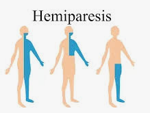
Hemiparesis or hemiplegia are the medical terms, for paralysis of one half of the body. Hemiplegia is a symptom of brain or spinal cord damage where as Hemiparesis refers to weakness in one leg, arm, or side of the face. Hemiplegia describes severe or complete unilateral loss of strength or paralysis. Both may result from a stroke and other conditions. Hemiparesis and hemiplegia may have the same cause, but depending on the severity and location of brain and spinal damage, you may develop one or the other.

Hemiparesis and hemiplegia are both inherently unpredictable, with symptoms that may change over time. Prompt intervention, including extensive physical therapy, can substantially improve the chances of a full recovery. Psychotherapy is also important, since the overwhelming nature of these injuries can interfere with a patient’s ability and willingness to stick with treatment.

Every patient is different, but there is no way to conclusively predict the prognosis of these disorders. Some people spontaneously recover, even with no treatment. Others make little progress, even with extensive treatment. In general, however, the more aggressively a patient works to recover some functionality, the more likely a full or partial recovery becomes.

Hemiparesis and Hemiplegia Causes

There are a number of conditions that can cause hemiparesis or hemiplegia. Examples of hemiparesis and hemiplegia causes include strokes, brain injuries, spinal cord injuries, infections, and a handful of other conditions affecting the central nervous system.

Both hemiparesis and hemiplegia are nervous system disorders, not caused by injury to the affected side of the body. For example, an injury to the spinal cord or brain interferes with the body’s ability to send or receive signals to the region of the body affected by the injury. So, while the left or right side of the body haven’t been damaged directly, the spinal cord injury may cause hemiparesis or hemiplegia.

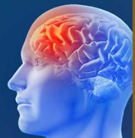
The most common cause of both injuries is a stroke. Strokes interfere with blood flow to the brain. If a region of the brain that affects movement or perception is affected, hemiparesis or hemiplegia may develop. The affected region is usually the opposite of the side of the brain affected, so an injury to the right side of the brain will affect the left side of the body.

Some other causes of hemiparesis and hemiplegia include:

  • Brain infections such as meningitis or encephalitis
  • Brain cancer or lesions
  • Damage to the neurons due to a degenerative disorder such as Parkinson’s
  • Traumatic injuries, such as a blow to the head during a car accident
  • Congenital disorders such as cerebral palsy

Rarely, psychological conditions can manifest as hemiparesis or hemiplegia. When this occurs, doctors must treat the psychological issue, not the physical symptoms.

Right Hemiplegia vs Left Hemiplegia

Left hemiplegia is the paralysis of limbs on the left side of the body, while right hemiplegia indicates paralysis on the right side of the body. Like hemiparesis, right or left hemiplegia may be caused by damage to the nervous system.

One common cause of left or right hemiplegia is an incomplete spinal cord injury. In an incomplete SCI, only part of the spinal cord is severed, so there may still be some function below the injury site. For example, an incomplete spinal cord injury in the cervical spinal cord might paralyze the left side of the body while leaving the right side functional — though this isn’t guaranteed because the specific nerves severed may alter the effects of the injury.

Right Hemiparesis vs Left Hemiparesis

As the name implies, right hemiparesis is weakness on the right side of the body, while left hemiparesis is weakness on the left side of the body. What causes this weakness in one side of the body and not the other varies, but damage to the nervous system from injury, infection, or degenerative conditions can result in hemiparesis.

In the case of degenerative conditions such as multiple sclerosis or amyotrophic lateral sclerosis (ALS), hemiparesis may progress into hemiplegia with time.

Did you ever thought why is only one side of the body affected?

Your spinal cord and brain have a left side and a right side. The halves are identical. Each half controls the movements on one side of the body.

An injury to one side of the spinal cord or brain may result in weakness or paralysis on the side of the body that half controls.

If you have multiple strokes on both sides of the brain, you may experience hemiparesis or hemiplegia on both sides of the body.

Symptoms of Hemiplegia & Hemiparesis

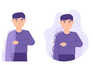
Hemiplegia is more severe than hemiparesis. It involves a complete loss of strength or paralysis (inability to move) on one side of the body. It may affect your arm, face, or leg only, or all of them.

Although the paralysis may not be extensive (whole body), it could affect your ability to:

  • breath
  • swallow
  • speak
  • control your bladder
  • control your bowel
  • move one side of your body

It’s common for hemiparesis to unevenly affect one side of the body. The disorder is on a continuum, with symptoms ranging from minor and sporadic to extensive and severe. signs include:

  •  Difficulties with motor skills, particularly standing.
  • Weakness in large areas on one side of the body. For example, a patient might be unable to lift his or her arms.
  • Pusher syndrome: a common byproduct of traumatic brain injuries, pusher syndrome causes a person with hemiparesis to shift their weight to the affected side, thereby undermining motor skills and making walking difficult and potentially painful. Doctors use a scale to assess the severity of pusher syndrome. This scale can help doctors make accurate prognostications, in addition to identifying the cause of symptoms.
  • Strain on the unaffected side of the body, which may overcompensate for injuries on the hemiparetic side.
  • Unusual sensations or tingling on the affected side.

Importantly, doctors will look for injuries on the affected side to rule out other causes, such as pinched nerves, muscle strains, or loss of blood flow.

Diagnosing hemiparesis and hemiplegia

A doctor will most likely use a number of diagnostic procedures and imaging tests to diagnose hemiparesis and hemiplegia.

These might include:

  • X-ray
  • magnetic resonance imaging (MRI)
  • computerized tomography (CT) scan
  • electromyography (EMG)
  • myelography (imaging test of your spinal cord)

Treatment for Hemiparesis and Hemiplegia

Though hemiparesis and hemiplegia produce different symptoms, they are substantially similar in cause and treatment. To treat either condition, a doctor will first look at what caused the symptoms. When an infection, growth, or other medical issue causes symptoms, a doctor may first address these symptoms. When a stroke is the culprit, the stroke itself cannot be reversed, but a number of treatments may help restore some or all functioning:

  • Physical, occupational, or exercise therapy to restore functioning and help the brain work around the injury.
  • Assistive devices such as walkers or wheelchairs.
  • Occupational therapy to support independence.
  • Psychotherapy to address the psychological impact of a severe injury.
  • Alternative treatments such as acupuncture and dietary changes.

Conclusion

Though the symptoms are different, the two conditions are essentially different versions of the same underlying problem. Mild to moderate nerve or brain damage will produce hemiparesis, while moderate to severe nerve or brain damage will result in hemiplegia. Moreover, the same injury can produce both symptoms at different times. This is especially true when nerves are compressed or the spinal cord is swollen, since changes in swelling or compression can also produce changes in symptoms.

People living with hemiplegia  and hemiparesis usually undergo a combination of rehabilitation therapy, which typically involves physical therapists, mental health professionals, and rehabilitation .

In addition to this if you or your loved ones are looking for medical opinion abroad feel free to email us the medical reports on query@gtsmeditour.com or you can WhatsApp us +91 9880149003

Happy to assist…!

Thank you..!


Is hemiplegia permanent?

Overview

brain

Hemiplegia (hemi = half; plegia = paralysis), as the name suggests, literally means paralysis of half of the body, is a type of paralysis of the muscles of the lower face, arm, and leg on one side of the body. hemiplegia is further classified as right or left hemiplegia. Hemiplegia is usually caused by brain damage localized to the cerebral hemisphere opposite the affected side. The most common cause of hemiplegia is stroke, other causes of hemiplegia include trauma eg. spinal cord injury; brain tumours; and brain infections.  hemiplegia can be temporary or permanent. Some causes of hemiplegia are treatable or even reversible with immediate medical care.

Hemiplegia may appear soon after birth (i.e., up to approximately two years of life), where it is known as congenital hemiplegia, or hemiplegic cerebral palsy. On the other hand, acquired hemiplegia presents later in life as a result of injury or other pathology. People living with hemiplegia usually undergo a combination of rehabilitation therapy, which typically involves physical therapists, mental health professionals, and rehabilitation therapists.

A person with hemiplegia can experience up to full paralysis on one side of their body and may have trouble speaking or breathing.

IMPORTANT: Hemiplegia is a key sign of a stroke, which is a life-threatening medical emergency. To recognize the symptoms of a stroke, remember to think FAST:

  • F is for face. Ask the person to smile. Look for a droop on one or both sides of their face, which is a sign of paralysis (facial hemiplegia) or muscle weakness.
  • A is for arm. A person having a stroke often has muscle weakness or paralysis on one side. Ask them to raise their arms. If they have new one-sided weakness or paralysis, one arm will stay higher while the other will sag and drop downward, or won’t raise up at all.
  • S is for speech. Strokes often cause a person to lose their ability to speak. They might slur their speech or have trouble choosing the right words.
  • T is for time. Time is critical, so don’t wait to get help! If possible, look at your watch or a clock and remember when symptoms start. Telling a healthcare provider about when the symptoms started can help them know what treatment options are best.

Conditions that cause hemiplegia

There are dozens of conditions and circumstances that can cause hemiplegia. Some of the most common causes include:

  • Strokes or transient ischemic attacks (TIAs).
  • Aneurysms and hemorrhages inside of your brain.
  • Concussions and traumatic brain injuries (TBIs).
  • Spinal cord injuries.
  • Injuries you experience during birth or very early childhood, causing conditions like hemiplegic cerebral palsy.
  • Congenital conditions (which you have when you’re born) such as alternating hemiplegia of childhood.
  • Facial paralysis conditions like Bell’s palsy.
  • Seizures and epilepsy.
  • Bleeding in between your brain and its outer layers (subdural hematomas or subarachnoid hemorrhages) or between your skull and your brain’s outer membrane (epidural hematomas).
  • Brain tumors (including cancers).
  • Nervous system diseases, especially autoimmune and inflammatory conditions, such as multiple sclerosis or progressive multifocal leukoencephalopathy (PML).
  • Infections that affect your nervous system, like encephalitis, meningitis or Ramsay Hunt syndrome.
  • Migraine headaches (when these involve hemiplegia, they’re known as hemiplegic migraines).

Hemiplegia symptoms

Hemiplegia can affect either the left or right side of your body. Whichever side of your brain is affected causes symptoms on the opposite side of your body.

People can have different symptoms from hemiplegia depending on its severity. Symptoms can include:

  • muscle weakness or stiffness on one side
  • muscle spasticity or permanently contracted muscle
  • Loss of motor function
  • trouble walking
  • poor balance
  • trouble grabbing objects
  • speech associated trouble.

Children with hemiplegia may also take longer to reach developmental milestones than their peers. They may also use only one hand when playing or keep one hand in a fist.

If hemiplegia is caused by a brain injury, the brain damage can cause symptoms that aren’t specific to hemiplegia, such as:

  • memory problems
  • trouble concentrating
  • speech issues
  • behavior changes
  • seizures

Diagnosis

Hemiplegia is often diagnosed after reviewing the individual’s past medical history and performing a detailed neurological examination assessing sensory and motor functions.

Additional tests can be conducted in order to determine the underlying cause of hemiplegia. Blood tests, including a

Complete blood count (CBC)

Erythrocyte sedimentation rate (ESR)

Coagulation tests (e.g., PT, aPTT) can help eliminate various underlying causes, such as infections or malignancies.

Imaging, including magnetic resonance imaging (MRI)

Computerized tomography (CT) scans of the head, are necessary in order to exclude most pathologies, including strokes, aneurysms, brain tumors, multiple sclerosis, and injuries.

Lastly, electroencephalography (EEG), which measures the electrical activity of the brain, can be used for diagnostic purposes (e.g., reveal the part(s) of the brain from which seizures originate).

Types of hemiplegia

The following are movement disorders that can cause hemiplegia symptoms.

Facial hemiplegia

People with facial hemiplegia experience paralyzed muscles on one side of their face. Facial hemiplegia may also be coupled with a slight hemiplegia elsewhere in the body.

Spinal hemiplegia

Spinal hemiplegia is also referred to as Brown-Sequard syndromeTrusted Source. It involves damage on one side of the spinal cord that results in paralysis on the same side of the body as the injury. It also causes loss of pain and temperature sensation on the opposite side of the body.

Contralateral hemiplegia

This refers to paralysis on the opposite side of the body that brain damage occurs in.

Spastic hemiplegia

This is a type of cerebral palsy that predominately affects one side of the body. The muscles on the affected side are constantly contracted or spastic.

Alternating hemiplegia of childhood

Alternating hemiplegia of childhood usually affects children younger than 18 months old. It causes recurring episodes of hemiplegia that affect one or both sides of the body.

Hemiplegia symptoms

Hemiplegia can affect either the left or right side of your body. Whichever side of your brain is affected causes symptoms on the opposite side of your body.

People can have different symptoms from hemiplegia depending on its severity. Symptoms can include:

  • muscle weakness or stiffness on one side
  • muscle spasticity or permanently contracted muscle
  • poor fine motor skills
  • trouble walking
  • poor balance
  • trouble grabbing objects

Children with hemiplegia may also take longer to reach developmental milestones than their peers. They may also use only one hand when playing or keep one hand in a fist.

If hemiplegia is caused by a brain injury, the brain damage can cause symptoms that aren’t specific to hemiplegia, such as:

  • memory problems
  • trouble concentrating
  • speech issues
  • behavior changes
  • seizures

Treatment

Treatment of hemiplegia involves a multidisciplinary approach. Healthcare professionals including physicians, physical therapists, occupational therapists, and psychologists must all contribute in order to assist affected individuals in regaining their functionality. Treatment typically aims at improving sensation and motor abilities so that individuals can perform their daily activities with as much independence as possible; as well as treating any underlying pathology causing the hemiplegia.

Physiotherapy

Working with a physiotherapist allows people with hemiplegia to develop their balance ability, build strength, and coordinate movement. A physiotherapist can also help stretch out tight and spastic muscles.

When hemiplegia isn’t temporary, healthcare providers often recommend rehabilitation in addition to other treatments. Rehabilitation can take place in a hospital (inpatient rehabilitation), clinic or office (outpatient rehabilitation), or at home. Rehabilitation generally includes:

  • Physical therapy: Focused on leg function, standing, walking and balance.
  • Occupational therapy: Focused on arm/hand function and other activities of daily life.
  • Prescription of equipment, to enhance safety and the ability to function inside and outside of your home.
  • Managing symptoms associated with hemiplegia, such as spasticity and depression.
  • Guidance and resources to address the consequences of hemiplegia, for example, returning to work or applying for disability benefits.

Modified constraint-induced movement therapy (MCIMT)

Modified constraint-induced movement therapy involves restraining the side of your body unaffected by hemiplegia. This treatment option forces your weaker side to compensate and aims to improve your muscle control and mobility.

One small studyTrusted Source published in 2018 concluded that including mCIMT in stroke rehabilitation may be more effective than traditional therapies alone.

Assistive devices

Some physical therapists may recommend the use of a brace, cane, wheelchair, or walker. Using an assistive deviceTrusted Source may help improve muscular control and mobility.

It’s a good idea to consult a healthcare professional to find which device is best for you. They may also recommend modifications you can make to your home such as raised toilet seats, ramps, and grab bars.

Mental imagery

Imagining moving the paralyzed half of your body may help activate the parts of the brain responsible for movement. Mental imagery is often paired with other therapies and is rarely used by itself.

One meta-analysis looking at the results of 23 studies found that mental imagery may be an effective treatment option for regaining strength when combined with physical therapy.

Electrical stimulation

A medical professional can help stimulate muscular movement by using electrical pads. The electricity allows muscles that you can’t move consciously to contract. Electrical stimulation aims to reduce imbalances in the affected side of the brain and improve brain plasticityTrusted Source

Hemiplegia is Preventive when you..

  • Pursue a healthy lifestyle (stop smoking, drink in moderation)
  • Eat a balanced diet
  • Attend scheduled check-up appointments
  • Avoid exposure to extremes of temperature
  • Try to avoid emotional stress
  • Avoid physical fatigue
  • Protect yourself against respiratory system infections

Conclusion

Hemiplegia is a severe paralysis on one side of your body caused by brain damage. It’s a non-progressive disorder and doesn’t get worse once it develops. With a proper treatment plan, it’s possible to improve the symptoms of hemiplegia.

If you’re living with hemiplegia, you can make the following changes to your lifestyle to aid your rehabilitation:

  • Stay active to the best of your ability.
  • Modify your home with assistive devices like ramps, grab bars, and handrails.
  • Wear flat and supportive shoes.
  • Follow your doctor’s recommendation for assistive devices.

Remember to think FAST

  • F is for face. Ask the person to smile. Look for a droop on one or both sides of their face, which is a sign of paralysis (facial hemiplegia) or muscle weakness.
  • A is for arm. A person having a stroke often has muscle weakness or paralysis on one side. Ask them to raise their arms. If they have new one-sided weakness or paralysis, one arm will stay higher while the other will sag and drop downward, or won’t raise up at all.
  • S is for speech. Strokes often cause a person to lose their ability to speak. They might slur their speech or have trouble choosing the right words.
  • T is for time. Time is critical, so don’t wait to get help! If possible, look at your watch or a clock and remember when symptoms start. Telling a healthcare provider about when the symptoms started can help them know what treatment options are best.

we provide free medical opinion abroad, we request you to provide us your latest medical reports on our email :query@gtsmeditour.com or whatsapp us on +91 9880149003.

Happy to assist..!

Thankyou..!


Is hemiparesis a stroke ?

Overview

hemi

Hemiparesis is the medical term for weakness on one side of the body. It is an early sign of stroke and transient ischemic attacks (TIAs). If you experience sudden hemiparesis in a limb or your face for an unknown reason, you should immediately go to the nearest emergency room (ER). Having this symptom unexpectedly can be an early sign of a stroke. You should also seek immediate medical help if you have any other signs of stroke, including balance issues, vision changes, face and arm drooping and speech difficulties. Hemiparesis is a common after-effect of stroke that causes weakness on one side of the body. This one-sided weakness can limit your movement and affect all basic activities, such as dressing, eating, and walking. People often confuse hemiparesis and hemiplegia. Both conditions occur as the result of a stroke.

Symptoms of hemiparesis

Common signs and symptoms of hemiparesis may include:

  • difficulty walking and standing
  • loss of balance and coordination
  • numbness or tingling on one side of the body
  • weakness in one arm or leg
  • weakness in one side of the face that causes drooping of an eyelid or corner of the mouth
  • numbness or weakness in the tongue that interferes with speech and swallowing
  • asymmetry in one side of the body (e.g. lifting both arms and having one of them not come all the way up)

Diagnosis

Complete blood count. This includes a check for your level of platelets, which are cells that help clot blood. A lab will also measure electrolyte levels in your blood to see how well your kidneys are working.

Clotting time. A pair of tests called PT (prothrombin time) and PTT (partial thromboplastin time) can check how quickly your blood clots. If it takes too long, it could be a sign of bleeding problems.

Computerized tomography (CT). Your doctor takes several X-rays from different angles and puts them together to show if there’s any bleeding in your brain or damage to brain cells. They may put dye in your vein first to look for an aneurysm, a thin or weak spot on an artery.

Magnetic resonance imaging (MRI). This uses powerful magnets and radio waves to make a detailed picture of your brain. It’s sharper than a CT scan and can show injuries earlier than a traditional CT.

Carotid ultrasound. This uses sound waves to find fatty deposits that may have narrowed or blocked the arteries that carry blood to your brain.

Echocardiogram. Sometimes a clot forms in another part of the body (often the heart) and travels to the brain. This imaging test of the heart can look for clots in the heart or enlarged parts of the heart.

Angiograms of your head and neck. Your doctor will put dye in your blood so they can see your blood vessels with X-rays. This can help find a blockage or aneurysm.

Hemiparesis treatment may include medical and nonmedical techniques:

  • Electrical stimulation. As part of this method, small electrical pads are placed on the weak muscles of the side that has hemiparesis. An electrical charge is sent to the body that helps the muscles contract. Some of these machines can be used at home once you learn how to use them.
  • Cortical stimulation. This is a bit similar to electrical stimulation, except that the electric current is sent to the tough membrane covering the brain (called the dura). While your brain is subjected to this current, you have to do rehabilitation exercises.
  • mCIT. Modified, constraint-induced therapy is a treatment method that forces you into using the weak side of the body. With regular practice, nerve function may improve in the region. mCIT works well when used with other therapies.

Treating Right Sided hemiparesis

A physical therapist can recommend the appropriate device. Braces, canes, walkers and wheelchairs can increase strength and movement. An ankle-foot orthosis brace can help control your ankle and foot. Repeated practice and regular activity will help increase control and flexibility and re-establish nerve circuitry.

 Possible complications or risks of not treating hemiparesis

The risks of not treating it can be high. In the worst cases, it can result in permanent brain damage, paralysis, loss of abilities or even death.

Conclusion/Takeover

A stroke happens when the flow of blood to part of your brain gets cut off. This means your brain can’t get oxygen, and without that, brain cells can be damaged in minutes. That’s why it’s important to get to the emergency room right away if you think you’re having a stroke. You might need a clot-busting drug within the first 3 hours.

The F-A-S-T test is an easy way to remember them:

  • Face: Smile. Does one side of your face sag?
  • Arms: Raise both and see if one droops.
  • Speech: Say a common phrase: Does it sound strange or slurred?
  • Time: Call Ambulance service right away if you notice any of these symptoms. Note what time they started.

For any medical assistance or  free opinion from our top doctors  feel free to email us your query at query@gtsmeditour.com or whats app us on +91 9880149003

Happy to help..!

Thank you..!


Blepharoplasty

Overview

ble

The eye is an important component of facial aesthetics, blepharoplasty often means removing excess skin, muscle and underlying fatty tissue. Blepharoplasty play a vital positive role in facial harmony and the perception of aging. Blepharoplasty is one of the most commonly performed facial cosmetic procedures. Symptoms such as tired-looking eyes, excess skin, droopy eyelids, or circles around the eyes may benefit from blepharoplasty. You might be a good candidate for this procedure if your vision is affected by sagging skin. Some people may complain that their vision when looking upwards is blocked by the hanging skin.

people who have blepharoplasty say they feel more self-confident and feel they look younger and more rested. For some people, surgery results may last a lifetime. For others, droopy eyelids can recur. Bruising and swelling generally lessen slowly in about 10 to 14 days. Scars from the surgical cuts may take months to fade.

Causes

Blepharoplasty might be an  ideal option for:

  • Baggy or droopy upper eyelids
  • Excess skin of the upper eyelids that partially blocks peripheral vision
  • Excess skin on the lower eyelids
  • Bags under the eyes

Blepharoplasty can be done at the same time as another procedure, such as a brow lift, face-lift or skin resurfacing.

Symptoms

Patient with below symptoms may require blepharoplasty

  • Tired-looking eyes
  • Excess skin
  • Droopy eyelids
  • Circles around the eyes

Risks involved

All surgery has risks, including reaction to anesthesia and blood clots. Besides those, rare risks of eyelid surgery include:

  • Infection and bleeding
  • Dry, irritated eyes
  • Difficulty closing the eyes or other eyelid problems
  • Noticeable scarring
  • Injury to eye muscles
  • Skin discoloration
  • Temporarily blurred vision or, rarely, loss of eyesight
  • The need for follow-up surgery.

Treatment

eye

Depending on your goals and the recommendation of your surgeon, blepharoplasty can involve your upper eyelids, lower eyelids or both.

Upper blepharoplasty

During an upper blepharoplasty, your surgeon will make cuts (incisions) in the natural crease of your upper eyelid. These incisions will be hidden when your eyes are open. Your surgeon will remove excess skin and protruding fat, and then they’ll close the incisions.

Lower blepharoplasty

During a lower blepharoplasty, your surgeon will make an incision just below your lower eyelash line. They’ll remove excess skin in your lower eyelid through this incision. They may also use an incision hidden inside your lower eyelid (transconjunctival incision) to correct lower eyelid issues and redistribute or remove excess fat.

There will be a stitch in each of your upper lids that’ll remain for about a week. The lower lids don’t require stitches if the incisions are made on the inside of your lids. It’s common for swelling and bruising to occur in your upper and lower lids. Expect to stay home from work and limit your activities for several days after surgery to allow your eyelids to heal.

Although the surgery is typically painless, you may have some swelling and bruising. Most people feel comfortable going out in public after 10 to 14 days. But it can take a few months to heal completely.

Recovery

During your blepharoplasty recovery, you can use cold compresses and antibiotic ointment to ease any swelling. Your surgeon will give you specific instructions that may include:

  • How to care for your eyes.
  • Medications to aid healing and reduce the potential for infection.
  • Specific concerns to look for at the surgical site.
  • When to follow up with your surgeon.

Some people experience dry eyes after surgery, but the condition rarely lasts more than two weeks. If you have dry eyes for more than two weeks, contact your healthcare provider.

Further for any kind of medical assistance or free medical opinion please share us your reports/complaints on our email : query@gtsmeditour.com or you can whatsapp us on +91 9880149003 

Happy to help always…:)

Thank you..!


Foot drop syndrome

Overview

Foot drop also known as drop foot, is a general term for difficulty lifting the front part of the foot. If you have foot drop, the front of your foot might drag on the ground when you walk. Foot drop can be unilateral (affecting one foot) or bilateral (affecting both feet)at the same time. It can strike at any age. In general, foot drop stems from weakness or paralysis of the muscles that lift the foot. Sometimes foot drop is temporary, but it can be permanent. If you have foot drop, Fortunately, there are several treatment options, which may include a combination of orthotics, physical therapy, and surgical procedures. However, it’s important to seek early treatment to improve the chances of recovery.

Causes

  • multiple sclerosis
  • amyotrophic lateral sclerosis (ALS), commonly known as Lou Gehrig’s disease
  • muscular dystrophy
  • spinal muscular atrophy
  • stroke
  • Charcot-Marie-Tooth disease (CMT), a neurological disorder
  • Parkinson’s disease
  • poliomyelitis, also known as polio

Foot drop can also be caused by injuries to the nerves that control the muscles that lift the foot. The affected nerves may be in the knee or in the lower spine.

Other causes include hip or knee replacement surgery and diabetes. Nerve damage in the eye, which can result in pain when moving the eye or even vision loss, can also cause difficulties with walking.

Other causes of foot drop include nerve compression or a herniated disc.

Symptoms

  • decreased muscle mass
  • frequent trips or falls
  • limpness of the foot
  • loss of sensation in the leg or foot
  • changes in gait, such as raising your leg higher or swinging your leg to the side when walking

Risk factors

The peroneal nerve controls the muscles that lift the foot. This nerve runs near the surface of the skin on the side of the knee closest to the hand. Activities that compress this nerve can increase the risk of foot drop. Examples include:

  • Leg crossing. People who habitually cross their legs can compress the peroneal nerve on their uppermost leg.
  • Prolonged kneeling. Occupations that involve prolonged squatting or kneeling — such as picking strawberries or laying floor tile — can result in foot drop.
  • Wearing a leg cast. Plaster casts that enclose the ankle and end just below the knee can exert pressure on the peroneal nerve.

Treatment

Treatment depends on the specific cause of foot drop. The most common treatment is to support the foot with lightweight leg braces and shoe inserts, called ankle-foot orthotics. Exercise therapy to strengthen the muscles and maintain joint motion also helps to improve gait. Devices that electrically stimulate the peroneal nerve during footfall are appropriate for a small number of individuals with foot drop. In cases with permanent loss of movement, surgery that fuses the foot and ankle joint or that transfers tendons from stronger leg muscles is occasionally performed.

Further any medical assistance or free medical opinion please feel free to email us on query@gtsmeditour.com or you whatsApp us on +91 9880149003