Global Treatment Services Pvt. Ltd.

Global Treatment Services

Endometriosis

Endometriosis a painful condition caused when tissues that line the uterus grow outside of uterus.The tissues that make up the lining of the uterus is called endometrium.Women with endometriosis have tissues similar to endometrium  on other organs of the body. Endometriosis usually occurs usually on other reproductive organs inside the pelvis or in the abdominal cavity and affects women of childbearing age.9 .Feb13Endometriosis


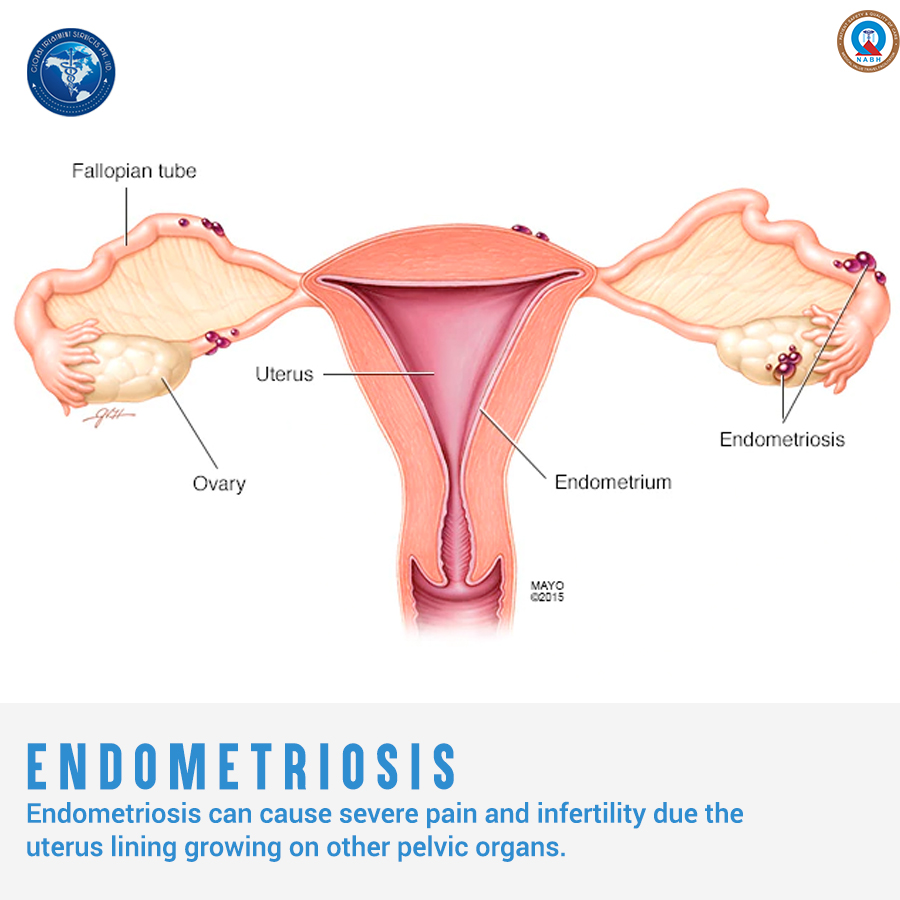
Understanding the condition

In a regular menstrual cycle,if fertilization has not happened , hormonal changes signal the uterus to prepare to shed its lining.The endometrial tissues build up and egg along with the uterine lining is shed in form menstrual bleeding.Whereas in patients with endometriosis,the misplaced endometrial tissues  also respond to the hormonal signals and by building up and breaking down just like the endometrium does, resulting in small bleeding inside of the pelvis.Unlike the cells in the uterus that leave the body , this blood has no way to escape.This leads to inflammation, swelling and scarring of the normal tissue surrounding the endometriosis implants.
Mostly,the endometrial tissues grow on ovaries, Fallopian tubes, bowel and outer walls of the uterus.But it can appear anywhere in the body.Though very rarely,it is found to develop on kidney, bladder and lungs. Endometriosis can be located on and even within an ovary, causing fibrous cysts called endometriomas. Blood become embedded in the normal ovarian tissue  surrounded by endometrioma.

Causes

Several ideas have been proposed to explain what causes the tissues on uterine lining to grow on other parts of the body.One idea explains endometriosis by retrograde menstruation.It is a sort of reverse menstruation in which the period blood and tissues travel out of the uterus  through Fallopian tubes to the abdominal cavity where it attaches and grows.But this idea fail to explain the occurrence of endometriosis at strange locations like thumb and knee.It is better explained by another theory that suggests that some cells outside uterus can transform into endometrial cells.  Another possible explanation is that the cells from the lining of the uterus travel  and implant via blood vessels or lymphatic system to reach other organs similar to how cancer cells spread. Also, endometriosis can spread as a result of direct transplantation during a cesarean section where in endometriosis cells attach to the abdominal incision and endometriosis is formed in the scar.

Symptoms

Symptoms of endometriosis vary in different women. Some women experience mild symptoms, but others can have moderate to severe symptoms. The severity of your pain is not the indicator of stage of the condition. Mild form of the disease may cause agonizing pain. It’s also possible to have a severe form but very little discomfort.For some,it is asymptomatic.

Pelvic pain is the most common symptom of endometriosis. You may also have the following symptoms:

  • painful periods especially excessive menstrual cramps or irregular periods
  • pelvic pain before and during menstruation
  • infertility
  • heavy menstrual bleeding or spotting between periods
  • Painful urination or  blood in the pee during menstrual period
  • uncomfortable bowel movements
  • pain after sexual intercourse
  • lower back pain that may occur at any time during your menstrual cycle
  • feeling sick, constipation, diarrhoea
  • Nausea and fatigue

Endometriosis and Pain,infertility

Due to the misplaced cells,the woman with endometriosis have bleeding from tissues outside the uterus as well during menstrual period.When blood comes into contact with other organs especially in the abdomen, it causes inflammation and irritation, creating pain. Also,the scar tissue developed from the endometriosis contribute to the pain.
Endometriosis seems to impair fertility  by causing distortion of the fallopian tubes making it incapable of picking up the egg after ovulation and by creating inflammation that can adversely affect the function of the ovary, egg, fallopian tubes or uterus.
Statistics show that  20 – 40% of women with infertility have endometriosis and this points to how much of a factor endometriosis is in infertility!

Diagnosis

Since endometriosis manifests itself in many ways and shares symptoms with other conditions, diagnosis can be difficult and often delayed. The condition is in most case diagnosed ,when the patient tries to evaluate infertility and  pelvic pain in one another reason.The only definitive way to diagnose endometriosis is by laparoscopy.Laparoscopy is a minor surgical procedure in which a laparoscope, a thin tube with a camera at the end, is inserted into the abdomen through a small incision. Laparoscopy helps to determine the location, extent and size of the endometrial growths.

Treatment

Treatment options available to women with endometriosis are:

  • Surgery
    As a treatment for endometriosis, surgery can be used to alleviate pain by removing the endometriosis, dividing adhesions or removing cysts.
    Conservative surgery: This aims to remove or destroy the deposits of endometriosis and is usually done via a laparoscopy (keyhole surgery). This is also used to diagnose the disease and can be used to improve fertility.  Although surgery can provide relief from symptoms, they can recur in time.
    Radical surgery: This refers to a hysterectomy or oophorectomy: Hysterectomy is the removal of the womb,  with or without removing the ovaries. If the ovaries are left in place then the chance of endometriosis returning is increased. Oopherectomy is the removal of the ovaries, either one or both.When both ovaries are removed, a woman will experience an instant and irreversible menopause.
    These procedures may be decided after taking many factors into consideration.
  • Hormone treatment
    These are treatments that are used to act on the endometriosis and stop its growth. They either put the woman into a pseudo-pregnancy or pseudo-menopause because during pregnancy the endometrium is thin and also inactive. Both states are reversed when the patient stops taking the hormones. In addition, testosterone derivatives are occasionally used to mimic the male hormonal state.All of these have a contraceptive effect, so are not used if the patient is trying to become pregnant.
  • Pain relief
    Pain medication avoids the use of hormones so it does not prevent the growth of endometriosis.When taken appropriately, pain medication can be extremely effective. Either painkillers, or drugs that modify the way the body handles pain, can be used.

Some complementary therapies may be beneficial in controlling the symptoms of endometriosis.These include acupuncture,homeopathy,yoga etc.

The course of treatment depends on several factors.The approach will depend on how severe the disease is and when you expect to become pregnant.Endometriosis is a debilitating and challenging condition. Fortunately, there are treatments available which can alleviate the symptoms and condition.

 For any queries regarding the procedure and treatment facilities,email us at query@gtsmeditour.com .

Read about:Crohn’s disease


Crohn’s disease

Crohn’s disease is a chronic inflammatory condition of the gastrointestinal tract and belongs to the group of  Inflammatory Bowel Diseases (IBD). It is named after Dr. Burrill B. Crohn, who first described the disease in 1932.While it primarily causes ulcerations in the small and large intestines, it can affect any part of the digestive system, from the mouth to the anus.What differentiate it from ulcerative colitis is that Crohn’s disease can affect any part of the digestive tract.Ulcerative colitis is limited to the colon, also called the large intestine.8.Feb12Crohnsdisease

When the immune system activates its immune cells and proteins to do battle, it causes inflammation at the site. In Crohn’s disease there is a loss of regulation of the immune system within the intestinal tract and it cycles between periods of great activity, ulceration, and periods of relative calm or remission.About 20% of people with Crohn’s have a blood relative who has IBD.It affects men and women equally.It can occur any time but mostly start in the ages 15-35.

Types and Symptoms

There are five  types of Crohn’s disease based on which part of the gastrointestinal tract it affects.The associated sypmtoms and complication differ for each types. It is  important to analyse the type and symptoms together to get an understanding of the type of Crohn’s disease.

Ileocolitis
This is the most common form of Crohn’s. ileocolitis affects the ileum(the end of the small intestine)  and the colon (large intestine). Symptoms: Diarrhea , cramping  in the right lower part or middle of the abdomen, weight loss.

Ileitis
This type affects only the ileum. Symptoms :same as ileocolitis. but in severe cases, complications may also include fistulas in right lower quadrant of abdomen.

Gastroduodenal Crohn’s disease
This type affects the stomach and the duodenum(the beginning of the small intestine). Symptoms: loss of appetite, weight loss, nausea and vomiting.

Jejunoileitis
This type is characterized by patchy areas of inflammation in the jejunum(the upper half of the small intestine ). Symptoms: mild to intense abdominal pain and cramps following meals, diarrhea. In severe cases, fistulas may be formed.

Crohn’s (granulomatous) colitis
This type affects the colon only. Symptoms: diarrhea, rectal bleeding, and disease around the anus like abscess, fistulas and  ulcers, skin lesions and joint pains.

Also Crohn’s disease can be classified on the basis of the nature of disease as follows.

  • Inflammatory in nature, it results in small erosions and breakdown of the lining of the bowel wall.
  • Fibrostenotic, scar tissue forming. The inflammation is healed by the body but the resulting scar tissue can cause obstructive types of symptoms where the bowel is not moving properly.
  • Penetrating type,  the inflammation goes through the wall of the bowel and has a tendency to form connections between loops of bowel or connections between the bowel and other organs or the skin.

Diagnosis

It’s important to diagnose Crohn’s disease early because Crohn’s disease is a progressive disease. And in some people the Crohn’s disease can progress very rapidly to complications such as narrowing of the small intestine leading to blockages, or perforation of the small intestine requiring surgery.

In general, there are two reasons to undergo Crohn’s tests and procedures:

  • To determine whether you have Crohn’s (to obtain or disprove a Crohn’s disease diagnosis)
  • To re-evaluate your Crohn’s disease — something your gastroenterologist will probably want to do on an ongoing, regular basis

Common tests for Crohn’s disease are
Blood Tests.Although blood tests alone cannot diagnose Crohn’s disease, they’re an important tool in diagnosis and monitoring of this disease. Several different kinds of blood tests are used.

  • Routine blood testsThese are used to detect infection, anemia, indicators of inflammation, and to identify deficiencies of vitamins or minerals.
  • Fecal blood test
  • Antibody blood tests

Imaging Tests
These are tests that take pictures of different parts of your body to provide a clearer picture of your condition. They show your doctor areas of disease activity and identify possible complications. These are only some of the imaging tests used for Crohn’s—there are others your doctor may recommend.

  • Conventional X-rays
    A standard X-ray of your abdominal area can show narrowing of the intestines or an intestinal blockage, possibly from inflammation or scarring. It may also be done to rule out certain Crohn’s complications.
  • Contrast X-rays
  • Computerized tomography (CT scan)
  • Leukocyte scintigraphy (white blood cell scan)
  • Endoscopy (includes colonoscopy)
  • Endoscopic ultrasound
  • Magnetic resonance imaging (MRI)

Treatment

Crohn’s is a chronic, life-long disease that requires constant treatment. Even though there is no cure for Crohn’s, there are many different medications available to treat it.The following are the categories of drugs prescribed to treat Crohn’s:

  • Antibiotics :Antibiotics may be used when infections occur, or to treat complications of Crohn’s disease.
  • Aminosalicylates (5-ASAs):To decrease inflammation in the lining of the intestines and are usually used to treat mild to moderate Crohn’s symptoms.
  • Corticosteroids (Steroids):To reduce inflammation by suppressing the immune system and are usually given to help with moderate to severe Crohn’s symptoms.
  • Immune modifiers (Immunomodulators):To suppress the body’s immune response so that it cannot cause ongoing inflammation.
  • Biologic therapies (Biologics):To uppress the immune system to reduce inflammation by targeting a specific pathway, and is usually given to people who have not responded to conventional therapy.

Regarding pregnancy,most women who have Crohn’s disease can have a normal pregnancy and healthy baby.However, some Crohn’s disease medicines can harm an unborn baby, so you should speak to your doctor if you are planning pregnancy.Women may also find it harder to get pregnant during a flare-up.Possible complications of the disease are

  • damage to your bowel that may require surgery
  • difficulty absorbing nutrients from food leading to problems like osteoporosis or anaemia.
  • bowel cancer :regular cancer screening is needed to check this.

So adequate screening and diagnosis is required for handling the disease and to prevent severe complications.

For any queries regarding the procedure and treatment facilities,email us at query@gtsmeditour.com .

Read about:Robotic or minimally invasive cardiac surgery

Robotic or minimally invasive cardiac surgery

Ever imagined what goes through the mind of a patient when he or she is told that they need to undergo a surgery?And then,after the patient has gathered enough courage and has submitted to it,the trauma it leaves behind is severe. Robotic cardiac surgery is a breakthrough development which provides greater advantages for heart patients for and post surgery.The use of minimally invasive procedures has not only changed the performance but also the strategic approaches to surgeries in general.Also,surgical robots have enhanced the ability and precision of the surgeon. 7.Feb11CardiacSurgery
Robotic cardiac surgery ,also called closed chest heart surgery, is done  through very small cuts in the chest rather than a median sternotomy (cutting through the breastbone).An incision in the right side of the chest between the ribs offers quicker recovery and better outcomes for many patients.
The surgeon uses a specially-designed computer console to control surgical instruments on thin robotic arms. This technology allows surgeons to perform certain types of complex heart surgeries with smaller incisions and precise motion control, offering patients improved outcomes. With the use of tiny instruments and robot-controlled tools, surgeons are able to do heart surgery in a way that is very much less invasive than open-heart surgery. The procedure is sometimes called da Vinci surgery after the manufacturer of the robot often used for this procedure.

Candidates for Robot Surgery

The treatment type recommended for your condition will depend on several factors like  the type and severity of heart disease, age, medical history and lifestyle etc.Several diagnostic tests are carried out to determine if you are appropriate for robotic cardiac surgery.This include a cardiac catheterization and chest x-ray. An echocardiogram and/or a computed tomography scan also may be required to provide more details about the medical condition.After reviewing the results of all these tests,doctors conclude whether to take robotic cardiac surgery or not.

Types of robotic cardiac surgery

  • Mitral valve repair and replacement
  • Tricuspid valve repair and replacement
  • Aortic valve replacement
  • Ablation of atrial fibrillation or Maze procedure for atrial fibrillation
  • Coronary artery bypass surgery
  • Saphenous vein harvest for coronary artery bypass surgery
  •  Atrial septal defect (ASD) repair
  • Patent foramen ovale (PFO) repair
  • Removal of cardiac tumors (Myxoma, Fibroelastoma)
  • Combined coronary artery bypass and angioplasty

Advantages

With open heart surgery ,splitting the sternum and spreading the rib cage to gain access to the heart contribute to significant morbidity. Also,cardiac surgery is different than other surgical procedures because the heart-lung machine adds further morbidity which is avoided in closed chest heart surgery.In addition,other advantages are

  • Less blood loss
  • Lower risk of infection
  • Reduced trauma and pain
  • Shorter time in the hospital, faster recovery and quicker return to normal activities
  • Smaller, less noticeable scars.Traditional surgery would leave a 20 cm scar for rest of the life.

Risks

As with any surgery,robotic assisted surgeries also pose some risks.Although the minimally invasive procedure removes many complications,some possible risks are

  • Heart attack
  • Stroke
  • Infection

In some cases, doctor may not be able to complete the surgery with the robot. In that case, open-heart surgery may be needed.

Technological advances have facilitated totally endoscopic/key hole  robotic cardiac surgery.In India several hospitals provide minimally invasive endoscopic surgeries.The success rate is very high ,more than 92%.The potential in this technology is enormous and the path is minimal.
For any queries regarding the procedure and treatment facilities,email us at query@gtsmeditour.com.

Read about: Meningitis,the deadly disease

Meningitis

Meningitis is deadly that it can kill in a matter of hours.Though some cases of meningitis improve without treatment,others can be life-threatening and require emergency treatment.
Meningitis occur when the fluid surrounding the meningis become infected causing inflamamtion of meningis. Meningis are three delicate protective membranes of brain and spinal cord.6.Feb8Meningitis

Causes

Meningitis is caused by bacterial,viral or fungal infection,viral infected ideas being the most common.It can also be caused by other factors such as cancer,drug irritations.Like any other infection,meningitis can be contagious too which can be transmitted through touch,sneezing etc.

Bacterial meningitis
Though it is rare,it can be fatal if not treated right away.Bacterial meningitis can cause stroke, hearing loss, and permanent brain damage.The disease can occur when certain bacteria invade the meninges directly or when  the bacteria spread to the meninges from a severe head trauma or a severe local infection, such as a serious ear infectionor nasal sinus infection (sinusitis).Several bacteria can first cause an upper respiratory tract infection and then travel to brain via bloodstream.

  • Pneumococcal meningitis  is the most serious form of bacterial meningitis,caused by the bacterium Streptococcus pneumoniae.It  also causes pneumonia, blood poisoning (septicemia), and ear and sinus infections.
  • Meningococcal meningitis is caused by the bacterium Neisseria meningitides. If diagnosed, people in close contact should be given preventative antibiotics.
  • Haemophilus influenzae meningitis.

Viral meningitis
Also called aseptic meningitis ,it is usually caused by common viruses that enter the body through the mouth and travel to the brain and surrounding tissues.Many of the enteroviruses viruses that cause meningitis are common, such as those that cause colds, diarrhea and the flu.Other viruses that cause meningitis include varicella zoster (chicken pox virus),, mumps, HIV, and herpes simplex type 2 (genital herpes).

Fungal meningitis
The most common of this is caused by the fungus cryptococcus neoformans(found mainly in dirt and bird droppings).  Fungal infectionns can affect brain.Although treatable, fungal meningitis often recurs in 50 % of the affected persons.
Meningitis can affect anyone .Children,people with weakened immune systems( including persons with HIV or taking immunosuppressant drugs) are at increased risk.

Symptoms

Significant signs of meningitis are

  • sudden fever
  • severe headache
  • nausea or vomiting
  • double vision, drowsiness
  • sensitivity to bright light
  • stiff neck.

In infants ,fever, lethargy, not waking for feedings, vomiting, body stiffness, unexplained irritability, and bulging fontanel (the soft spot on the top of the head) are important signs of meningitis and should be watched for.
Distinctive rashes called meningitis rash are seen in some forms of the disease.This is one late sign of bacterial meningitis.These are faint rashes and may not be easily visible in dark skinned people.Rash will become more obvious as  infection worsen and spots grow darker. Meningococcal meningitis damages capillary cells leading to  capillary damage and mild blood leaks.
Meningitis can also cause septicaemia.It is the blood poisoning caused by the same germs that cause meningitis.It is the more life threatening form of the disease. Septicaemia can occur with or without meningitis.

Diagnosis

Early diagnosis is vital, as symptoms can appear suddenly and escalate to brain damage, hearing and/or speech loss, blindness, or even death.
Diagnostic tests include:

  • series of physical examination tests designed to assess motor and sensory function, nerve function, hearing and speech, vision, coordination and balance, mental status, and changes in mood or behavior.
  • screening of blood, urine, and body secretions can help detect and identify brain and/or spinal cord infection.
  • Cerebrospinal fluid  is tapped to detect the presence of bacteria, blood, and viruses. Analysis of the fluid can also reveal infections in the brain and/or spinal cord, acute and chronic inflammation, and other diseases.

Brain imaging  is used to detect signs of brain inflammation, internal bleeding or hemorrhage.Following are noninvasive painless imaging procedures routinely used

  • Computed tomography, also known as a CT scan to produce rapid, clear, two-dimensional images of organs, bones, and tissues.
  • Magnetic resonance imaging (MRI) can help identify brain and spinal cord inflammation, infection, tumors, and other conditions.

Furthermore, electroencephalography(EEG) can identify abnormal brain and diagnose patterns that may suggest specific viral infections such as herpes virus and to detect seizures.

Treatment

Early treatment of bacterial meningitis involves antibiotics that can cross the blood-brain barrier . Appropriate and immediate antibiotic treatment for most types of meningitis can greatly reduce the risk casualties. Anticonvulsants to prevent seizures and corticosteroids to reduce brain inflammation may be prescribed.Infected sinuses may need to be drained.  Corticosteroids such as prednisone  are usually prescribed  to relieve brain pressure, swelling and to prevent hearing loss that is common in Haemophilus influenza meningitis.Viral meningitis can ease without treatment over a few weeks.Fungal meningitis is treated with intravenous antifungal medications.

Vaccination

Vaccinations offer protection against certain causes of meningitis.These include the:

  • meningitis B vaccine – offered to babies aged 8 weeks, followed by a second dose at 16 weeks, and a booster at 1 year
  • 6-in-1 vaccine – offered to babies at 8, 12 and 16 weeks of age
  • pneumococcal vaccine – offered to babies at 8 weeks, 16 weeks and 1 year old
  • Hib/MenC vaccine – offered to babies at 1 year of age
  • MMR vaccine – offered to babies at 1 year and a second dose at 3 years and 4 months
  • meningitis ACWY vaccine – offered to teenagers, sixth formers and “fresher” students 

Outcome generally depends on the the severity of the illness and how quickly treatment is given.  In most cases, meningitis can make a full recovery although the process may be slow. Individuals may need long-term therapy, medication, and supportive care in more serious cases where disease has caused permanent damages like brain and nerve damage,blindness,memory loss,seizures,lack of muscle control etc

Bronchiectasis

Bronchiectasis is serious!Bronchiectasis is a form of chronic obstructive pulmonary disease (COPD) .It is a lung condition in which the airways to the lungs are damaged making it abnormally wide,scarred and thickened.This extra space allows bacteria and mucus to build up causing recurrent infections and  leading to the blockages of airways.5.feb7BronchiectasisBronchiectasis in Detail

When we breathe, air is carried into lungs through our airways, also called bronchi. The bronchi divide into thousands of smaller airways called bronchioles which contain tiny glands that produce mucus.Mucus is a slimy substance that helps to keep airways moist and traps the dust and germs that are inhaled.The mucus is moved away by tiny hairs, called cilia, which line the airways.
In bronchiectasis, airways are scarred and inflamed with thick mucus, also called phlegm or sputum,airways slowly lose their ability to clear out mucus. When mucus can’t be cleared, it builds up and creates an environment in which more bacteria are breathed in.Bacteria grows in the excess mucus, leading to repeated, serious lung infections.Each infection causes more damage to your airways ,the breathing tubes become baggy and holes form in the lungs.. Over time, the airways lose their ability to move air in and out preventing enough oxygen from reaching your vital organs.This can ultimately lead to the loss of lung function over time, as well as serious health problems such as respiratory failure,heart failure and atelectasis.

Causes

Bronchiectasis can be congenital resulting from an abnormal lung development before birth. Bronchiectasis usually begins with some type of  lung damage which might have been caused by a common childhood infection such as whooping cough or measles, or from a lung-related medical condition such as  cystic fibrosis or primary ciliary dyskinesia.There are two main categories of this condition. One is related to having  cystic fibrosis and is known as CF bronchiectasis. CF is a hereditary condition that causes an abnormal production of mucus.The other category is non-CF bronchiectasis, which isn’t related to CF.

Bronchiectasis can develop in the following conditions:

  • Humoral immunodeficiency (low levels of infection-fighting proteins in the blood)
  • Inflammatory bowel disease (Crohn’s disease and ulcerative colitis)
  • Rheumatologic diseases (rheumatoid arthritis and Sjögren’s disease)
  • Alpha1-antitrypsin deficiency (genetic cause of COPD in some people)
  • Chronic obstructive pulmonary disease or COPD
  • HIV infection
  • Allergic bronchopulmonary aspergillosis (a type of allergic lung inflammation)
  • pneumonia
  • Whooping cough or measles
  • Tuberculosis
  • Fungal infection

Symptoms

The most common signs and symptoms of bronchiectasis are:

  • A daily cough that occurs over months or years
  • Daily production of large amounts of sputum (spit). Sputum, which you cough up and spit out, may contain mucus (a slimy substance), trapped particles, and pus.
  • Shortness of breath and wheezing (a whistling sound when you breathe)
  • Chest pain
  • Clubbing (the tissue beneath the nail thickens and the fingertips become rounded and bulbous)
  • Fatigue which is profound

Diagnosis

If your doctor listens to your lungs with a stethoscope, he or she may hear abnormal lung sounds.

Some tests that aid in the diagnosis include:

  • Chest x-ray: A chest x-ray may show some signs of the disease, but other tests (such as a CT) are usually needed to confirm or refute the diagnosis.
  • Chest CT scan: The diagnosis is usually made based on the appearance of a CT scan.
  • Pulmonary function tests can help with the diagnosis as well as the severity.
  • A bronchoscopy may be done to rule out a foreign body or lung cancer.

Treatment

The goals of treatment are to:

  • Treat any underlying conditions and lung infections.
  • Remove mucus (a slimy substance) from your lungs. Maintaining good hydration helps with mucus removal.
  • Prevent complications.

Early diagnosis and treatment of the underlying cause of bronchiectasis may help prevent further lung damage. In addition, any disease associated with the bronchiectasis, such as cystic fibrosis or immunodeficiency, also should be treated.
Medicines
Your doctor may prescribe antibiotics, bronchodilators, expectorants, or mucus-thinning medicines to treat bronchiectasis.
Antibiotics
Antibiotics are the main treatment for the repeated lung infections that bronchiectasis causes. Oral antibiotics often are used to treat these infections.For hard-to-treat infections, your doctor may prescribe intravenous (IV) antibiotics. These medicines are given through an IV line inserted into your arm. Your doctor may help you arrange for a home care provider to give you IV antibiotics at home.Expectorants and Mucus-Thinning MedicinesYour doctor may prescribe expectorants and mucus thinners to help you cough up mucus.
Expectorants help loosen the mucus in your lungs. They often are combined with decongestants, which may provide extra relief. Mucus thinners, such as acetylcysteine, loosen the mucus to make it easier to cough up.
Hydration
Drinking plenty of fluid, especially water, helps prevent airway mucus from becoming thick and sticky. Good hydration helps keep airway mucus moist and slippery, which makes it easier to cough up.
Chest Physical Therapy
CPT also is called physiotherapy (FIZ-e-o-THER-ah-pe) or chest clapping or percussion. This technique is generally performed by a respiratory therapist but can be done by a trained member of the family. It involves the therapist pounding your chest and back over and over with his or her hands or a device. Doing this helps loosen the mucus from your lungs so you can cough it up.You can sit with your head tilted down or lie on your stomach with your head down while you do CPT. Gravity and force help drain the mucus from your lungs.
Some people find CPT hard or uncomfortable to do. Several devices can help with CPT, such as:

  • An electric chest clapper, known as a mechanical percussor.
  • An inflatable therapy vest that uses high-frequency air waves to force mucus toward your upper airways so you can cough it up.
  • A small handheld device that you breathe out through. It causes vibrations that dislodge the mucus.
  • A mask that creates vibrations to help break loose mucus from your airway walls.

Some of these methods and devices are popular with patients and doctors, but little information is available on how well they actually work. Choice usually is based on convenience and cost.
Several breathing techniques also are used to help move mucus to the upper airway so it can be coughed up. These techniques include forced expiration technique (FET) and active cycle breathing (ACB).FET involves forcing out a couple of breaths and then doing relaxed breathing. ACB is FET that involves deep breathing exercises.
Other Treatments
Depending on your condition, your doctor also may recommend bronchodilators, inhaled corticosteroids, oxygen therapy, or surgery.
Bronchodilators
Bronchodilators relax the muscles around your airways. This helps open your airways and makes breathing easier. Most bronchodilators are inhaled medicines. You will use an inhaler or a nebulizer to breathe in a fine mist of medicine.Inhaled bronchodilators work quickly because the medicine goes straight to your lungs. Your doctor may recommend that you use a bronchodilator right before you do CPT.
Inhaled Corticosteroids
If you also have wheezing or asthma with your bronchiectasis, your doctor may prescribe inhaled corticosteroids (used to treat inflammation in the airways).
Oxygen Therapy
Oxygen therapy can help raise low blood oxygen levels. For this treatment, you’ll receive oxygen through nasal prongs or a mask. Oxygen therapy can be done at home, in a hospital, or in another health facility.
Surgery
Your doctor may recommend surgery if no other treatments have helped and only one part of your airway is affected. If you have major bleeding in your airway, your doctor may recommend surgery to remove part of your airway or a procedure to control the bleeding.In very rare instances of severe bronchiectasis, your doctor may recommend that you receive a lung transplant replacing your diseased lungs with a healthy set of lungs.

To prevent bronchiectasis, it’s important to prevent the lung infections and lung damage that can cause it.Childhood vaccines for measles and whooping cough prevent infections related to these illnesses. These vaccines also reduce complications from these infections, such as bronchiectasis.Avoiding toxic fumes, gases, smoke, and other harmful substances also can help protect your lungs.Proper treatment of lung infections in children also may help preserve lung function and prevent lung damage that can lead to bronchiectasis.Stay alert to keep children (and adults) from inhaling small objects (such as pieces of toys and food that might stick in a small airway). If you think you, your child, or someone else has inhaled a small object, seek prompt medical care.In some cases, treating the underlying cause of bronchiectasis can slow or prevent its progression.


Uterine didelphys

We have two kidneys,one brain,one heart. Aren’t the numbers right?Now,can you guess how many uterus a woman can have?Most of you would say ‘one’.But there can be ‘two’ too!In this post,we are discussing one of the  congenital uterus anomaly-uterine didelphys. There are two uterus ,two cervices involved and in many cases two vaginas too.

Formation and malformation

As aforementioned,uterine didelphys is an anomaly and so a result of malformation.4.feb6uterinedidelphys

Now let’s look at the formation of uterus,females fetus ofcourse. The development of the normal female reproductive tract is a complex process. It starts in the womb.The mesonephros , principal excretory organ during early embryonic life (4—8 weeks), appears in humans during the 4th week of gestation and degenerates after 8th week. Draining function of this transient kidney is handled by mesonephric ducts(Wolffian ducts).The ‘indifferent’ or ‘bipotential’ gonad (meaning it appears same for both sexes at this stage) differentiates to the ovary.Two paramesonephric ducts called Müllerian ducts form from coelomic epithelium extending from the vaginal plate to lie beside the developing ovary. The wall between these paired tubes breaks down in its lower aspect (fuse from the vaginal plate end), forming the primordial body of the uterus .And the unfused lateral portion of the paramesonephric ducts form the uterine tubes.
Thus,the mesonephros, Wolffian and Müllerian ducts differentiate in an orchestrated manner to form the uterus, vagina and lower urinary tract. Any disordered differentiation can result in congenital abnormalities affecting the female reproductive tracts, renal tract and lower intestines.When the Müllerian ducts do not fuse completely resulting in complete duplication of uterine horns as well as duplication of the cervix, with no communication between them,it is called uterine didelphys or double uterus.

Other anomalies due to the incomplete fusion are

  • Uterus bicornis
  • Uterus bicornis unicollis rudimentary horn
  • Uterus Arcuatus
  • Atresia of cervix
  • Atresia of vagina

Symptoms and complications

In most women,the condition is asymptomatic.In others,it remains undetected untill menarche,the time when menstrual cycles start in a girl.They can discover this condition as putting a tampon into one of the vaginas does not cause bleeding to stop completely.Since women with uterine didelphys also has vaginal septum or partition in varying degrees,some may present with following symptoms.
Dyspareunia or painful intercourse
Dysmenorrhea or painful periods.This is accompanied by heavy monthly menstrual periods .
In extremely rare cases, genital neoplasms(tumor of the genital system) and renal anomalies are reported.

The condition is in most cases diagnosed when women undergo frequent miscarriages.It usually do not hinder woman becoming pregnant.One of the uteri is usually bigger and better than the other, which is also composed of stronger tissues and muscles, which can support implantation which occurs once she conceives. That is why, women with double uterus can carry their babies only in the stronger one.
The pregnancy in these women is classified as high risk .The pregnancy complications associated are reccurent miscarriages,late miscarriage,pre term delivery and still births.Term deliveries of untreated didelphys uterus is approximately 45%.
Despite these complications, there are many women that did not exhibit any gestational challenges.Some percentage of women can go on to have full term and healthy babies.The delivery would normally require a C section.Also a very few cases are known where these women delivered vaginally.
For those of women who do not face pregnancy problems,contraception may pose an issue.Often the two uteri are of unequal size, so fitting an intrauterine device to each may not be possible.They will need to use contraceptive pills which can work like for any other normal woman.

Diagnosis

  • Hysterosalpingography for Double Uterus Diagnosis: This method of examination make use of a dark colored dye which is filled into the uterus which is followed by taking X rays of the reproductive organs. As the uterus is filled with colored dye, a clear image of the shape of uterus can be observed in the images taken by the X-rays.However, these methods rely on the clinician’s subjective interpretation rather than strict diagnostic criteria
  • Ultrasound Test is Conducted to Diagnose Double Uterus: The use of 3D ultrasound is becoming more commonly used for diagnosis as it is not only noninvasive, but it also overcomes the limitation of 2D ultrasound by providing a coronal view that enables examination of both the endometrial cavity and uterine fundus, thus giving all the information needed for morphological classification.Modern ultrasounds make use of advanced transducers that are capable of producing a 3-D image of the tissue. It is more effective than traditional ultrasound.
  • Sonohysterogram: This is a special type of ultrasound for diagnosing double uterus in which the uterus is filled with a fluid before carrying out ultrasound scan. Usually a tube is first inserted into the uterus through which the fluid is filled into the uterus. It gives a better view of the vagina to the doctor and helps him in proper diagnoses of condition.
  • Magnetic Resonance Imaging (MRI): It is an advanced method of imaging which is more effective than ultrasound for double uterus. It consists of tunnel shaped MRI machine that is open at both the ends. The person to be examined is made to lie on a table that is moved into this tunnel. This tunnel shaped machine produces a combination of radio waves and magnetic waves to produce a 3 dimensional image of the complete body.

Treatment

It is the degree of the müllerian malformation that bears the most important consequences on reproductive outcome rather than the type of malformation encountered.Generally, women with this condition should make sure to work closely with a doctor during pregnancy to watch for signs of pre term or other risks to the baby. These women will likely need an obstetrician who specializes in high-risk pregnancies.
Before conceiving, a woman with a double uterus should discuss her plans to become pregnant with her physician. Doctors may perform surgery to unify the uterus or to remove an underdeveloped uterus if a woman is having health problems.
Surgery is rarely performed for the condition, though. It’s usually reserved for women who’ve had repeated pregnancy problems. A physician may also help such women take additional steps to lower their risk of complications during pregnancy, labor, and delivery.


Lupus

It is estimated that more than 5 million people are living with lupus worldwide.Lupus is a complex disease which can cause inflammation throughout the body damaging any part including skin,joints,organs-kidneys,heart,lungs- and bodily systems-blood vessels and nervous system.about 90% lupus sufferers are women of child bearing age.The illness is usually aggressive in children and teenagers than in adults.And it is a harsh reality that 20% of lupus diagnosis happens to be in children.3.feb5Lupus
In lupus,immune system is hyper active and produces abnormal antibodies invading bodily tissues and organs.This condition is thought to be triggered by a combination of genetic and environmental factors.Lupus when referred generally means Systemic Lupus Erythematosus (SLE) which is the most common form of  lupus. SLE in its severe cases involve complications to major organ system.Before we analyse SLE in detail, let us have a brief look at three other types of lupus.

Cutaneous lupus erythematosus
This is the form of lupus limited to the skin but is worsened by exposure to sun. Discoid rash which is the most prevalent rash appears raised, scaly and red, but not itchy.In another form namely ‘Butterfly rash’ ,rash appears over the cheeks and across the bridge of the nose.It is to be noted that Butterfly rash happens to be the significant symptom of SLE. Also,10% of people who have cutaneous lupus will develop systemic lupus.

Drug-induced lupus erythematosus
Although ,the symptoms of drug-induced lupus resemble systemic lupus,it rarely affects major organs.It is a lupus-like disease and symptoms generally vanish in about six months once after the  drug intake is  stopped.Lupus inducing drugs include Hydralazine—Treatment for high blood pressure or hypertension,Procainamide—Treatment for irregular heart rhythms and Isoniazid—Treatment for tuberculosis.

Neonatal lupus
A condition which  affects infants of  whose mothers are lupus patients, but rarely.It is  due to the antibodies of mother acting upon the infant in the womb .Consequently,the baby at birth  may have skin rash, liver problems, or low blood cell counts. Fortunately,symptoms disappear completely  within months leaving no lasting effects. Some infants  can also have a serious heart defect.

Systemic Lupus Erythematosus (SLE)


Symptoms

Lupus range from mild to life-threatening forms.As mentioned earlier,butterfly rash or malar rash is one of the striking feature of Lupus in general.Lupus begin to manifest with a fever, vascular headaches, epilepsy, or psychoses.Several people will have to suffer from cardiovascular disease, strokes, disfiguring rashes, and painful joints.People also experience extreme fatigue, hair loss, cognitive issues and devastating physical impairments. For others, there may be no visible symptoms.
Symptoms also exhibit relapses(symptoms flare up and become worse) and remissions (symptoms settle down).For some,symptoms are seen to be constant.
In the mild variant,sufferers are usually young women.They experience photosensitivity and hypertension.
Symptoms are limited to arthritis-like joint pain,fatigue,sun light induced skin rashes,mild anemia and problems with blood platelet regulation.Moderate lupus inflame skin and other parts of the body,including lungs,heart and kidneys.
Severe SLE may  result in

  • Kidney disease called lupus nephritis.
    It can be so damaging that dialysis or kidney transplant may be required.
  • Major blood disorders; Inflammation in the brain’s blood vessels can cause high fevers, seizures, and behavioral changes.
  • Neuropsychiatric symptoms such as mild cognitive dysfunction, organic brain syndrome,peripheral neuropathies, sensory neuropathy, transverse myelitis, and paralysis and stroke.
  • Terrible psychological disorders like personality change, paranoia,mania,schizophrenia,epilepsy , psychosis
  • Hardening of the arteries or coronary artery disease—the buildup of deposits on coronary artery walls—can lead to a heart attack.
  • Problems with the lung, heart and pancreas.
  • Serious form of lupus rash results in “bullous” lupus rash.
  • Raynaud’s phenomenon-fingers and toes turning white or blue and feeling numb when a person is cold or stressed

Diagnosis

Diagnosis is challenging as there is no single test specifically for lupus.Tests are usually a combination of blood and urine tests,physical examination findings ,symptom analysis and medical history.

Complete blood count:For WBC,RBC and platelet counts.
Urine analysis: To check for higher levels of protein and RBCs or if  kidney is affected
Blood clotting test:To test for clotting problems.
Biopsy:Tests to check for signs of inflammation by removing a small piece of tissue from different parts of body.(For eg: skin biopsy,kidney biopsy)
Antinuclear antibody (ANA) test. A positive result for the presence of these antibodies  indicates a stimulated immune system. As most people with lupus have a positive ANA test ,if tested positive for ANA, more-specific antibody testing may be advised by rheumatologist.

As symptoms gradually present over time and also varies from time to time,diagnosis may require series of repeated tests.

Treatment

Currently,there is no complete cure for lupus.Treatments are generally used to ease the symptoms and prevent organ damage.Mild forms can turn into severe SLE due to flare ups leading to irreversible damage to the organs.Although still treatable,close monitoring is required to reduce the chances of organ damage.
Treatment must be tailored for individuals.Also,treatment for different forms are different.

Treatment for mild SLE

Nonsteroidal Anti-inflammatory Drugs (NSAIDs):For joint pain and muscle pain are classic early symptoms of mild SLE.
Anti-malarial Agents:The malaria drug hydroxychloroquine (HCQ) for both the muscle and bone pain of SLE as well as the rashes and other sunlight-induced skin problems caused by SLE
Corticosteroids:Low dose corticosteroids are often effective in treating the musculoskeletal inflammation, fatigue, occasional low-grade fever.
The bad news is that for those with mild SLE, the side effects of these conventional treatments outweigh their benefits
Emerging Therapies: Hormone Manipulation
Although it doesn’t mean that estrogen cause lupus,there is evidence that estrogen and prolactin somehow regulate the severity of lupus.This suggests that controlling these hormones through drugs can be groundbreaking treatment for SLE.

Treatment for severe SLE

New Cytotoxic Agents
Cytotoxic agents such as cyclophosphamide, azathioprine and cyclosporine are only used for severe SLE, as they can cause many serious side effects
Stem Cell Restoration
With stem cell therapy, doctors target bone marrow with radiation or various drugs and then transplant healthy stem cells. 
Inhibition of the Idiotypic Network
Intravenous immunoglobulin (IVIg) is a substance that has the ability to regulate lupus activity and neuropsychiatric mainifestations.
There are a few  ongoing experimental therapies targeting various stages of the immunoinflammatory response like inhibition of costimulatory pathways, manipulation of the complement system27 and manipulation of cytokines.Until recently,SLE was considered to be fatal as many people would die of an associated complication within a few years of being diagnosed.Latest statistics points that 80% of the diagnosed lived more than 10 years and enjoyed a normal life span possibly as a result of early diagnosis and advancement in treatments.As we have obtained a better understanding of the regulation of the immune system ,we are in the process of developing more targeted therapies for SLE  that promises to lead to safer and more effective SLE treatments.


Gullain-Barré Syndrome

Are you or someone you know diagnosed with Gullain-Barré Syndrome? You may want to read up here to find more about it.IMG-GBS
Gullain-Barré Syndrome (GBS) is a serious neurological condition in which the immune system attack peripheral nervous system.As you might have guessed,the name originates from two scientists who first described it,George Guillan and Jean Alexandre Barré. Although this autoimmune disease cause lingering symptoms for a very long time,individuals can achieve spontaneous treatment induced recovery.Also,GBS is neither contagious nor hereditary.

Understanding GBS and variants

The peripheral nervous system -network of nerves outside brain and spinal cord- controls the ability to feel and move by transmitting electrochemical impulses.Neurons in these network have nerve extensions called axons,coated by myelin sheath.In GBS,the myelin sheath is damaged/removed(demyelinating neuropathy) or axon is degenerated (Axonal neuropathy) hindering the messages being delivered to the brain properly and thus muscles become unable to respond to brain commands.

It is not clearly discovered how immune system attacks healthy nerve cells.As GBS is usually preceded by a microbial infection,most established theory is the “molecular mimicry/innocent bystander theory”.According to this theory,some nerve cells resemble molecules of certain microorganisms making it vulnerable to attack when the immune system fights off the infection.
Until last three deacdes,GBS was thought to be only demyelinationg polyneuropathy.But several variants have been identified.

Acute Variants
Acute Inflammatory demyelinationg polyneuropathy (AIDP)
Acute motor axonal neuropathy (AMAN) and Acute motor-sensory axonal neuropathy (AMSAN)
Miller Fischer Syndrome

Chronic Variants
Paraproteinamic demyelinating neuropathy(PDN)
Multi focal motor neuropathy(MMN)
Lewis-Sumner syndrome(MADSAM)
chronic axonal neuropathy
Sub acute inflammatory demyelinating polyradiculoneuropathy(SIDP)

Symptoms

The onset of GBS is generally rapid from muscle weakness,tingling to paralysis.It is also accompanied by loss of bladder control,severe pain in the lower back,inability to breathe and difficulty moving eyes,face,talking or swallowing.
In AIDP,the paralysis is ascending indicating that it travels from limps towards upper part of the body. AMAN is characterized by symmetrical weakness and respiratory failure. ASMAN typically appears in adults as severe motor and sensory dysfunction.Miller-Fischer syndrome presents itself with ataxia-loss of muscle coordination, areflexia-stimuli non responsive muscles and ophthalmoplegia-progressive paralysis of movement muscles in eyes.
In most severe cases,people with GBS can be fully paralysed. Paralysis may eventually affect diaphragm or chest muscles preventing proper breathing becoming fatal.

Diagnosis

Lumbar puncture
Cerebrospinal fluid is tapped to test for levels of protein.
People with GBS have higher levels of specific proteins without any increase in WBCs.

Electromyogram (EMG)
In this effective nerve function test,electrical activity from muscles is recorded to check if the muscle weakness is caused by nerve damage.

Nerve conduction velocity (NCV)
NCV records the speed at which signals travel along the nerves. These signals are surely slowed in GBS.

Treatment

It is critical that patients are immediately moved for treatments.Prognosos is that 70% of affected individualy fully recover in 6 weeks provided the treatment was started in two weeks from the onset of symptoms.Treatment is usually followed by rehabilitative care to regain muscle strength and mobility.

Plasmapherisis(plasma exchange)
Plasma from the patient is extracted using a machine to remove the antibodies attacking the nerve cells and then the blood cells is returned back to the person.

Intravenous immunoglobulin
The immunoglobulins developed from a pool of donors which contains healthy antibodies are administered to the patient.This helps to commission off the harmful antibodies.

It is also very important to combine treatment with rehabilitative care.As the recovery can be non uniform,stronger muscles tend to take over the functions of weaker muscles.This process is called substitution.During the treatment,specific exercises may be needed to prevent substitution,blood stagnation and sludging. After the recovery,rehabilitation therapy facilitates the individual to resume physical strength to carry out daily activities normally.

Narcolepsy 

1.feb1NarcolepsyCan you recall a time when you dozed off during work? Probably, it was on the day after you partied whole night long. You were really exhausted and managed only a few hours of sleep. But, can you imagine a situation in which you fall into sleep instantly every now and then? While driving or eating? Isn’t it stressful? This is what happens to people with Narcolepsy.
Narcolepsy is a chronic neurological disorder that affects the brain’s ability to control sleep-wake cycles. It is characterized by sleep attacks, sleep paralysis, hallucination and in some cases episodes of cataplexy (explained in a later section). People with narcolepsy are overwhelmingly drowsy throughout much of the day. They also experience intermittent, uncontrollable episodes of falling asleep during the daytime and uneven interrupted sleep during night. 

Narcolepsy explained

Narcolepsy can affect greatly affect daily activities ,ofcourse adversely. These sudden sleep attacks may occur during any type of activity at any time of the day. Sleep invades even in the middle of an activity like driving, eating, talking or dancing. Yes, you read that right! So, if left undiagnosed or untreated, this neurological condition can interfere with overall psychological, social and cognitive function and development which can in turn inhibit academic, work and social life.
In a normal sleep cycle, we initially enter early stages of sleep followed by deeper sleep stages and ultimately REM (rapid eye movement) sleep. It takes around 60-90 minutes. Dreams happen during REM sleep. Now during this stage, brain keeps muscles “limp” and as a result, people are incapacitated to act out of their dreams.
In contrast, narcoleptic people enter REM sleep rapidly in about 15 minutes of falling asleep without experiencing non-rapid eye movement sleep (NREM), both at night and during the day. The boundary between awake and sleep is blurred. Consequently, aspects of REM sleep intrude on wakefulness, while wakefulness intrudes on their sleep. 

Who is affected?

It can affect irrespective of the gender. Typical onset of narcolepsy is between the ages of 10-25; but can occur anytime in life, even in early childhood. Statistics show that about 1,35,000-2,00000 in US have narcolepsy. In India, it is fewer than a million. But in reality, the numbers may be higher since people with narcolepsy are often misdiagnosed with other conditions such as psychiatric disorders and emotional problems. It is estimated that up to 50% of patients with Narcolepsy may be undiagnosed.  

Causes of Narcolepsy

The cause of narcolepsy is not clearly established. Some experts suggest that narcolepsy may be due to the deficiency of certain chemicals in brain which signals sleep-wake cycles called hypocretin.        Current research points that narcolepsy involves multiple factors, which causes the neurological dysfunction and REM sleep disturbances, working together to cause a lack of hypocretin. 

Symptoms of Narcolepsy 

Narcolepsy is a lifelong condition. But symptoms can partially improve over time, but never disappear completely. All narcoleptics experience excessive daytime sleepiness, but all of the symptoms are seen only in 10-25 % of affected individuals. 

Excessive daytime sleepiness (EDS)
This most obvious symptom is the inability to stay awake and alert during the day regardless of how much sleep an individual gets at night. The real scare is that the sleepiness is not normally drowsiness, but ‘sleep attacks’ where the individual falls off to sleep suddenly.

Sleep disruption and insomnia
Narcoleptic sleep is disrupted by frequent awakening, also called fragmented sleep. Sleep may be also disrupted by insomnia, vivid dreams, sleep apnea, acting out while dreaming and rapid leg movements.

Cataplexy
Cataplexy is a sudden and uncontrollable, brief loss of voluntary muscle control (actually the ‘muscle paralysis of REM sleep) often triggered by experiencing strong and intense emotions such as laughter, stress, anger, fear or excitement. Cataplexy attacks may vary in severity from partial cataplexy, which involve the slurring of speech to full cataplexy where an individual collapse to the ground, awake but unable to move, speak or keep eyes open.
In about 10% of narcoleptics, this is the first symptom to appear and could be misdiagnosed as a seizure disorder. But unlike fainting or seizure disorder, people remain conscious even during the most severe attack.

Sleep Paralysis
While falling asleep or waking up, sleep paralysis causes temporary inability to move or speak. It usually lasts for only a few seconds or minutes. It resembles cataplexy except that it occurs on the edges of sleep.

Hallucinations
Very vivid and frightening images can accompany sleep paralysis and usually occur when people are falling asleep or waking up. Most often, the content is primarily visual, but any of the other senses can be involved too.

Automatic behavior
Sometimes, narcoleptics experience temporary sleep episodes that can be very brief, lasting no more than a few seconds. Person automatically continues any activity they were doing, for example, eating or talking for a few seconds or minutes without consciousness.
They cannot recall their actions and their performance is almost always impaired. If episodes occur during driving, individuals may get lost or have an accident. 

Types of Narcolepsy 

Type 1 narcolepsy (Narcolepsy with cataplexy).
It is diagnosed by low levels of hypocretin or reporting cataplexy/excessive sleepiness on a special nap test. 

Type 2 narcolepsy (Narcolepsy without cataplexy)
These people experience severe day time sleepiness but usually do not have muscle weakness triggered by emotions. 

Diagnosis

On average it takes 10 years from onset of symptoms to an official diagnosis of narcolepsy. Prior to diagnosis, it is not uncommon for people to be misdiagnosed with other sleep disorder, depression, psychiatric conditions, ADHD and even epilepsy.
If doctor suspects narcolepsy, he or she will most likely recommend the following sleep studies: 

Polysomnography (PSG)
During the PSG, a technician places multiple sensor, which are connected to a computer, on the patient’s head, neck, chest and legs to record brain waves, oxygen levels, eye movements, muscle tone and heat and breathing rates. Also, a video is recorded for monitoring the body movement. 

Multiple Sleep Latency Test (MSLT)
MSLT is a day time sleep study conducted after a PSG to assess the quality of night time sleep and the degree of sleepiness during the day. The MSLT involves several naps scheduled at 2-hour intervals beginning a couple of hours after you wake up in the morning. It documents how readily a person can fall asleep, REM patterns, eye movements, heart rate, chin movements and certain brain activities. 

Lumbar Puncture
The lumbar puncture is used to test the level of Hypocretin-I. It involves a spinal tap to collect the fluid. This test is specific to narcolepsy, as there are no other conditions known that can cause a decrease in hypo cretin levels. 

Treatment 

While is no cure for narcolepsy, individuals with narcolepsy can lead full and productive lives with a combination of pharmacological treatment and lifestyle adjustments.
Every individual with narcolepsy is different and medications that work best for one individual may not work for best for another. Therefore, it is necessary to talk to your doctor or health care professional about the option for specific specialized treatments.
Medications for narcolepsy can include, sodium oxybate (Xyrem), stimulants, antidepressants, and selective serotonin reuptake inhibitors (SSRIs) and serotonin/norepinephrine reuptake inhibitors (SNRIs). 

Life style changes
Improve sleep hygiene: 
Sleep hygiene is the practice of healthy sleep habits that optimize nighttime sleep quality.
Exercise: Daily exercise performed more than 3 hours before bedtime promotes better quality sleep.
Rise: When it’s time to wake up, let in as much natural light as possible. Natural light assists internal regulation of our sleep-wake cycle so getting outdoors during the day is important too.
Listen to your body: Scheduled daytime naps are effective ways recharging your energy level.
Avoid smoking: especially at night 

Due to narcolepsy being poorly understood and largely invisible, many people experience feelings of isolation, loss of self-confidence, depression and anxiety. Additionally, the diagnosis of narcolepsy may affect education and career choice, as not all activities and routines fit into the type of schedule an individual with narcolepsy may require. Children and adolescents with narcolepsy may struggle with emotional regulation and display aggressive behaviors that lead to social exclusion and feelings of self-loathing and shame.  

With adequate medications and life style changes, narcolepsy can be brought under control. It is believed, Thomas Alwa Edison was narcoleptic. Late night show host Jimmy Kmmel is narcoleptic. What more assurance is required to prove that narcoleptics can be successful in any direction they choose! But make sure to use proper medications. 


Pages:12