Global Treatment Services Pvt. Ltd.

Global Treatment Services

Retinal Detachment : causes, Symptoms & Treatments

Retinal detachment occurs when the retina becomes separated from the nerve tissues and blood supply underneath it. While painless, visually this has a clouding effect that has been likened to a gray curtain moving across the field of vision.

Retinal detachment is a treatable condition, but it must be taken care of promptly, or it can cause vision loss and in the worst cases, blindness.

What Causes Retinal Detachment?

Retinal detachment usually happens due to an exceptionally thin or damaged retina allowing eye fluid to enter it. As fluid gets in, it pushes the retina away from the supportive tissue underneath it, causing separation, and eventually, detachment.

The detached portion of the retina is no longer able to properly transmit light signals to the brain.  Vision can also be disrupted by retinal blood vessels that leak fluid into the inner portion of the eye where vitreous, or gel-like like fluid would normally be. If the retinal detachment progresses into the macula, or central part of the retina, the impact on vision can become more severe.

Image result for retinal detachment

Symptoms of Retinal Detachment

An eye care professional can determine retinal detachment through a number of retinal and pupil response tests, ranging from simple visual acuity testing to an ultrasound of the eye.

For the patient, the gray curtain mentioned above occurs after retinal detachment has already begun. Before this happens, there are signs and symptoms that can alert one to the possible onset of retinal detachment, including:

  • Eye floaters accompanied by eye flashes
  • Sudden onset of blurred vision
  • Shadows or blind spots in the field of vision

Treatment for Retinal Detachment

Surgery has proven a highly successful treatment for retinal detachment, provided the condition has been detected early enough. To ensure that treatment can be effective, anyone experiencing the symptoms above should be given medical attention within 24 hours.

Typical surgical procedures include:

  • Laser surgery: Repairs tears in the retina that are the underlying cause of separation
  • Cryopexy: Applies intense cold to the underlying tissue, causing a scar to develop that holds the retina in place
  • Pneumatic retinoplexy: a tiny gas bubble is placed in the eye that floats the retina back into place; usually accompanied by laser surgery to ensure the retina stays in correct position permanently.
  • Scleral buckle: suturing a silicone “buckle” to the eye that indents the wall of the eye into a position that allows the retina to reattach.

Pernicious Anemia : Causes, Symptoms & Treatments

What is pernicious anemia?

Anemia is a condition in which the body does not have a sufficient number of red blood cells or hemoglobin. Hemoglobin is present within red blood cells and is important for carrying oxygen to all tissues of the body. In males, anemia is typically defined as hemoglobin level of less than 13.5 gram/100ml, while in women, a hemoglobin level of less than 12.0 gram/100ml is considered to be indicative of anemia. These definitions may vary slightly depending on the source and the laboratory reference used. Pernicious is a term that means destructive, injurious or deadly.

Anemia can result from disruptions in the production of red blood cells or hemoglobin as well as from an increased destruction of red blood cells or loss of blood.

Pernicious anemia is a disease where large, immature, nucleated cells (megaloblasts, which are forerunners of red blood cells) circulate in the blood, and do not function as blood cells; it is a disease caused by impaired uptake of vitamin B-12 due to the lack of intrinsic factor (IF) in the gastric mucosa. It was termed “pernicious” because before it was learned that vitamin B-12 could treat the anemia, most people that developed the disease died from it.

Pernicious anemia is due to an inability to absorb vitamin B-12 (also known as cobalamin or Cbl) from the gastrointestinal tract. Humans get vitamin B-12 from animal products; both meat and dairy products are dietary sources of vitamin B-12. The body is able to store vitamin B-12 for a long time, so inadequate dietary intake must persist for years before a true deficiency of vitamin B-12 is reached. Therefore, the symptoms of pernicious anemia usually do not appear for years. While pernicious anemia is most commonly diagnosed in adults with an average age of 60, a rare, congenital (inborn) type of pernicious anemia has been described.

As with other causes of anemia, symptoms related to decreased oxygen-carrying capacity of the blood can include tiredness and shortness of breath. Vitamin B-12 deficiency also interferes with the function of the nervous system, and symptoms due to nervous system damage may be apparent even before the anemia is discovered.

Pernicious anemia facts

  • Pernicious anemia is defined as a type of vitamin B12 deficiency that results from impaired uptake of vitamin B-12 due to the lack of a substance known as intrinsic factor (IF) produced by the stomach lining.
  • Pernicious anemia is a condition caused by too little vitamin B12 in the body. It is one form of vitamin B12 deficiency anemia.
  • Vitamin B12 helps the body make healthy red blood cells and helps keep nerve cells healthy. It is found in animal foods, including meat, fish, eggs, milk, and other dairy products.
  • The most common cause of pernicious anemia is the loss of stomach cells that make intrinsic factor. Intrinsic factor helps the body absorb vitamin B12 in the intestine. The loss of parietal cells may be due to destruction by the body’s own immune system.
  • Pernicious anemia can cause permanent damage to nerves and other organs if it goes on for a long time without being treated. It also raises the risk for developing stomach cancer.
  • Common signs and symptoms of vitamin B12 deficiency, seen in pernicious anemia are:
    • Feeling tired and weak
    • Tingling and numbness in hands and feet
    • A bright red, smooth tongue
  • Pernicious anemia is diagnosed using family history and medical history, a physical exam, and diagnostic tests and procedures.
  • Pernicious anemia is easy to treat with vitamin B12 pills or shots as well as dietchanges. Life-long treatment is needed.
  • Complications caused by untreated pernicious anemia may be reversible with treatment.
  • Doctors don’t know how to prevent pernicious anemia that is caused by the immune system destroying stomach cells.
  • Eating foods high in vitamin B12 and folic acid can help prevent vitamin B12 deficiency caused by a poor diet.

What causes pernicious anemia?

Pernicious anemia is considered to be an autoimmune disease, in which the body’s own immune system mistakenly damages its own tissues. It is believed that the decreased absorption of vitamin B-12 from the gastrointestinal tract in pernicious anemia results from the presence of an autoantibody against intrinsic factor (IF), a protein made in the stomach that is necessary for the absorption of vitamin B-12. Normally, vitamin B-12 binds to intrinsic factor in the stomach, and this facilitates its absorption by the small intestine further along in the digestive process. Along with the autoimmune process that attacks the IF protein and lowers IF levels in stomach secretions, another autoimmune reaction against the stomach lining cells also occurs, resulting in a form of inflammation known as chronic atrophic gastritis.

Pernicious anemia is sometimes associated with other autoimmune diseases such as Graves’ disease, Hashimoto’s thyroiditis and vitiligo (depigmentation or blanching of skin areas)

What are the symptoms of vitamin B-12 deficiency/ pernicious anemia?

As discussed previously, true pernicious anemia results from an autoimmune condition that impairs absorption of dietary vitamin B-12, resulting in vitamin B-12 deficiency. Vitamin B-12 deficiency of any cause, including pernicious anemia, will result in anemia and neurologic symptoms. Because the body has large stores of vitamin B-12, a deficiency takes many years to establish.

Vitamin B-12 deficiency affects the nervous system, leading to a variety of symptoms. Sometimes, these may be apparent before symptoms related to the anemia. Neurological symptoms vary and may be nonspecific (meaning that these are symptoms that can be caused by a number of different conditions). Feelings of numbness, tingling, weakness, lack of coordination, clumsiness, impaired memory, and personality changes can all occur. Both sides of the body are usually affected, and the legs are typically more affected than the arms. A severe deficiency can result in more serious neurological symptoms, including severe weakness, spasticity, paraplegia, and fecal and urinary incontinence.

Symptoms of anemia are due to the reduced oxygen-carrying capacity of the blood. Shortness of breath, fatigue, dizziness, and pale skin can all occur with anemia. In anemia, the heart is placed under stress since it has to work harder to deliver enough oxygen to body tissues. This can result in heart murmurs, fast heartbeats, arrhythmias, an enlarged heart (cardiomegaly), or even heart failure. It is important to note that not all people who have vitamin B-12 deficiency and neurological symptoms also will have anemia.

A deficiency of vitamin B-12 also can alter the surface of the tongue, making it appear shiny or smooth.

Finally, sometimes pernicious anemia is diagnosed in a patient with no symptoms. In these cases, it is usually found incidentally when blood tests are ordered for another reason.


  •  Related image


What is the treatment for pernicious anemia and vitamin B-12 deficiency?

The symptoms of pernicious anemia and vitamin B-12 deficiency can be treated by replenishing the vitamin B-12 supply in the body. If a condition other than pernicious anemia is responsible for vitamin B-12 deficiency, treatment also must be directed at the underlying condition. Symptoms of vitamin B-12 deficiency may be improved after just a few days of medical treatment.

Vitamin B-12 is typically given as an intramuscular injection (shot). An injection of 1000 micrograms (1 mg) of vitamin B-12 is generally given every day for one week, followed by 1 mg every week for four weeks and then 1 mg every month thereafter.

Alternative treatments for pernicious anemia include high-dose oral vitamin B-12, since a lower-efficiency absorption system for vitamin B-12 exists in the intestine that does not require the presence of IF. However, the oral dose required for this type of therapy (1 to 2 milligrams/day) is more than 200 times higher than the minimum daily vitamin B-12 requirement for adults and is significantly higher than that available in most standard multivitamins and B-12 supplements. Nasal spray and sublingual (under the tongue) preparations of vitamin B-12 also are available and are under investigation.

Myelofibrosis: Causes, Symptoms & Treatments

Myelofibrosis (MF) is a rare kind of blood cancer that starts in your marrow, a spongy tissue inside your bones that makes blood cells. The disease causes scars called fibrosis, which affects how many blood cells your body can make.

MF is long-lasting and usually gets worse slowly. You may be able to live with it for years without a problem. But some people’s myelofibrosis grows more quickly and causes symptoms that need to be treated.

What Causes It?

A problem with one of your genes causes you to make stem cells that don’t work the way they should. These are the cells that make blood in your bone marrow. With MF they get inflamed, and scar tissue forms.

About 90% of people who get this kind of cancer have a change in one of three genes: JAK2, CALR, or MPL. These genes change during your lifetime, but we don’t know why. Maybe you were exposed to toxic chemicals or radiation. In most cases, you don’t inherit these gene problems from your parents, and you don’t pass them on to your children.

These faulty genes make copies of themselves. The bad versions spread through your marrow and try to stop your body from making normal blood cells.

We don’t know how to prevent MF. But there’s research under way to find out more about it.

Image result for myelofibrosis causes, symptoms & treatments

Who Gets MF?

Most people are diagnosed around age 60. Both men and women can get it. Young adults or small children can get myelofibrosis, but it’s rare. Girls are affected twice as often as boys when it happens in childhood.You may get MF by itself. Or it could happen if you have another type of cancer that spreads to your marrow. Blood cancers like leukemia or myeloma could also bring it on.

Long-term exposure to radiation or toxic chemicals like benzene could make you more likely to get MF. But that doesn’t happen often.

How Does MF Affect You?

Blood cells: You have three types. They travel from your marrow to the rest of your body. Each has a special job to do. But if MF slows production, that can’t happen.

Red blood cells bring oxygen to your organs and tissues like muscles. If you have too few (your doctor will call this anemia), you might feel weak, short of breath, lightheaded, or really tired. You might have bone pain.

White blood cells help you fight off infections. If you have too many, your body can’t defend you from illness like it’s supposed to.

Platelets make your blood clot when you get a cut so you can form a scab and heal. Without enough working platelets, it may be hard for you to stop bleeding.

Organs: Because your marrow has problems making blood cells, organs like your spleen, liver, or lungs may start the process instead. You could also make blood cells in your spinal cord or lymph nodes — small glands in your groin, neck, and armpits.

All that extra blood can cause organs to get too large, especially your spleen. You might feel pain or fullness in your belly if that happens. This can be serious, so you need to get it looked at right away.

See your doctor regularly to check your blood for any problems. About 20% of people with MF could get acute myeloid leukemia, a kind of cancer that’s harder to treat.

Coagulopathy: Causes, Symptoms & Treatments

What is coagulopathy?

Coagulopathy is a condition in which the blood’s ability to clot is impaired. This condition can cause prolonged or excessive bleeding, which may occur spontaneously or following an injury or medical and dental procedures. Coagulopathy can be a primary medical condition or a complication of some other disorder.

The normal clotting process depends on the interplay of various proteins in the blood. Coagulopathy may be caused by reduced levels or absence of blood-clotting proteins, known as clotting factors or coagulation factors. Genetic disorders, such as hemophilia and von Willebrand’s disease, can cause a reduction in clotting factors. Coagulopathy may also occur as a result of dysfunction or reduced levels of platelets (small disk-shaped bodies in the bloodstream that aid in the clotting process). The goal of the clinical evaluation for coagulopathy is to identify the root cause for the bleeding problem.

If you have coagulopathy, your health care provider may help you manage your symptoms with medications or replacement therapy. In replacement therapy, the reduced or absent clotting factors are replaced with proteins derived from human blood or created in the laboratory. This therapy may be given either to treat bleeding that has already begun or to prevent bleeding from occurring.

Coagulopathy may cause uncontrolled internal or external bleeding. Left untreated, uncontrolled bleeding may cause damage to joints, muscles, or internal organs and may be life threatening.

What are the symptoms of coagulopathy?

Symptoms of coagulopathy include excessive bleeding that may occur spontaneously or following an injury, surgery, or other incident. You may observe excessive bleeding in the mouth or nose or from a cut in the skin. You may experience additional symptoms that could indicate that bleeding is occurring in joints, internal organs, or the brain. The symptoms may vary widely in severity depending on the underlying cause of coagulopathy.

Related image

Common symptoms of coagulopathy

You may experience coagulopathy symptoms daily or just once in a while. At times, any of these symptoms can be severe. The following symptoms may occur spontaneously:

  • Abnormal menstrual bleeding
  • Bleeding of the mouth and gums
  • Easy bleeding or bruising
  • Frequent nosebleeds

Symptoms of coagulopathy following injury or procedures

Coagulopathy reduces the blood’s ability to clot. Although some bleeding may be expected after a minor procedure or injury, excessive or extended bleeding is a symptom of coagulopathy. Seek medical attention if heavy bleeding occurs or the bleeding cannot be stopped. Excessive or extended bleeding may occur after:

  • Dental procedures
  • Loss of a tooth
  • Minor trauma or cut
  • Receiving an injection (shot)
  • Surgery

Symptoms of joint bleeding caused by coagulopathy

Coagulopathy may cause bleeding in your elbows, knees, or other joints. The following symptoms may indicate that bleeding is occurring in your joints:

  • Joint feels hot to the touch
  • Joint pain
  • Joint swelling
  • Joint tightness
  • Reduced mobility (range of motion of the joint)

Serious symptoms that might indicate a life-threatening condition

In some cases, coagulopathy may cause bleeding in your brain or internal organs, which can be life threatening. someone you are with, have any of these life-threatening symptoms of internal bleeding including:

  • Bloody or pink-colored urine (hematuria)
  • Bloody stool (the blood may be red, black, or tarry in texture)
  • Confusion or loss of consciousness for even a brief moment
  • Convulsions or seizures
  • Gradual difficulty walking and speaking, memory loss, tingling, or weakness of extremities
  • Loss of vision or changes in vision
  • Repeated vomiting

What causes coagulopathy?

Blood clotting requires healthy platelet levels and approximately 20 proteins called clotting factors, or coagulation factors. Coagulopathy occurs when your body is missing or has low levels of one or more of these proteins. It also may occur when platelet levels are low. Reduced or missing clotting factors may be caused by a variety of inherited or acquired disorders.

Genetic causes of coagulopathy

Inherited or genetic disorders causing coagulopathy include:

  • Antithrombin III deficiency
  • Factor II deficiency
  • Factor V deficiency
  • Factor VII deficiency
  • Factor X deficiency
  • Factor XII deficiency
  • Hemophilia A or B
  • Protein C deficiency
  • Protein S deficiency
  • von Willebrand’s disease (hereditary bleeding disorder)

Other causes of coagulopathy

Chronic disease, severe infection, and the use of certain medications can all affect the body’s blood-clotting ability. Examples of conditions that may lead to coagulopathy include:

  • Anticoagulant drug use, such as heparin or warfarin (Coumadin)
  • Disseminated intravascular coagulation (DIC; serious disorder in which the proteins that control blood clotting become abnormally active)
  • Idiopathic thrombocytopenic purpura or thrombotic thrombocytopenic purpura (rare conditions in which small blood clots form throughout the body, resulting in low platelet levels)
  • Long-term use of antibiotics
  • Severe liver disease
  • Viral infection
  • Vitamin K deficiency

What are the risk factors for coagulopathy?

A number of factors increase the risk of developing coagulopathy. Not all people with risk factors will get coagulopathy. Risk factors for coagulopathy include:

  • Heavy menstrual periods in women, in the case of von Willebrand’s disease
  • Male gender, in the case of hemophilia
  • Parent or sibling with coagulopathy disorder

How is coagulopathy treated?

Treatment for coagulopathy begins with seeking medical care from your health care provider. To determine whether you have coagulopathy and, if so, its underlying cause, your health care provider will likely draw blood samples for laboratory testing.

The overall goal of coagulopathy treatment is to improve the blood’s ability to clot, but the specific treatment largely depends on the underlying cause. Therapy to improve the blood’s ability to clot may include replacing the missing clotting factors or increasing platelet levels through transfusion.

Medications used to treat coagulopathy

When appropriate, your health care provider may prescribe one or more of the following medications to treat coagulopathy:

  • Aminocaproic acid (Amicar), which prevents the breakdown of blood clots and may be used to limit bleeding in the mouth during dental procedures
  • Birth control (oral contraceptive) pills, which may help control excessive menstrual bleeding in women with coagulopathy
  • Desmopressin acetate (DDVAP or Stimate), which may be used to release blood-clotting proteins stored in the body in cases of mild to moderate coagulopathy
  • Plasma-derived factor concentrates and recombinant factor concentrates, which replace missing clotting factors

What are the potential complications of coagulopathy?

Getting prompt treatment when bleeding begins is important to prevent or limit further injury or damage. Complications of untreated coagulopathy can be serious, even life threatening in some cases. You can help minimize your risk of serious complications by following the treatment plan you and your health care professional design specifically for you. Complications of coagulopathy include:

  • Anemia (low red blood cell count)
  • Deep internal bleeding
  • Joint deformity and destruction

Hemochromatosis: Causes, Symptoms & Treatments

Signs, symptoms, and diagnosis

Signs and symptoms are often mild. Nowadays, people with hemochromatosis are less likely to develop symptoms, as diagnosis tends to happen earlier than it did in the past.

If the condition advances, the main symptoms include:

  • Abdominal pain
  • Females may stop menstruating
  • High blood sugar levels
  • Hypothyroidism, or low thyroid function
  • Loss of libido, or sex drive, and male impotence
  • Reduction in size of testicles
  • Skin becomes bronzed, like a suntan
  • Tiredness or fatigue
  • Weakness
  • Weight loss

In time, the following conditions may develop:

  • Arthritis
  • Cirrhosis of the liver
  • Diabetes
  • Enlargement of the liver
  • Heart disease
  • Pancreatitis

Around 75 percent of patients who have started to show symptoms will have abnormal liver function, 75 percent will experience tiredness and lethargy, and 44 percent will have pain in the joints. A bronze skin color will be visible in 70 percent of patients with symptoms.

Risk factors and causes

There are some known risk factors for hemochromatosis.

Genetic factors: Having two copies of a mutated “high iron” or, HFE gene, is the greatest risk factor for hereditary hemochromatosis. The person inherits one copy of the mutated HFE gene from each parent. H refers to high, and FE means iron.

Family history: A person with a parent, child, brother, or sister with hemochromatosis is more likely to have it.

Ethnicity: People of British, Scandinavian, Dutch, German, Irish, and French ancestry have a higher risk of having the HFE gene mutation and of developing hemochromatosis.

Gender: Men are significantly more likely to develop hemochromatosis than women, and they tend to experience signs and symptoms between the ages of 40 and 60 years, while women are more likely to develop it after menopause.

This is because women lose iron during menstruation and pregnancy. A woman’s risk increases after the menopause or a hysterectomy. Among every 28 people with hemochromatosis, 18 are male and 10 are female.

Some researchers have suggested that genetic hemochromatosis may be linked to autoimmune thyroiditis, either playing a role in the development of thyroiditis, or overlapping with it.

Image result for iron overload causes, symptoms & treatments

Causes

Most people absorb around 10 percent of the iron they consume. When there are sufficient stores of iron, the body reduces iron absorption through the intestine to prevent levels from rising too far.

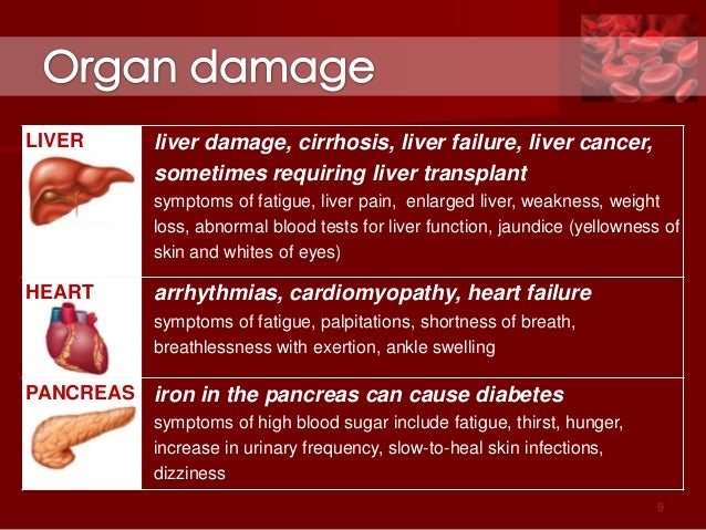
People with hereditary hemochromatosis may absorb up to 30 percent of the iron they consume. At this rate, the body cannot expel the extra iron fast enough, so it builds up. The body stores the excess in the tissues of major organs, mainly in the liver, and also the heart and the pancreas.

People with hemochromatosis may eventually build up between 5 and 20 times the amount of iron they should have. Over time this excess iron can destroy organs, resulting in organ failure and chronic diseases, such as cirrhosis, heart disease, and diabetes.

Primary hemochromatosis: A genetic mutation

Every living organism has genes. Genes are a set of instructions that decide what the organism is like, how it survives, and how it behaves in its environment. A mutation in one gene can change the way the body works.

[genetic test]
Genetic tests can show whether a person is likely to develop hemochromatosis and enable an early diagnosis.

HFE is the gene that controls the amount of iron we absorb. The two common mutations in the HFE gene are C282Y and H63D. In the U.S., most people with inherited hemochromatosis have inherited two copies of C282Y, one from the mother and the other from the father. Around 31 percent of people with two copies of C282Y develop symptoms by their early fifties.

A person who inherits just one gene with the C282Y mutation is not certain to develop iron overload syndrome, although they will probably absorb more iron than normal, and they will be a carrier.

If both parents are carriers, there is a 1 in 4 chance of inheriting two mutated genes, one from each parent. However, some people with two copies of the C2H2Y mutation never experience symptoms.

Some individuals may inherit one C282Y and one H63D mutation. A small proportion of these people will develop hemochromatosis symptoms.

Inheriting two copies of H63D is rare. Having two copies of the H63D mutation may increase the risk of developing hemochromatosis, but this is not confirmed.

Men with HFE defects can develop symptoms from the age of 40, but in women, symptoms normally appear after the menopause.

Treatment and complications

Venesection, or phlebotomy, is a regular treatment to remove iron-rich blood from the body. It is like donating blood, but the aim is to reduce iron levels to normal.

How much blood is taken and how often depends on the patient’s age, overall health, and the severity of the iron overload. Normally, this happens weekly until levels return to normal.

When iron levels build up again, the patient will need to repeat the treatment.

Phlebotomy cannot reverse the symptoms of cirrhosis, but it can improve symptoms such as nausea, abdominal pain, and fatigue. It may improve heart function and joint pain.

The amount of blood that can be taken depends on whether the patient is male or female, and their overall health condition. One unit a week is normally taken. One unit is one pint or around 473 milliliters.

Medications can include a drug that binds the excess iron before being excreted from the body.

The patient should have regular blood tests, and treatment normally continues for life.

Life expectancy is normal if hemochromatosis is diagnosed and treated early, before too much additional iron accumulates.

Complications

If hemochromatosis is left untreated, the accumulation of iron can lead to complications.

These include:

  • Cirrhosis, or permanent scarring of the liver, which can lead to serious and life-threatening complications
  • A higher chance of liver cancer in individuals with both cirrhosis and hemochromatosis
  • Diabetes and associated complications, such as kidney failure, blindness, and heart problems
  • Congestive heart failure, if too much iron builds up in the heart and the body cannot circulate enough blood to meet its needs
  • Irregular heart rhythms, or arrhythmias, leading to chest pain, palpitations and dizziness.
  • Endocrine problems, such as hypothyroidism and hypogonadism
  • Arthritis, osteoarthritis, and osteoporosis
  • Bronze or gray skin color due to the deposits of iron in skin cells

Other complications can include depression, gall bladder disease, and some cancers. Autopsies have indicated that some people with neurodegenerative diseases, such as Alzheimer’s, have had high iron levels.

To reduce iron absorption, people with the condition have been encouraged to avoid red meat and other foods rich in iron, but reducing the intake of dietary iron has not been shown to lower iron levels. However, patients are advised not to take iron tablets or use vitamin C supplements, as vitamin C enhances iron absorption.

A high sugar consumption may also increase absorption of iron. Calcium-rich foods and eggs and tea may inhibit iron absorption.

People with the condition should limit the alcohol they drink, because it will increase the risk of liver damage.

Polycythemia Vera: Causes, Symptoms & Treatments

What Is Polycythemia Vera?

It’s a blood cancer that begins in the marrow of your bones, the soft center where new blood cellsgrow. If you have polycythemia vera, your marrow makes too many red blood cells, which causes your blood to get too thick. That can make you more likely to have blood clots, a stroke, or a heart attack.

This disease develops very slowly, usually over many years. Although it can be life-threatening if you don’t get any treatment, most people have a good chance of living a long life when they get the right care.

Most people find out they have this condition when they’re 60 or older. But it can happen at any age. It’s more common among men than women.

Although you might have some warning signs like dizziness or feeling tired and weak, lots of things can cause those problems. If you have polycythemia vera, the first sign may be when a routine blood test shows that you have a high number of blood cells.

The treatment you get depends on your age and your particular case. If you don’t have a lot of symptoms, your doctor may just want to check you regularly without any treatment.

It’s natural to worry when you learn that you have cancer. But keep in mind that everyone is different and all cancers aren’t the same. With the support of your doctor, family, friends, and other people who have polycythemia vera, you’ll be in the best position to manage it.

Causes

You don’t “catch” polycythemia vera like you do a cold or the flu. It’s something you get because you have a gene called JAK2 that doesn’t work right. This gene is supposed to make sure your bone marrow doesn’t make too many blood cells.

Your bone marrow makes three types of blood cells: red, white, and platelets. Red blood cells carry oxygen, white fight infections, and platelets clot the blood to stop bleeding.

Most people with polycythemia vera have too many red blood cells. But the disease may also cause you to have too many white blood cells and platelets.

It’s rare, but parents can pass this broken gene to children. Most likely, the problem in your JAK2 gene happened over the course of your life.

Symptoms

At first, you may not notice any problems. When you do start having symptoms, you may have:

  • Headaches
  • Double vision or seeing dark or blind spots that come and go
  • Itching all over your body, especially after you’ve been in warm or hot water
  • Sweating, especially at night
  • Reddened face that looks like sunburn or blushing
  • Weakness
  • Dizziness
  • Weight loss
  • Shortness of breath
  • Tingling or burning in your hands or feet
  • Painful swelling of a joint

You may also feel pressure or fullness below the ribs on your left side. Those symptoms come from an enlarged spleen, which happens to some people. The spleen is an organ that helps filter your blood.

Without treatment, the extra red blood cells in your veins can cause blood clots that reduce your blood flow. This makes you more likely to have a stroke and heart attack. It can also cause pain (angina) in your chest.

Related image

Getting a Diagnosis

If your doctor thinks you might have polycythemia vera, you’ll get a physical exam, including a check of your spleen. He’ll also see if your face is unusually red.

  • Do you get a lot of headaches?
  • Have you lost weight lately?
  • Do you sometimes feel dizzy or weak?
  • Have you had shortness of breath?
  • Do you sweat a lot at night?

You may get some blood tests, too. These include:

Complete blood count (CBC). Your doctor takes a sample of your blood and sends it to a lab, where a machine counts the number of red blood cells, white blood cells, and platelets. An unusually high number of any of these could be a sign of polycythemia vera.

Blood smear. In this test, your doctor will look at a sample of your blood through a microscope. It’s a way of checking for other diseases that are sometimes linked to polycythemia vera.

EPO level. This test measures how much of the hormone EPO you have in your blood. EPO tells your bone marrow to make blood cells. People who have polycythemia vera have very low levels of it.

Treatment

Polycythemia vera varies from person to person. If you don’t have many symptoms, you might not need to start treatment yet. Your doctor will keep a close watch on your health.

If you do need treatment, the goal is to lower the number of red blood cells and prevent blood clots and other complications. Your options include:

Phlebotomy. This is often the first treatment for polycythemia vera. In this therapy, your doctor removes blood from your vein. It’s a lot like donating blood. The goal is to lower the number of your blood cells. After it’s done, your blood will be thinner and flow better. You’ll usually feel better, too. Some symptoms will get better, such as headaches or dizziness.

Your doctor will decide how often you need phlebotomy. For some people, it’s all they need for many years.

Low-dose aspirin keeps platelets from sticking together. That makes you less likely to get blood clots, which can cause heart attacks or strokes. Most people with polycythemia vera get this treatment.

Medicine to lower blood cells. If you need more than phlebotomy and aspirin, your doctor may prescribe hydroxyurea, a pill that lowers your red blood count and relieves symptoms.

Another drug,interferon alfa, helps the immune system cut back on making blood cells. The drug ruxolitinib(Jakafi) is approved for use in people who aren’t helped by hydroxyurea or can’t handle its side effects.

If you have a lot of itching that doesn’t go away, your doctor may prescribe antihistamines.

Thrombophilia : Causes, Symptoms & Treatments

Thrombophilia is a condition where the blood has an increased tendency to form clots. Blood clots can cause problems such as deep vein thrombosis (DVT) or pulmonary embolism. There are different types of thrombophilia – some are inherited and some are acquired, meaning they usually develop in adult life. Often thrombophilia is mild. Many people with thrombophilia do not have problems from their condition. Blood tests can diagnose the problem. Thrombophilia does not always require treatment but some people need to take aspirin or warfarin. If you have thrombophilia, be aware of the symptoms of a blood clot and get treatment immediately if you have symptoms.

Thrombophilia refers to a groups of conditions where the blood clots more easily than normal. This can lead to unwanted blood clots (called thromboses) forming within blood vessels. These blood clots can cause problems such as deep vein thrombosis (DVT) or pulmonary embolism. See separate leaflets called Deep Vein Thrombosis and Pulmonary Embolism for more details.
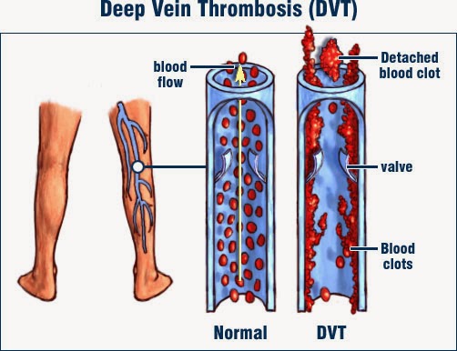
Image result for thrombophilia causes, symptoms & treatments

The body has a natural clotting process in the blood, which is altered in thrombophilia.

The normal clotting process is called haemostasis. It helps to stop bleeding if you have an injury such as a cut. When a blood vessel is injured, the clotting process is triggered. This is called the clotting cascade. It is a chain reaction of different chemicals in the blood which are called clotting factors. The clotting cascade makes the blood solidify into a clot, which sticks to the injured part of the blood vessel. Small particles in the blood, called platelets, also help to form the clot.

There are also natural chemicals in the blood which act against the clotting system, to stop the blood clotting too much.

Thrombophilia occurs if the normal balance of the clotting system is upset. There may be too much of a clotting factor, or too little of a substance that opposes clotting.

Thrombophilia can cause unwanted blood clots (thromboses). This does not mean that every person with thrombophilia will develop a blood clot. But, it means that you have a higher risk than normal of having clots. The extra risk will depend on the type of thrombophilia that you have.

A blood clot that forms within a blood vessel is known medically as a thrombus. Thrombosis is the process that occurs to form a thrombus. A thrombus can block a blood vessel – this blockage is now also known as a thrombosisThromboses is the plural version of thrombosis (that is, more than one).

Thrombophilias can be classified into inherited or acquired. The inherited ones are genetic and may be passed on from parent to child.

Acquired thrombophilias are not inherited, meaning they have nothing to do with your genes. Usually, acquired thrombophilias become apparent in adulthood. They can happen as a result of other medical problems that have developed, or they might be due to problems with the immune system.

It is possible to have a mixed thrombophilia, due partly to genetic and partly to non-genetic factors.

The different types of thrombophilia are explained in more detail later in this leaflet.

There are no symptoms unless the thrombophilia results in a blood clot (thrombosis).

Many people with thrombophilia do not develop a blood clot and have no symptoms at all.

What are the symptoms of blood clots?

Blood clots can form in arteries and veins. Arteries are blood vessels that take blood away from the heart to the organs and tissues of the body. Veins are blood vessels that bring blood back to the heart, from the rest of the body.

Diagram of leg showing veins and a Deep Vein Thrombosis

A blood clot in a vein is the most common problem with thrombophilia – this is called venous thrombosis. Possible symptoms are:

  • Pain and swelling in a leg. This occurs if you have a blood clot in a large vein in a leg. This is commonly known as a deep vein thrombosis (DVT). A DVT can occur in any vein in your body but most often affects a leg vein.
  • The blood clot may travel to the heart and on into a lung, causing a pulmonary embolism. Possible symptoms are chest pain, pain on deep breathing, shortness of breath or, rarely, collapse.
  • Some types of thrombophilia can cause a blood clot in an unusual site such as the brain, gut or liver. This can cause symptoms in the head or the tummy (abdomen). A blood clot in the liver veins is called Budd-Chiari syndrome.

A blood clot in an artery can occur with some types of thrombophilia. This is called arterial thrombosis. Depending on which artery is affected, a blood clot in an artery can cause a stroke, a heart attack or problems with the placenta during pregnancy. So the possible symptoms of arterial thrombosis due to thrombophilia are:

  • Having a stroke at a relatively young age.
  • Repeated miscarriages.
  • Pregnancy problems: pre-eclampsia, reduced fetal growth or, rarely, fetal death (a stillbirth, or intrauterine death).
  • A heart attack.

It is important to remember that all of these conditions can be due to causes other than thrombophilia. For example, smoking, high blood pressure, diabetes and high cholesterol are the main risk factors for developing problems such as heart attack and stroke.

For this reason, not everyone with a stroke or heart attack is tested for thrombophilia, as it is not a common cause.

Thrombophilia is diagnosed by blood tests.

Tests are done some weeks or months after having a DVT or pulmonary embolism, as the presence of these conditions can affect the results. Usually you have to wait until you have been off blood-thinning medication (anticoagulants), such as warfarin, for 4-6 weeks. If you have recently been pregnant, the tests may have to be delayed by eight weeks. This is because the results in pregnancy can be much harder to understand.

A sample of blood is taken and a number of different tests will be done on it, to check different parts of the clotting process. Usually, the tests are done in two stages. The first test is a thrombophilia screen which is some basic clotting tests. If the results of this suggest that thrombophilia is possible then another blood sample will be taken for more detailed tests.

You may be referred to a doctor specialising in blood conditions (a haematologist). The doctor will usually ask about your history and family history. This will help with interpreting the test results.

The first step is for you and your doctor to consider how much risk there is of you developing a blood clot. This risk depends on a combination of things, such as:

  • What type of thrombophilia you have (some are more high-risk for blood clot than others).
  • Your age, weight, lifestyle and other medical conditions.
  • Whether you are pregnant or have recently given birth.
  • Whether you have already had a blood clot.
  • Your family history – whether any close relatives have had a blood clot.

This information will help your doctor to assess how much risk you have of developing a blood clot and what type of blood clot could occur. Then you and your doctor can discuss the pros and cons of taking treatment and, if needed, what type of treatment to take.

Possible treatments for thrombophilia are:

Low-dose aspirin

Low-dose aspirin inhibits the action of platelets, so can help to prevent blood clots. It may also help prevent miscarriage or pregnancy problems, in some types of thrombophilia.

Anticoagulant treatment

Anticoagulation is often called thinning the blood. However, it does not actually thin the blood. It alters certain chemicals in the blood to stop blood clots forming so easily – in effect, it slows down the clotting process. It doesn’t dissolve a blood clot either (as some people incorrectly think). The body’s own healing mechanisms can then get to work to break up any existing blood clot.

Anticoagulation can greatly reduce the chance of a blood clot from forming. Anticoagulant medication is commonly used to treat a venous thrombosis (such as a DVT) or a pulmonary embolism.

In thrombophilia, anticoagulant medication may be advised if:

  • You have had a blood clot, to prevent another one.
  • You have not had a blood clot but have a high risk of developing one.
  • You have a temporary situation that puts you at high risk of a blood clot. This may be the case if you are pregnant, within six weeks after childbirth, or are immobile for a long period.

Anticoagulant medicines are either given by injection (eg, heparin) or can be taken as a tablet. Warfarin is the most commonly used tablet anticoagulant medicine. Other anticoagulants taken in tablet form include apixaban, edoxaban, dabigatran and rivaroxaban.

Warfarin is the usual anticoagulant. However, it takes a few days for warfarin tablets to work fully. Therefore, heparin injections (often given just under the skin) are used alongside warfarin in the first few days (usually five days) for immediate effect if you currently have a blood clot. If you are starting warfarin and don’t have a blood clot (that is, it is just to prevent one), you won’t need heparin injections first.

The aim is to get the dose of warfarin just right so the blood will not clot easily. Too much warfarin may cause bleeding problems. To get the dose right, you will need a regular blood test, called International Normalised Ratio (INR), whilst you take warfarin. The dose is adjusted on an individual basis according to the result of this blood test. The INR is a blood test that measures your blood clotting ability. You need the tests quite often at first but then less frequently once the correct dose is found.

An INR of 2.5 is usually the aim if you take warfarin to prevent a blood clot in thrombophilia or to treat a DVT or pulmonary embolism. However, anywhere in the range 2-3 is usually OK. If you have had recurrent DVTs, or have had a pulmonary embolism whilst on warfarin, you might need a higher INR (even ‘thinner’ blood). INR blood tests can usually be done in an outpatient clinic, or sometimes by your GP. You may be advised to take warfarin on a lifelong basis to prevent blood clots if you have thrombophilia. Or, you may have short-term treatment for your current DVT or pulmonary embolism (usually 3-6 months).

Heparin is an injectable anticoagulant. Standard heparin is given intravenously (IV), which means directly into a vein – usually in the arm. This type of heparin is given in hospital and monitored with blood tests.

Low molecular weight heparin (LMWH) is injected into the skin on the lower tummy (abdomen). It can be given at home, either by a district nurse, or you can be taught to self-inject (or a carer can do it for you). It does not need blood tests to monitor it. Different doses are used for prevention (prophylaxis) and treatment of an existing blood clot. There are different brands of heparin injection; the common ones you might see used are Clexane® and Fragmin®.

Treatment in pregnancy

If you are pregnant or planning a pregnancy, discuss this with your doctor, and tell your midwife and doctor specialising in pregnancy and childbirth (obstetrician) about the thrombophilia. Treatment for thrombophilia may be different in pregnancy because:

  • Some women with certain types of thrombophilia are advised to take low-dose aspirin while pregnant, to help prevent miscarriage or pregnancy problems.
  • The pregnancy itself increases the risk of a venous thrombosis – this applies to the whole pregnancy and especially to the six weeks after childbirth. So you may be advised to start anticoagulant treatment while pregnant or after childbirth. This will depend on the type of thrombophilia and your medical history.
  • If you were taking warfarin, you will normally be advised to change to heparin instead. This is because heparin is safer for the unborn baby (there is a significant chance that warfarin could cause fetal abnormalities). Both heparin and warfarin are safe for breast-feeding.

Prevention of DVT

Certain situations can temporarily put you at high risk of having a blood clot, and in these situations you may be advised to take extra treatment for a while. Examples are pregnancy and after childbirth, severe illness, major surgery, or anything which immobilises you, such as travel or an operation. Special stockings such as flight socks or compression stockings may also be advised to help prevent a DVT.

General advice for people with thrombophilia

  • If you are having any medical treatment or surgery, tell your doctor/nurse/pharmacist about the thrombophilia.
  • Be aware of the warning symptoms of a blood clot – obtain medical help immediately if you suspect one (see above for symptoms).
  • Avoid lack of fluid in the body (dehydration) by drinking adequate amounts of fluid. Dehydration can contribute to blood clots forming.
  • Keep active, and avoid being immobile for long periods – immobility helps cause blood clots in the legs (DVTs).
  • Caution with medication: some medications increase the risk of a blood clot. For example, the combined oral contraceptive (COC) pill or patch and hormone replacement therapy (HRT). You may be advised to avoid certain medications, or to change to one which does not affect blood clotting.
  • Keep to a healthy weight – being overweight or obese increases the risk of blood clots in legs.
  • To keep blood vessels healthy (arteries in particular), do not smoke. This is important if you have thrombophilia of a type that can cause blood clots in arteries, as smoking also promotes arterial blood clots.

Hemoglobin Deficiency : Causes, symptoms & Treatments

Hemoglobin C, S-C, and E diseases are inherited conditions characterized by abnormally shaped red blood cells and chronic anemia that is caused by excessive destruction of red blood cells. 

Hemoglobin C, S, and E are abnormal forms of hemoglobin (the protein in red blood cells that carries oxygen). These abnormal forms of hemoglobin result from inheriting an abnormal gene.

Hemoglobin C disease occurs mostly in blacks. One copy of the gene that causes hemoglobin C disease is present in 2 to 3% of blacks in the United States. However, people must inherit two copies of the abnormal gene to develop the disease. In general, symptoms are few. Anemia varies in severity. People who have this disease, particularly children, may have episodes of abdominal and joint pain, an enlarged spleen, and mild jaundice, but they do not have severe crises, as occur in sickle cell disease.

Hemoglobin S-C disease occurs in people who have one copy of the gene for sickle cell disease and one copy of the gene for hemoglobin C disease. Hemoglobin S-C disease is more common than hemoglobin C disease, and its symptoms are similar to those of sickle cell disease but milder. However, people may have blood in the urine.

Hemoglobin E disease affects primarily people of Southeast Asian descent. This disease causes anemia but none of the other symptoms that occur in sickle cell disease and hemoglobin C disease.

Diagnosis and Treatment

Blood tests, including hemoglobin electrophoresis, are done for diagnosis. In electrophoresis, an electrical current is used to separate the different types of hemoglobin and thus detect abnormal hemoglobin.

Treatment varies depending on the symptoms and their severity. Some people do not need treatment, but when treatment is needed, it is similar to that of sickle cell disease.

Anemia from excessive bleeding results when loss of red blood cells through bleeding exceeds production of new red blood cells.

  • When blood loss is rapid, blood pressure falls, and people may be dizzy.


  • When blood loss occurs gradually, people may be tired, short of breath, and pale.
  • Stool, urine, and imaging tests may be needed to determine the source of bleeding.
  • The cause of bleeding is corrected, and transfusions and iron supplements are given if needed.

Excessive bleeding is the most common cause of anemia. When blood is lost, the body quickly pulls water from tissues outside the bloodstream in an attempt to keep the blood vessels filled. As a result, the blood is diluted, and the hematocrit (the percentage of red blood cells in the total blood volume) is reduced. Eventually, increased production of red blood cells by the bone marrow may correct the anemia. However, over time, bleeding reduces the amount of iron in the body, so that the bone marrow is not able to increase production of new red blood cells to replace those lost.

Image result for blood related diseases

Rapid blood loss

The symptoms may be severe initially, especially if anemia develops rapidly from a sudden loss of blood, such as from an injury, surgery, childbirth, or a ruptured blood vessel. Losing large amounts of blood suddenly can create two problems:

  1. Blood pressure falls because the amount of fluid left in the blood vessels is insufficient.
  2. The body’s oxygen supply is drastically reduced because the number of oxygen-carrying red blood cells has decreased so quickly.
  3. Either problem may lead to a heart attack, stroke, or death.

Chronic blood loss

Far more common than a sudden loss of blood is long-term (chronic) bleeding, which may occur from various parts of the body. Although large amounts of bleeding, such as that from nosebleeds and hemorrhoids are obvious, small amounts of bleeding may not be noticed. For example, a small amount of blood may not be visible in the stool. This type of blood loss is described as occult. If a small amount of bleeding continues for a long time, a significant amount of blood may be lost. Such gradual bleeding may occur with common disorders, such as ulcers in the stomach or small intestine and diverticulosis, polyps, or cancers in the large intestine. Other sources of chronic bleeding include kidney or bladder tumors, which may cause blood to be lost in the urine, and heavy menstrual bleeding.

Symptoms and Diagnosis

Symptoms are similar to those of other types of anemia and vary from mild to severe, depending on how much blood is lost and how rapidly. When the blood loss is rapid—over several hours or less—loss of just one third of the blood volume can be fatal. Dizziness upon sitting or standing after a period of lying down (orthostatic hypotension) is common when blood loss is rapid. When the blood loss is slower—over several weeks or longer—loss of up to two thirds of the blood volume may cause only fatigue and weakness or no symptoms at all, if the person drinks enough fluids.

Computer Vision Syndrome: Causes, Symptoms & Treatments

These days, many of us have jobs that require us to stare at computer screens for hours at a time. That can put a real strain on your eyes.Eye problems caused by computer use fall under the heading computer vision syndrome (CVS). It isn’t one specific problem. Instead, it includes a whole range of eye strain and pain. Research shows that between 50% and 90% of people who work at a computer screen have at least some symptoms.Working adults aren’t the only ones affected. Kids who stare at tablets or use computers during the day at school can have issues, too, especially if the lighting and their posture are less than ideal.

How Do Computers Affect Vision?

Image result for computer vision syndrome causes, symptoms & TREATMENTS

CVS is similar to carpal tunnel syndrome and other repetitive motion injuries you might get at work. It happens because your eyes follow the same path over and over. And it can get worse the longer you continue the movement.

When you work at a computer, your eyes have to focus and refocus all the time.  They move back and forth as you read. You may have to look down at papers and then back up to type. Your eyes react to changing images on the screen to create so your brain can process what you’re seeing. All these jobs require a lot of effort from your eye muscles. And to make things worse, unlike a book or piece of paper, the screen adds contrast, flicker, and glare.

You’re more likely to have problems if you already have eye trouble, if you need glasses but don’t have them, or if you wear the wrong prescription for computer use.

Computer work gets harder as you age and the lenses in your eyes becomes less flexible. Somewhere around age 40, your ability to focus on near and far objects will start to go away. Your eye doctor will call this condition presbyopia.

Image result for computer vision syndrome causes, symptoms & TREATMENTS

Symptoms:

There’s no proof that computer use causes any long-term damage to the eyes. But regular use can lead to eye strain and discomfort.

You may notice:

  • Blurred vision
  • Double vision
  • Dry, red eyes
  • Eye irritation
  • Headaches
  • Neck or back pain

If you don’t do anything about them, it could affect more than your eyes. You could also have issues with your work performance.

Image result for computer vision syndrome causes, symptoms & TREATMENTS

Treatments:

A few simple changes to your workspace can improve your symptoms and prevent new problems:

Syncope: Causes, Symptoms & Treatments

What is syncope?

[Woman fainting]
Fainting can sometimes be a sign of a more serious illness, or nothing to worry about at all.

Sometimes syncope may be just that – a fainting episode with no medical importance. On some occasions, however, it may be caused by a serious illness, condition or disorder. Every case of fainting should be treated as a medical emergency until the cause is known and signs and symptoms have been treated. Anybody who has recurring fainting episodes should contact their doctor.

If oxygen levels are below 16% at atmospheric pressure most people faint due to hypoxia. If oxygen levels fall below 11% individuals may die by suffocation. The amount of oxygen in the air depends on its partial pressure – inhaling pressurized gas while scuba diving which is below 16% oxygen does not cause hypoxia (because the air is pressurized).

Syncope due to hypoxia may also be caused by malfunctioning lungs, problems with blood circulation, or carbon monoxide poisoning. Some people faint at the sight of blood, or when receiving an injection or seeing somebody having one.

In Victorian England (19th Century) fainting in women was a commonplace stereotype, as well as modern portrayals of that period. Some believe that the respiratory effects of tight corsets worn at the time may have been a contributory factor. However, during Victorian times aristocratic women were encouraged to display a feminine frailty by fainting at dramatic moments.

Children sometimes play a game (fainting game) in which they deliberately restrict blood flow to the brain in order to trigger syncope – this is dangerous and may cause brain damage, and even death.

Near-syncope vs syncope

  • Pre- or near-syncope (a pre- or near-syncoptic episode) – this is when the person can remember events during the loss of consciousness, such as dizziness, blurred vision, muscle weakness, as well as the fall before hitting their head and losing consciousness.
  • Syncope (a syncoptic episode) – this is when the individual may remember the feelings of dizziness and loss of vision, but not the fall.

Related image

Symptoms of syncope

The hallmark sign is evident to anyone around – the patient passes out, faints, suddenly loses consciousness.

The following signs and symptoms may precede a fainting episode:

  • A feeling of heaviness in the legs
  • Blurred vision
  • Confusion
  • Feeling warm or hot
  • Lightheadedness, dizziness, a floating feeling
  • Nausea
  • Sweating
  • Vomiting
  • Yawning.

When a person faints, the following signs may be evident:

  • The individual may be falling over
  • The patient may be slumping
  • The person may be unusually pale
  • There may be a drop in blood pressure
  • There may be a weak pulse.

Image result for Sleep syncope causes, symptoms treatments

What to do if somebody feels faint

You feel faint:

  • Don’t stay standing up. Find somewhere to either sit or lie down.
  • If you manage to sit down place your head between your knees.
  • When you do get up, do so slowly.

If you see somebody fainting:

  • Place the patient on his/her back, facing up
  • If the individual is breathing, raise their legs about 12 inches (30 centimeters) above heart level to restore blood flow to the brain
  • Try to loosen all belts, ties, collars and restrictive clothing
  • When the person comes round do not let them get up too quickly
  • If they remain unconscious for more than about a minute, put the patient into the recovery position and get emergency medical help
  • Check the patient’s airway for any obstruction and check for vomiting
  • Check for breathing, coughing or movement (signs of circulation). If you cannot detect any, start CPR (cardiopulmonary resuscitation). Continue administering CPR until either help arrives or the patient starts breathing on his/her own
  • If the individual was injured and is bleeding after the fall, apply direct pressure to control the bleeding.

Treating syncope

If the fainting is found to be caused by an underlying health condition, that will need to be treated, which should help prevent syncoptic episodes.

  • Treating neurocardiogenic syncope – in the majority of cases no further treatment is needed. Avoid the triggers, such as long periods standing still, dehydration, and being in hot and stuffy places. This may help prevent future episodes.
  • Injections and blood – if the sight or thought of injections or blood make a person feel faint, they should tell the doctor or nurse beforehand. The health care professional can then make sure the patient is in a safe position (lying down) before any procedure begins.
  • Beta-blockers – this medication is primarily used for the treatment of high blood pressure (hypertension). However, it may help patients whose neurocardiogenic syncope interferes with their quality of life. The side effects of beta-blockers may include fatigue, cold extremities (hands and feet), slow heartbeat (pulse rate), nausea, and diarrhea. Although rare, the following side effects are also possible – erectile dysfunction(impotence, problems either getting or sustaining an erection), nightmares, and sleep disturbances.
Pages:123