Global Treatment Services Pvt. Ltd.

Global Treatment Services

Pancreatic Cancer : Causes, Symptoms & Treatments

Pancreatic Cancer

Pancreatic cancer is a disease in which malignant (cancerous) cells form in the tissues of the pancreas. The pancreas is a gland located behind the stomach and in front of the spine. The pancreas produces digestive juices and hormones that regulate blood sugar. Cells called exocrine pancreas cells produce the digestive juices, while cells called endocrine pancreas cells produce the hormones. The majority of pancreatic cancers start in the exocrine cells.

Symptoms:

Early pancreatic cancers usually cause few symptoms, most of which are vague. Because signs and symptoms of most pancreatic cancer may be mistaken for less-serious digestive problems, the disease is rarely detected before it has spread to nearby tissues or distant organs via the bloodstream or lymphatic system. Symptoms that may arise include:

  • Significant weight loss accompanied by abdominal pain — the most likely warning signs.
  • Vague but gradually worsening abdominal pain that may decrease when leaning forward and increase when lying down. Pain is often severe at night and may radiate to the lower back.
  • Digestive or bowel complaints such as diarrhea, constipation, gas pains, bloating, or belching.
  • Nausea, vomiting, and loss of appetite.
  • Jaundice, which is usually painless and is indicated by yellowish discoloration of the skin and eye whites, very dark urine, and light-colored stools.
  • Sudden onset of glucose tolerance disorder, such as diabetes.
  • Black or bloody stool, indicating bleeding from the digestive tract.
  • Overall weakness.
  • Enlarged liver and gallbladder.
  • Itching.
  • Blood clots in the legs.
  • Mental status changes, such as a new onset of depression.

  Causes:

A few rare types of pancreatic cancer cause hormonal imbalances that produce their own symptoms, which might include:

  • Episodes of weakness, sweating, rapid heartbeat, irritability, or skin flushing related to low blood sugar
  • Severe watery diarrhea
  • A new, unusual skin rash
  • Severe gastrointestinal symptoms, such as stomach pain and watery diarrhea, which do not respond to antacids or ulcer medications

If you experience any of these symptoms, call your doctor for a full physical exam.

How Is Pancreatic Cancer Diagnosed?

To diagnose pancreatic cancer, a doctor will order certain imaging tests, such as a pancreatic ultrasound or a CT scan of the abdomen. Endoscopic ultrasonography (EUS) uses an ultrasound device connected to the end of a small flexible tube that is inserted into the mouth and is about 85% to 90% accurate in diagnosing pancreatic cancer. If necessary, endoscopic retrograde cholangiopancreatography (ERCP) is used. With ERCP, detailed images are obtained by inserting an endoscope into the mouth to the pancreas, injecting a dye, and then taking X-rays. A tissue sample for biopsy can also be extracted through the scope. If a biopsy confirms cancer, further tests are done to determine how far the disease has advanced. Laparoscopy, may be used. In this technique, a small tube with a small video camera and light source is introduced into the abdominal cavity. The tumor can then be seen. Occasionally, exploratory surgery is needed. The surgeon can then study the tumor directly, determine if nearby lymph nodes are cancerous, and take tissue samples for microscopic exam.

What Are the Treatments for Pancreatic Cancer?

Pancreatic cancer is very hard to control. But if it is caught early and the cancer hasn’t spread beyond the pancreas, it can be treated with surgery. This offers the best outcome for pancreatic cancer. The surgery is called a “Whipple procedure,” or pancreaticoduodenectomy, and is named after Dr. George Hoyt Whipple, the surgeon who pioneered it. If possible, the surgeon removes the malignant tumor, leaving as much of the normal pancreas as possible to allow continued pancreatic function. Less often, the entire pancreas must be removed. If a patient undergoes a total pancreatectomy, a lifelong regimen of replacement enzymes and hormones, including insulin, must be administered.

Unfortunately, pancreatic cancers have subtle, and vague symptoms. Thus the disease is often diagnosed after it has advanced and spread. However, even at later stages, treatment can improve quality of life by controlling uncomfortable symptoms and complications of the disease.

Depending on the type and stage of pancreatic cancer, patients may be given chemotherapy treatments alone or in combination with radiation. Whether or not the tumor is removed surgically, or has spread to lymph nodes, these therapies may be given after surgery in an effort to extend survival time. These treatments may also be given before surgery to shrink the tumor and make it possible for surgery to be performed or used as a means of relieving symptoms, such as pain. Prescription medications, usually narcotics, are given to help manage pain associated with advanced pancreatic cancer.


Mitral Valve Stenosis: Causes, Symptoms & Treatments

Mitral valve stenosis is when the mitral valve opening in the heart is narrowed, usually due to rigidity or scarring.The mitral valve is located on the left side of your heart, between two chambers: the atrium and the ventricle.Mitral valve stenosis commonly leads to shortness of breath, especially during exercise or when lying down.

The mitral valve is located on the left side of your heart, between two chambers: the atrium (upper) and the ventricle (lower). Blood is pumped from the left atrium, through the mitral valve, and into the left ventricle on its way to the aorta. The healthy mitral valve allows blood to pass through, but prevents it from flowing back.

Mitral valve stenosis, also known as mitral stenosis, is when the mitral valve opening is narrowed. This means that not enough blood can flow through it. This can lead to a variety of issues, including fatigue, difficulty breathing, blood clots, and heart failure.

 Scarring caused by rheumatic fever is the leading cause of mitral valve stenosis.
Mitral valve stenosis is typically caused by rheumatic fever. This is usually a childhood disease, and it results from the body’s immune response to an infection with the streptococcal bacteria. It is a serious complication of strep throat or scarlet fever.

The body’s organs most affected by the acute rheumatic fever are the joints and the heart. The joints can become very inflamed and can lead to temporary and sometimes chronic disability. The lining of the heart (endocarditis), the heart muscle (myocarditis), and the membrane surrounding the heart (pericarditis) can become inflamed.

When the mitral valve (or any of the heart valves) becomes involved, this leads to a chronic heart condition called rheumatic heart disease. The clinical signs and symptoms of this condition might not occur until 5 to 10 years after the episode of rheumatic fever.

The factors that make some people more susceptible than others to mitral valve stenosis are unclear, but it is estimated that women are more at risk than men.

A congenital heart defect may cause mitral valve stenosis in a baby. Babies born with this condition usually need surgery.

In rare cases, calcium may build up and lead to narrowing of the mitral valve. Other, more rare causes for mitral valve stenosis include:

  • tumors
  • blood clots
  • radiation treatments
  • calcium buildup on valves
  • congenital heart defects

What Are the Symptoms of Mitral Valve Stenosis?

Mitral valve stenosis commonly leads to shortness of breath, especially during exercise or when lying down.

Other common symptoms include:

  • cough, with or without blood
  • chest pain, or chest discomfort
  • fatigue
  • swelling in ankles and/or feet
  • respiratory infections
  • plum-colored cheeks

If mitral valve stenosis is severe, you may feel your heart fluttering or beating rapidly.

Rarely, you may feel discomfort in your chest. Your chest might feel tight or constricted, or you may feel pain that radiates outward from your chest.

In some cases, mitral valve stenosis may not cause any symptoms, or the symptoms may appear only during exercise. You might develop symptoms when your body undergoes stress such as during an infection or pregnancy.

In addition to the common symptoms, children with this issue might experience slower growth.

How Is Mitral Valve Stenosis Treated?

Treatment for mitral valve stenosis can vary greatly, depending on your symptoms and the severity of the condition. If you have no symptoms and only mild mitral valve stenosis, you might not need any treatment.

Drugs and Medication

If your mitral valve stenosis is causing symptoms, your doctor might prescribe medications. Although these do not actually fix the problem with your mitral valve, they can help treat your symptoms. Types of medications your doctor might prescribe include:

  • anticoagulants (blood thinners)
  • diuretics (to reduce fluid buildup through increased urine output)
  • antiarrhythmics (medications to treat abnormal heart rhythms)
  • beta-blockers (medications to slow your heart rate)

Valvuloplasty

Your doctor may choose to perform a mitral balloon valvuloplasty. If you need more treatment than just medication, but your mitral valve is not damaged enough to require heart surgery, this procedure is an option. This involves threading a tube (catheter) with a balloon attached to it through a vein and into your heart. Once in the mitral valve, the doctor inflates the balloon to expand the valve. In some cases, you may need to undergo this procedure more than once.

Surgery

Surgery may become necessary. Your doctor might be able to surgically repair your existing mitral valve to make it function properly. If that isn’t possible, you may need to have your mitral valve replaced with a new one. The replacement might be biological or mechanical.

Finger Infection: Causes & Symptoms

What Causes a Finger Infection?

Bacteria cause most types of finger infections. The exception to this is the herpetic whitlow, which is caused by a virus. How the infection starts and is found in a particular location is what makes each specific type of infection unique. Usually, some form of trauma to the finger or hand is the initial event. This may be a cut, animal bite, or puncture wound.

  • Paronychia: The offending bacteria are usually staphylococcal and streptococcal organisms. Rarely, a fungus causes this infection, which usually begins as a hangnail. Often a person will attempt to bite off the piece of nail that is at the corner of the nail, leading to local infection. This infection can also be caused by aggressively shoving back or trimming cuticles. This results in an open wound that allows the bacteria on the skin and the bacteria in the mouth to infect the wound. The infection can then spread to the surrounding tissue next to the nail and cuticle.
  • Felon: This bacterial infection of the finger pad, caused by the same organisms that cause paronychia, is usually the result of a puncture wound. The wound allows the introduction of bacteria deep into the fingertip pad. Because the fingertip has multiple compartments, the infection is contained in this area.
  • Herpetic whitlow: The offending viral organism is the herpessimplex virus type I or II. This is the same virus that causes oral or genital herpes infections. People in certain occupations are more at risk for this infection. These include dentists, hygienists, physicians, nurses, or any other person who may have contact with saliva or body fluids that contain the virus. People with oral or genital herpes may also infect their own fingers.
  • Cellulitis: The most common causes of this bacterial infection are staphylococcal and streptococcal organisms. This infection is usually the result of an open wound that allows the bacteria to infect the local skin and tissue. The infection can also spread to the hand and fingers by blood flow carrying the organisms.
  • Infectious flexor tenosynovitis: This bacterial infection is usually the result of penetrating trauma that introduces bacteria into the deep structures and tendon sheaths, which allows the spread along the tendon and associated sheath.
  • Deep space infection: This bacterial infection is usually the result of a puncture wound or deep cut that introduces the bacteria to the deep tissue. The collar button abscess is associated with the web space between the fingers. The deep structures of the hand create many potential compartments for this infection to invade. 

What Are Risk Factors for Finger Infections?

There are several risk factors for developing finger infections, including the following:

  • Puncture wounds
  • Deep cuts
  • Any open wound
  • Penetrating trauma
  • Picking hangnails
  • Nail biting
  • Aggressive manicuring or cuticle trimming

Some individuals may be at higher risk for finger infections, including

  • people with diabetes;
  • people with compromised immune systems;
  • people in certain occupations, such as dentists, hygienists, physicians, and nurses; and
  • people in occupations involving extended exposure of the hands to water (such as dishwashers).

What Are Symptoms and Signs of a Finger Infection?

Each of the main finger infections has specific signs and symptoms that make identification unique and may cause confusion if not properly evaluated.

  • Paronychia: Symptoms and signs of paronychia include redness and swelling of the area next to the fingernail. A visible collection of pus may be seen under the skin and nail. This fluid may be leaking out of the wound. The area will be tender and painful to the touch. The drainage from the area is usually a cloudy white-yellow color.
  • Felon: Symptoms of a felon include a swollen and painful fingertip. The swelling usually develops over several days and is located in the pad area of the fingertip. The area will have a throbbing pain and be painful to the touch. The area is usually red, and a visible collection of pus may be seen under the skin. The swollen area may have a portion that feels soft as if it contains fluid. As the swelling continues, the area may become tense or hard to the touch.
  • Herpetic whitlow: Symptoms of herpetic whitlow include redness and tenderness of the fingertip area. A burning or itching sensation may be present in the area. There may be mild swelling but not as extensive as in the felon. There may be a single or many open wounds in the area affected. These open wounds often occur in clusters after the formation of a small blister-like lesion. The fluid in these lesions is usually clear in appearance but may be slightly cloudy. The affected individual may also have a low-grade fever and have swollen and tender lymph nodes in the area.
  • Cellulitis: Symptoms of cellulitis include skin area redness and warmth to the touch. The area may be slightly swollen and tender. This is usually a superficial infection so the deep structures should not be involved. The motion of the fingers and hand should not be difficult or painful. If painful or difficult, this may indicate a deep space infection of some type.
  • Infectious flexor tenosynovitis: Four major signs often are found with infectious flexor tenosynovitis.
    • First is tenderness over the flexor or palm side of the finger. This pain is found over the tendons in the finger.
    • Second is uniform swelling of the finger.
    • Third is pain on extending or straightening of the finger.
    • Fourth, the finger will be held in a slightly flexed or partially bent position.

These signs are called Kanavel cardinal signs. All four signs may not be present at first or all at once.

  • Deep space infections: The deep space infection that arises in the web space of the fingers is also called a collar button abscess. Symptoms and signs of deep space infections include pain and swelling in the space between the fingers. The area may also be red and warm to the touch. As the abscess becomes larger, the fingers will be slightly spread apart by the increasing pressure. The central area may have a soft spot that represents a collection of pus under the skin. 

Premature Ventricular Contractions: Causes, symptoms & treatments

Premature ventricular contractions (PVCs) are extra, abnormal heartbeats that begin in one of your heart’s two lower pumping chambers (ventricles). These extra beats disrupt your regular heart rhythm, sometimes causing you to feel a flip-flop or skipped beat in your chest. Premature ventricular contractions are very common — they occur in most people at some point.

Premature ventricular contractions are also called:

  • Premature ventricular complexes
  • PVCs
  • Ventricular premature beats
  • Extrasystoles

If you have occasional premature ventricular contractions, but you’re an otherwise healthy person, there’s generally no reason for concern, and no treatment is needed. If you have frequent premature ventricular contractions or underlying heart disease, you may need treatment to help you feel better and treat underlying heart problems.

Symptoms:

Premature ventricular contractions often cause no symptoms. But you may feel an odd sensation in your chest, such as:

  • Flip-flops
  • Fluttering
  • Pounding or jumping
  • Skipped beats or missed beats
  • Increased awareness of your heartbeat

When to see a doctor

If you feel flip-flops, a sensation of skipped heartbeats or odd feelings in your chest, talk to your doctor. You’ll want to identify the source of these symptoms. Premature ventricular contractions may be the problem, but other conditions also may be to blame, including other rhythm problems, serious heart problems, anxiety, anemia or infections.

Causes:

Your heart is made up of four chambers — two upper chambers (atria) and two lower chambers (ventricles). The rhythm of your heart is normally controlled by the sinoatrial node (SA node) — or sinus node — an area of specialized cells located in the right atrium.

This natural pacemaker produces the electrical impulses that trigger the normal heartbeat. From the sinus node, electrical impulses travel across the atria to the ventricles, causing them to contract and pump blood out to your lungs and body.

Premature ventricular contractions are abnormal contractions that begin in the ventricles. These extra contractions usually beat sooner than the next expected regular heartbeat. And they often interrupt the normal order of pumping, which is atria first, then ventricles. As a result, the extra, out-of-sync beats are usually less effective in pumping blood throughout the body.

Why do extra beats occur?

The reasons aren’t always clear. Certain triggers, heart diseases or changes in the body can make cells in the ventricles electrically unstable. Underlying heart disease or scarring may also cause electrical impulses to be misrouted.

Premature ventricular contractions may be associated with:

  • Chemical changes or imbalances in the body
  • Certain medications, including common asthma medications
  • Alcohol or illegal drugs
  • Increased levels of adrenaline in the body that may be caused by caffeine, tobacco, exercise or anxiety
  • Injury to the heart muscle from coronary artery disease, congenital heart disease, high blood pressure or heart failure

Risk factors:

The following stimulants, conditions and triggers may increase your risk of premature ventricular contractions:

  • Caffeine, tobacco and alcohol
  • Exercise
  • High blood pressure (hypertension)
  • Anxiety
  • Underlying heart disease, including congenital heart disease, coronary artery disease, heart attack, heart failure and a weakened heart muscle (cardiomyopathy)

Mitral Valve Stenosis: Causes, Symptoms & Treatments

What is mitral valve stenosis?

Mitral valve stenosis is a heart problem in which the mitral valve doesn’t open as wide as it should. The valve becomes stiff or scarred, or the valve flaps become partially joined together.

See a picture of mitral valve stenosis .

Mitral valve stenosis can lead to heart failure; a stroke; an infection in the heart (endocarditis); or a fast, slow, or uneven heartbeat (arrhythmia). Fortunately, mitral valve stenosis can be treated.

How does the mitral valve work?

Your heart has four chambers and four valves. The valves have flaps, or leaflets. The flaps open and close to keep blood flowing in the proper direction through your heart.The mitral valve connects the heart’s upper left chamber (left atrium) to the lower left chamber (left ventricle). When the heart pumps, blood forces the flaps open, and blood flows from the left atrium to the left ventricle. Between heartbeats, the flaps close tightly so that blood does not leak backward through the valve.With mitral valve stenosis, not as much blood can flow into the left ventricle. More blood stays in the left atrium, and blood may back up into the lungs.

See a picture of a normal mitral valve .

card_02.jpg

What causes mitral valve stenosis?

Nearly all cases of mitral valve stenosis are caused by rheumatic fever. This fever results from an untreated strep infection, most often strep throat. But many people who have mitral valve stenosis don’t realize that they had rheumatic fever.

What are the symptoms?

Mitral valve stenosis is a lifelong disease, but symptoms may not happen for many years. When symptoms first appear, they usually are mild. You may only have a few symptoms, even if your mitral valve is very narrow. An early symptom is shortness of breath when you are active. This shortness of breath may seem normal to you.

Symptoms later in the disease may include:

  • Shortness of breath even when you have not been very active or when you are resting.
  • Feeling very tired or weak.
  • Pounding of the heart (palpitations).

How is mitral valve stenosis diagnosed?

Mitral valve stenosis may not be diagnosed until you’ve had the disease for some time. If you don’t have symptoms, the first clue might be a heart murmur your doctor hears during a routine checkup.

Your doctor will ask you questions about your past health and do a physical exam. If your doctor thinks you might have the disease, he or she may do more tests. These may include:

  • An echocardiogram. This ultrasound test lets your doctor see a picture of your heart, including the mitral valve.
  • An electrocardiogram (EKG or ECG). This test can check for problems with your heart rhythm.
  • A chest X-ray. This shows your heart and lungs and can help your doctor find the cause of symptoms such as shortness of breath.

These tests also help your doctor find what caused the stenosis and how severe it is.

How is it treated?

Treatment depends on how severe the disease and your symptoms are.

  • You’ll probably need only regular checkups if you have mild or moderate stenosis.
  • You may need medicines to treat complications.
  • You may need your mitral valve repaired or replaced if you have severe symptoms, your valve is very narrow, or you are at risk for other problems, such as heart failure.

You will likely need regular echocardiograms so your doctor can check for any changes in your mitral valve and heart.

You can make lifestyle changes to keep your heart healthy. Your doctor may advise you to:

  • Quit smoking and stay away from secondhand smoke.
  • Follow a heart-healthy diet and limit sodium.
  • Be active. If your stenosis is mild, you’ll probably be able to do your usual activities, get mild exercise, and play some sports. But if your stenosis is moderate or severe, you may need to avoid intense exercise. But your doctor can help you choose an activity or exercise that is safe for you.
  • Stay at a healthy weight, or lose weight if you need to.

Underactive Thyroid Diseases: Causes, Symptoms & Treatmennts

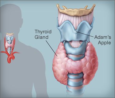
Hypothyroidism, also called underactive thyroid disease, is a common disorder. With hypothyroidism, your thyroid gland does not make enough thyroid hormone.The thyroid gland is located in the front lower part of your neck. Hormones released by the gland travel through your bloodstream and affect nearly every part of your body, from your heart and brain, to your muscles and skin.

Thyroid

The thyroid controls how your body’s cells use energy from food, a process called metabolism. Among other things, your metabolism affects your body’s temperature, your heartbeat, and how well you burn calories. If you don’t have enough thyroid hormone, your body processes slow down. That means your body makes less energy, and your metabolism becomes sluggish.

What Causes Hypothyroidism?

The most common cause of hypothyroidism is Hashimoto’s thyroiditis. “Thyroiditis” is an inflammation of the thyroid gland. Hashimoto’s thyroiditis is an autoimmune disorder. With Hashimoto’s, your body produces antibodies that attack and destroy the thyroid gland. Thyroiditis may also be caused by a viral infection.

Other causes of hypothyroidism include:

Radiation therapy to the neck area. Treating certain cancers, such as lymphoma, requires radiation to the neck. Radiation damages the cells in the thyroid. This makes it more difficult for the gland to produce hormone.

Radioactive iodine treatment. This treatment is commonly prescribed to people who have an overactive thyroid gland, a condition known as hyperthyroidism. However, radiation destroys the cells in the thyroid gland. This usually leads to hypothyroidism.

Use of certain medications. Certain medicines to treat heart problems, psychiatric conditions, and cancer can sometimes affect the production of thyroid hormone. These include amiodarone (Cordarone), lithium, interferon alpha, and interleukin-2.

Thyroid surgery . Surgery to remove the thyroid will lead to hypothyroidism. If only part of the thyroid is removed, the remaining gland may still be able to produce enough hormone for the body’s needs.

Too little iodine in the diet. The thyroid needs iodine to produce thyroid hormone. Your body doesn’t make iodine, so you need to get it through your diet. Iodized table salt is rich in iodine. Other food sources of iodine include shellfish, saltwater fish, eggs, dairy products, and seaweed.

Symptoms of Hypothyroidism

Symptoms of hypothyroidism may be vague and can often mimic other conditions. They may include:

  • Changes in the menstrual cycle
  • Constipation
  • Depression
  • Dry hair and hair loss
  • Dry skin
  • Fatigue
  • Greater sensitivity to cold
  • Slow heart rate
  • Swelling of the thyroid gland (goiter)
  • Unexplained weight gain or difficulty losing weight
  • Carpal tunnel syndrome

Babies with hypothyroidism may have no symptoms. If symptoms do occur, they can include:

  • Cold hands and feet
  • Constipation
  • Extreme sleepiness
  • Hoarse cry
  • Little or no growth
  • Low muscle tone (floppy infant)
  • Persistent jaundice (yellowing of the skin and whites of the eyes)
  • Poor feeding habits
  • Puffy face
  • Stomach bloating
  • Swollen tongue

Make an appointment with your health care provider if you or your baby has any of these symptoms. It is important to note that these symptoms can be due to other medical conditions.

How Is Hypothyroidism Treated?

If you have hypothyroidism, your doctor will prescribe a synthetic (man-made) thyroid hormone T4. You take this pill every day. Certain other medications can interfere with how your body absorbs synthetic thyroid hormone. Make sure you doctor knows about all the medicines, herbs, and supplements you take, including over-the-counter products.

You will need regular blood tests to check your thyroid hormone levels. Your doctor may need to adjust your medication dose from time to time.

Complications of Hypothyroidism

Untreated, hypothyroidism may cause:

  • Heart problems
  • Infertility
  • Joint pain
  • Obesity

Thyroid problems in a pregnant woman can affect the developing baby. During the first three months of pregnancy, the baby receives all thyroid hormone from its mother. If the mother has hypothyroidism, the baby does not get enough thyroid hormone. This can lead to problems with mental development.

Extremely low levels of thyroid hormone can cause a life-threatening condition called myxedema. Myxedema is the most severe form of hypothyroidism. A person with myxedema can lose consciousness or go into a coma. The condition can also cause the body temperature to drop very low, which can cause death.

Irritable Bowel Syndrome(IBS): causes & treatments

IBS is a mix of belly discomfort or pain and trouble with bowel habits: either going more or less often than normal (diarrhea or constipation) or having a different kind of stool (thin, hard, or soft and liquid).It’s not life-threatening, and it doesn’t make you more likely to get other colon conditions, such as ulcerative colitis, Crohn’s disease, or colon cancer. But IBS can be a long-lasting problem that changes how you live your life. People with IBS may miss work or school more often, and they may feel less able to take part in daily activities. Some people may need to change their work setting: shifting to working at home, changing hours, or even not working at all.

What Are the Symptoms of IBS?

People with IBS have symptoms that can include:

  • Diarrhea (often described as violent episodes of diarrhea)
  • Constipation
  • Constipation alternating with diarrhea
  • Belly pains or cramps, usually in the lower half of the belly, that get worse after meals and feel better after a bowel movement
  • A lot of gas or bloating
  • Harder or looser stools than normal (pellets or flat ribbon stools)
  • A belly that sticks out

Stress can make symptoms worse.

Some people also have urinary symptoms or sexual problems.

There are four types of the condition. There is IBS with constipation (IBS-C) and IBS with diarrhea (IBS-D). Some people have an alternating pattern of constipation and diarrhea. This is called mixed IBS (IBS-M).  Other people don’t fit into these categories easily, called unsubtyped IBS, or IBS-U.

What Are the Causes?

While there are several things known to trigger IBS symptoms, experts don’t know what causes the condition.

Studies suggest that the colon gets hypersensitive, overreacting to mild stimulation. Instead of slow, rhythmic muscle movements, the bowel muscles spasm. That can cause diarrhea or constipation.

Some think that IBS happens when the muscles in the bowels don’t squeeze normally, which affects the movement of stool. But studies don’t seem to back this up.

Another theory suggests it may involve chemicals made by the body, such as serotonin and gastrin, that control nerve signals between the brain and digestive tract.

Other researchers are studying to see if certain bacteria in the bowels can lead to the condition

Because IBS happens in women much more often than in men, some believe hormones may play a role. So far, studies haven’t borne this out.

How Is IBS Treated?

Nearly all people with IBS can get help, but no single treatment works for everyone. You and your doctor will need to work together to find the right treatment plan to manage your symptoms.

Many things can trigger IBS symptoms, including certain foods, medicines, the presence of gas or stool, and emotional stress. You’ll need to learn what your triggers are. You may need to make some lifestyle changes and take medication.

Periodontal (gum) disease: Causes, symptoms & treatments

What is gum disease?

Periodontal disease (gum disease) is a chronic bacterial infection of the gums and bone supporting the teeth. It is a major cause of tooth loss in adults. However, because gum disease is usually painless, you may not even know you have it. Gum disease is caused by plaque bacteria which form a sticky film on the tooth surface. These plaque bacteria release toxins that can damage the gums. As the amount of plaque increases the saliva in your mouth can begin to mineralise the plaque to form tartar (calculus). These hard deposits on the roots of the teeth only serve to promote more plaque formation and further damage to the gums.

Types of Gum disease

There are two main types of gum disease- gingivitis and periodontitis. Gingivitis is very common and may affect up to 70% of the population. Periodontitis affects around 30% of adults over age of 30. Both these infections can be treated and controlled, but the earlier they are diagnosed and managed, generally the better the prognosis for your teeth.

A patient with generalised gingivitis. Note the red, swollen, inflammed gum tissues around the teeth. These gums bleed easily on brushing.

Diagnosis and treatment

Diagnosis and treatment of periodontitis in its early stages will significantly enhance the prognosis for the dentition. If you have not had a periodontal screening examination with your dentist in the last 12 months, make it a priority. Remember, most patients who present with periodontitis are completely unaware of a gum problem being present in their mouth because it rarely causes pain until it becomes very severe, by which time it may be too late to save your teeth.

What are the warning signs of gum disease?

Periodontal disease is typically painless and often the signs are subtle. Some of the symptoms may include:

  • gums that bleed easily with brushing or flossing
  • red, swollen or tender gums
  • pus between gums and teeth
  • gums that pull away from the teeth
  • persistent bad breath
  • loose or separating teeth
  • a change in the way your teeth fit together when you bite


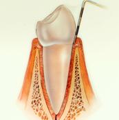
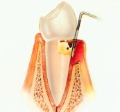
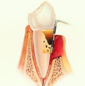
Normal healthy gumsHealthy gums and bone anchor teeth firmly in place. PeriodontitisUnremoved plaque hardens into calculus (tartar), As plaque and calculus continue to build up, the gums begin to recede( pull away) from the teeth, and pockets form between the teeth and gums. Advanced PeriodontitisThe gums recede further, destroying more bone and the periodontal ligament. Even healthy teeth may become loose and need to be extracted
An increasing amount of research also suggests that there is evidence linking chronic periodontal disease to conditions such as coronary heart disease, diabetes and respiratory disease. Moreover, current research suggests that if you have periodontal disease during pregnancy you may be significantly more likely to have a baby born prematurely or with a low birth weight. Keeping your gums healthy will ensure a better chance of good general health.

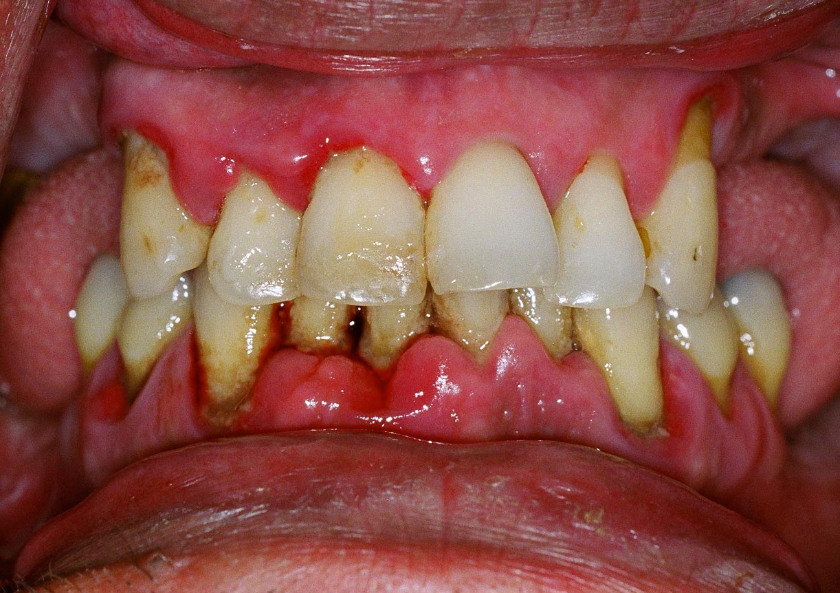
A case of advanced Periodontitis. Very swollen gums, loose teeth, staining and heavy plaque and calculus deposits on all the teeth.

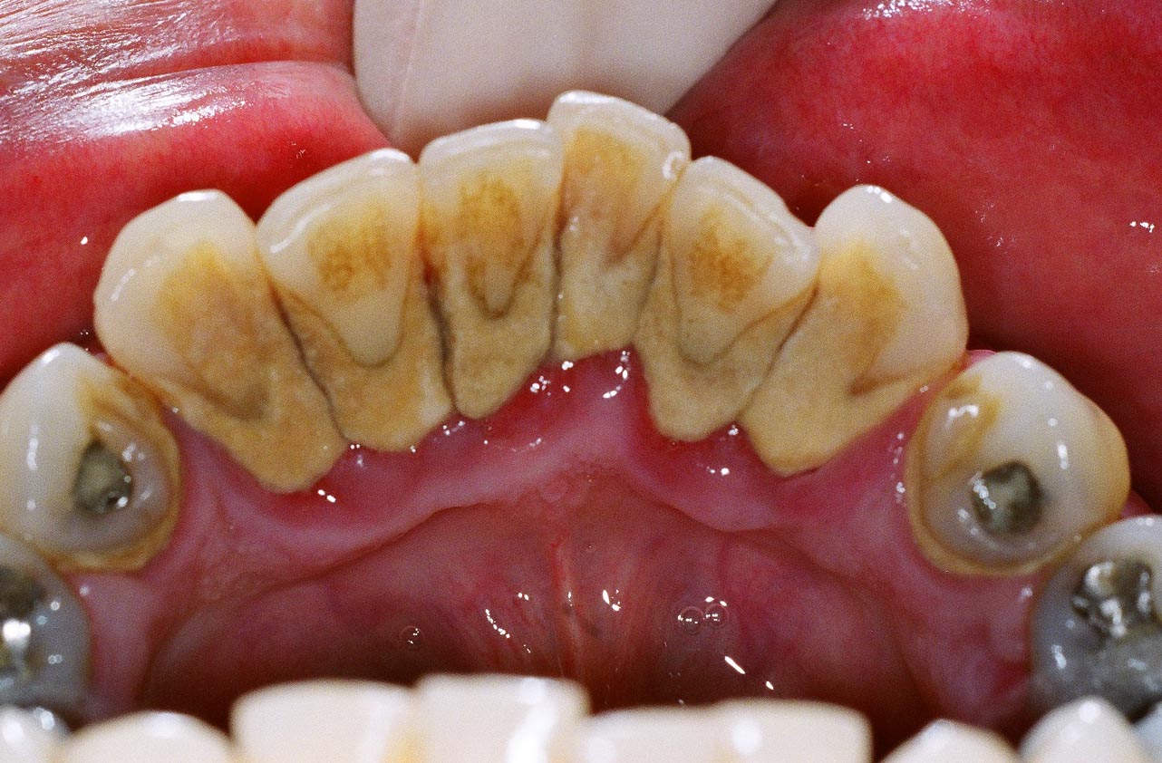
Heavy calculus (tartar) deposits on the inside aspect of the lower incisor teeth in a patient with periodontitis.

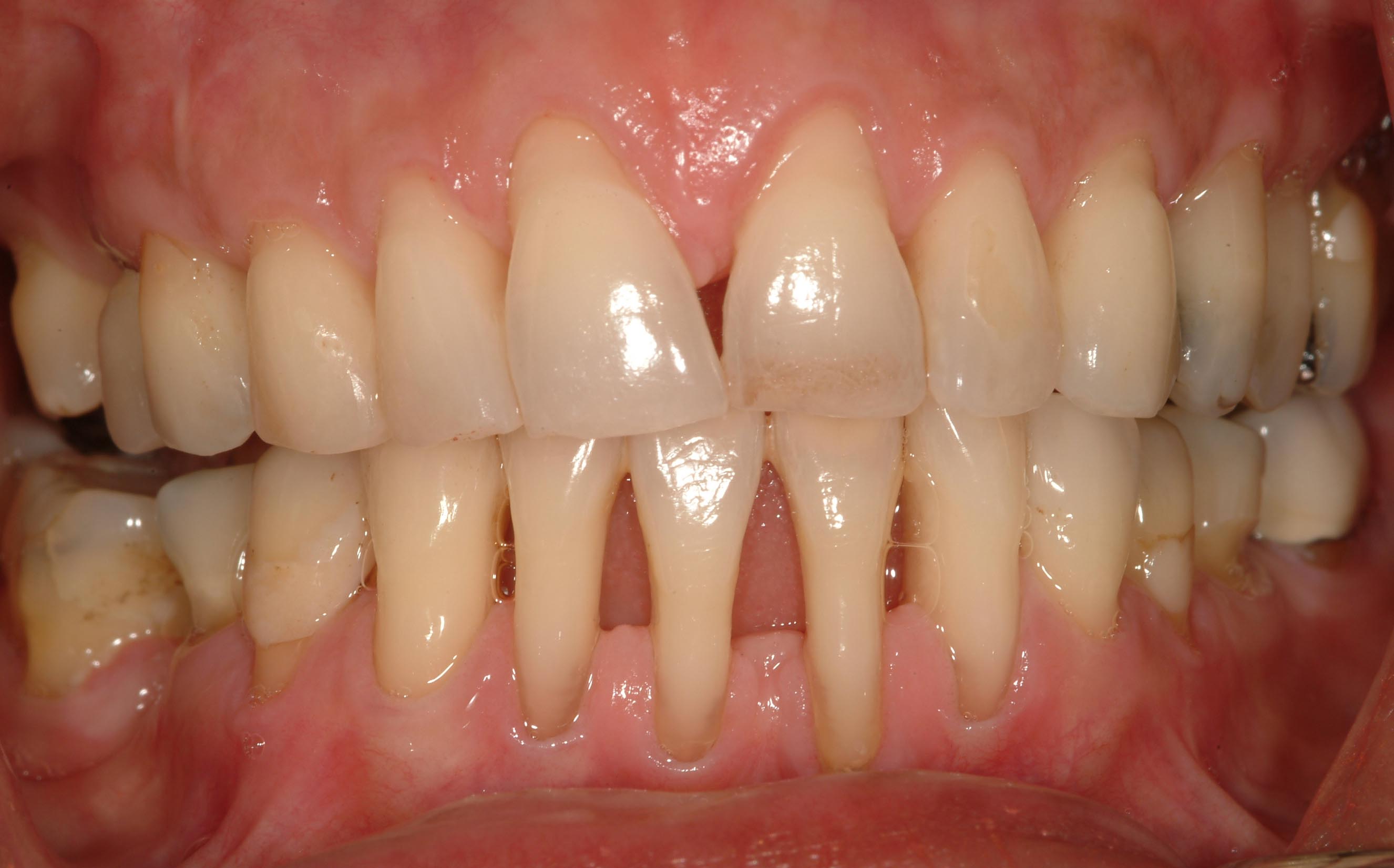
Patient with advanced periodontitis showing severe loss of bone and gum recession.

Risk Factors for Gum Disease

The main cause of periodontal disease is bacterial plaque. Oral bacteria form a sticky, colourless film on your teeth. These bacteria release various toxic by-products which irritate your gum tissues. While plaque bacteria are the main cause of periodontal disease there are other factors, such as those outlined below, that can also affect the health of your gums.

Smoking and Tobacco Use

The use of tobacco is linked with many serious illnesses such as cancer, lung disease and heart disease, as well as numerous other health problems. Smoking also increases the risk of periodontal disease and recent research has shown that tobacco use may be one of the most significant risk factors in the development and progression of periodontal disease. Smoking also reduces the ability of your gums to heal properly following treatment, which may mean a less successful treatment outcome. Patients who smoke are also more prone to developing post operative infections following treatment. For these reasons all patients are encouraged to quit smoking as part of their periodontal therapy.

Genetics

Recent studies suggest that approximately 30% of the population may be genetically more susceptible to developing gum disease. Thus, some individuals are more prone to periodontal disease. Often the susceptibility to periodontal infection can be traced though families. Patients with a susceptibility to periodontal disease may require more intensive maintenance and more frequent recall for hygiene treatment.

Pregnancy & Puberty

At certain times such as puberty, menopause, menstruation and pregnancy your gum tissues may be more susceptible to irritation and infection. During these particular times  your body experiences specific hormonal changes which can affect your gum tissues. Your gums may become more sensitive and at times react strongly to the hormonal fluctuations. Recent research also suggests that pregnant women with chronic gum disease are up to seven times more likely to deliver pre-term, low-birth-weight babies.

Grinding or Clenching your Teeth

Tooth grinding typically occurs at night while you are asleep and is also know as bruxism. Your jaw joint may click on opening or may be painful at times. If you wake up with frequent headaches and show wear on your teeth, you may be grinding your teeth. Tooth grinding may worsen your gum health if you have pre-existing periodontal disease.

Diabetes

Diabetes is one of the main risk factors in periodontal disease. Diabetics are generally more prone to developing infections, including periodontal disease. Periodontal disease is often more severe in diabetics and can be more difficult to treat, depending you the level of diabetic control.

Stress

Chronic stress can have a negative effect of you body’s immune system. This may in turn make you more susceptible to periodontal disease as it can make it more difficult for the body to fight off infection.

Medications

Certain drugs, such as antidepressants, oral contraceptives and some heart medicines, can have an effect on your oral health. You should always inform your dental professional of the medications you are taking so that these can be taken into consideration.

Poor Nutrition

A diet low in important vitamins and nutrients can compromise the body’s immune system and make it harder for the body to fight off infection. One of the more common conditions is iron deficiency. Deficiencies such as this can worsen the condition of your gums.

Other Systemic Diseases

Certain diseases may worsen your gum disease by interfering with the way your body’s defences deal with infection. Some examples include uncontrolled diabetes, radiation therapy, chemotherapy, corticosteroid use.

Maintaining Periodontal Health

After your gum disease has been diagnosed and treated, ongoing supportive treatment to maintain your periodontal heath will be recommended.

Supportive Periodontal Therapy

This consists of structured and regular hygiene treatment that is aimed at preventing and reducing the possibility of the recurrence and progression of periodontal disease. Each patient will have specific maintenance requirements which will need to be professionally tailored to their needs. The Oral Hygiene Spa at the Institute of Dental Implants & Periodontics provides the highest level of periodontal maintenance under the specialist supervision of a Periodontist.

Good Oral Hygiene Habits

As part of your periodontal treatment you will be given careful oral hygiene instruction and shown the right ‘tools’ to use. It is important that you regularly remove plaque bacteria through brushing, flossing, and regular professional cleaning. Good oral hygiene will leave you with a fresh, healthy mouth and reduce your risk of further gum disease. There are other risk factors that may compromise your periodontal health such as stress, diabetes, genetics, and pregnancy. These other factors may be transient, but should be discussed with your dental health professional.

Corneal collagen cross linking: Treatment

Corneal collagen cross-linking is a technique which uses UV light and a photosensitizer to strengthen chemical bonds in the cornea. The goal of the treatment is to halt progressive and irregular changes in corneal shape known as ectasia. These ectatic changes are typically marked by corneal thinning and an increase in the anterior and/or posterior curvatures of the cornea, and often lead to high levels of myopia and astigmatism. The most common form of ectasia is keratoconus and less often ectasia is seen after laser vision correction such as LASIK.

Image result for collagen cross linking causes, symptoms & treatments

Cross-linking of collagen refers to the ability of collagen fibrils to form strong chemical bonds with adjacent fibrils. In the cornea, collagen cross-linking occurs naturally with aging due to an oxidative deamination reaction that takes place within the end chains of the collagen. It has been hypothesized that this natural cross-linkage of collagen explains why keratoectasia (corneal ectasia) often progresses most rapidly in adolescence or early adulthood but tends to stabilize in patients after middle-age.

In addition to the cross-linking that occurs commonly with corneal maturation, there are several other pathways that can lead to crosslinkage. Glycation refers to a reaction seen predominantly in diabetics that can lead to the formation of additional bonds between collagen. In the pathway most relevant to this topic, oxidation has been shown to be able to trigger corneal crosslinkage through the release of oxygen free radicals.


Ocular Hypertension: Causes, Symptoms & Treatments

The term ocular hypertension usually refers to any situation in which the pressure inside the eye, called intraocular pressure, is higher than normal. Eye pressure is measured in millimeters of mercury (mm Hg). Normal eye pressure ranges from 10-21 mm Hg. Ocular hypertension is an eye pressure of greater than 21 mm Hg.

Although its definition has evolved through the years, ocular hypertension is commonly defined as a condition with the following criteria:

  • An intraocular pressure of greater than 21 mm Hg is measured in one or both eyes at two or more office visits. Pressure inside the eye is measured using an instrument called a tonometer.
  • The optic nerve appears normal.
  • No signs of glaucoma are evident on visual field testing, which is a test to assess your peripheral (or side) vision.
  • To determine other possible causes for your high eye pressure, an ophthalmologist (a medical doctor who specializes in eye care and surgery) assesses whether your drainage system (called the “angle”) is open or closed. The angle is seen using a technique called gonioscopy. This technique involves the use of a special contact lens to examine the drainage angles (or channels) in your eyes to see if they are open, narrowed, or closed.
  • No signs of any ocular disease are present. Some eye diseases can increase the pressure inside the eye.

Ocular hypertension should not be considered a disease by itself. Instead, ocular hypertension is a term that is used to describe individuals who should be observed more closely than the general population for the onset of glaucoma. For this reason, another term to refer to a person with ocular hypertension is “glaucoma suspect,” or someone whom the ophthalmologist is concerned may have or may develop glaucoma because of elevated pressure inside the eyes. An eye exam may show a glaucoma-damaged optic nerve.

As mentioned above, increased intraocular pressure can result from other eye conditions. However, within this article, ocular hypertension primarily refers to increased intraocular pressure without any optic nerve damage or vision loss. Glaucoma occurs when increased intraocular pressure, optic nerve damage, and vision loss are present.

Ocular Hypertension Causes

Elevated intraocular pressure is a concern in people with ocular hypertension because it is one of the main risk factors for glaucoma.

High pressure inside the eye is caused by an imbalance in the production and drainage of fluid in the eye (aqueous humor). The channels that normally drain the fluid from inside the eye do not function properly. More fluid is continually being produced but cannot be drained because of the improperly functioning drainage channels. This results in an increased amount of fluid inside the eye, thus raising the pressure.

Another way to think of high pressure inside the eye is to imagine a water balloon. The more water that is put into the balloon, the higher the pressure inside the balloon. The same situation exists with too much fluid inside the eye—the more fluid, the higher the pressure. Also, just like a water balloon can burst if too much water is put into it, the optic nerve in the eye can be damaged by too high of a pressure.

Medical Treatment:

The goal of medical treatment is to reduce the pressure before it causes glaucomatous loss of vision. Medical treatment is always initiated for those people who are believed to be at the greatest risk for developing glaucoma and for those with signs of optic nerve damage.

How your ophthalmologist chooses to treat you is highly individualized. Depending on your particular situation, you may be treated with medications or just observed. Your doctor will discuss the pros and cons of medical treatment versus observation with you.

  • If you are experiencing symptoms like halos, blurred vision, or pain, or if your intraocular pressure has recently increased and then continues to increase on subsequent visits, your ophthalmologist will most likely start medical treatment.

Your intraocular pressure is evaluated periodically using these guidelines:

  • If your intraocular pressure is 28 mm Hg or higher, you are treated with medicines. After 1 month of taking the drug, you have a follow-up visit with your ophthalmologist to see if the medicine is lowering the pressure and there are no side effects. If the drug is working, then follow-up visits are scheduled every 3-4 months.
  • If your intraocular pressure is 26-27 mm Hg, the pressure is rechecked in 2-3 weeks after your initial visit. On your second visit, if the pressure is still within 3 mm Hg of the reading at the initial visit, then follow-up visits are scheduled every 3-4 months. If the pressure is lower on your second visit, then the length of time between follow-up visits is longer and is determined by your ophthalmologist. At least once a year, visual field testing is done and your optic nerve is examined.
  • If your intraocular pressure is 22-25 mm Hg, the pressure is rechecked in 2-3 months. At the second visit, if the pressure is still within 3 mm Hg of the reading at the initial visit, then your next visit is in 6 months and includes visual field testing and an optic nerve examination. Testing is repeated at least yearly.


Pages:12