Global Treatment Services Pvt. Ltd.

Global Treatment Services

Sudden Loss of vision: Causes, Symptoms & treatments

Loss of vision is considered sudden if it develops within a few minutes to a couple of days. It may affect one or both eyes and all or part of a field of vision. Loss of only a small part of the field of vision (for example, as a result of a small retinal detachment) may seem like blurred vision. Other symptoms, for example eye pain, may occur depending on the cause of vision loss.

Causes:

Sudden loss of vision has three general causes:

  • Clouding of normally transparent eye structures

  • Abnormalities of the retina (the light-sensing structure at the back of the eye)

  • Abnormalities of the nerves that carry visual signals from the eye to the brain (the optic nerve and the visual pathways)

Light must travel through several transparent structures before it can be sensed by the retina. First, light passes through the cornea (the clear layer in front of the iris and pupil), then the lens, and then the vitreous humor (the jellylike substance that fills the eyeball). Anything that blocks light from passing through these structures, for example, a corneal ulcer or bleeding into the vitreous humor, can cause loss of vision.

Most of the disorders that cause total loss of vision when they affect the entire eye may cause only partial vision loss when they affect only part of the eye.

An Inside Look at the Eye

When the Visual Pathways Are Damaged

Nerve signals travel along the optic nerve from each eye. The two optic nerves meet at the optic chiasm. There, the optic nerve from each eye divides, and half of the nerve fibers from each side cross to the other side. Because of this arrangement, the brain receives information via both optic nerves for the left visual field and for the right visual field. Damage to an eye or the visual pathway causes different types of vision loss depending on where the damage occurs.

Common causes

The most common causes of sudden loss of vision are

  • Blockage of a major artery of the retina (central retinal artery occlusion)

  • Blockage of an artery to the optic nerve (ischemic optic neuropathy)

  • Blockage of a major vein in the retina (central retinal vein occlusion)

  • Blood in the jellylike vitreous humor near the back of the eye (vitreous hemorrhage)

  • Eye injury

Sudden retinal artery blockage can result from a blood clot or small piece of atherosclerotic material that breaks off and travels into the artery. The artery to the optic nerve can be blocked in the same ways and can also be blocked by inflammation (such as may occur with giant cell [temporal] arteritis). A blood clot can form in the retinal vein and block it, particularly in older people with high blood pressure or diabetes. People with diabetes are also at risk of bleeding into the vitreous humor.

Sometimes what seems like a sudden start of symptoms may instead be sudden recognition. For example, a person with long-standing reduced vision in one eye (possibly caused by a dense cataract) may suddenly become aware of the reduced vision in the affected eye after covering the unaffected eye.

Less common causes:

Less common causes of sudden loss of vision (see Table: Some Causes and Features of Sudden Loss of Vision) include stroke or transient ischemic attack (TIA), acute glaucoma, retinal detachment, inflammation of the structures in the front of the eye between the cornea and the lens (anterior uveitis, sometimes called iritis), certain infections of the retina, and bleeding within the retina as a complication of age-related macular degeneration.

Cleft lip and cleft palate : causes and treatments

Cleft lip and cleft palate are facial and oral malformations that occur very early in pregnancy, while the baby is developing inside the mother. Clefting results when there is not enough tissue in the mouth or lip area, and the tissue that is available does not join together properly.

A cleft lip is a physical split or separation of the two sides of the upper lip and appears as a narrow opening or gap in the skin of the upper lip. This separation often extends beyond the base of the nose and includes the bones of the upper jaw and/or upper gum.

A cleft palate is a split or opening in the roof of the mouth. A cleft palate can involve the hard palate (the bony front portion of the roof of the mouth), and/or the soft palate (the soft back portion of the roof of the mouth).

Cleft lip and cleft palate can occur on one or both sides of the mouth. Because the lip and the palate develop separately, it is possible to have a cleft lip without a cleft palate, a cleft palate without a cleft lip, or both together.

What Causes a Cleft Lip and Cleft Palate?

In most cases, the cause of cleft lip and cleft palate is unknown. These conditions cannot be prevented. Most scientists believe clefts are due to a combination of genetic and environmental factors. There appears to be a greater chance of clefting in a newborn if a sibling, parent, or relative has had the problem.

Another potential cause may be related to a medication a mother may have taken during her pregnancy. Some drugs may cause cleft lip and cleft palate. Among them: anti-seizure/anticonvulsant drugs, acne drugs containing Accutane, and methotrexate, a drug commonly used for treating cancer, arthritis, and psoriasis.

Cleft lip and cleft palate may also occur as a result of exposure to viruses or chemicals while the fetus is developing in the womb.In other situations, cleft lip and cleft palate may be part of another medical condition.

Who Treats Children With Cleft Lip and/or Palate?

Due to the number of oral health and medical problems associated with a cleft lip or cleft palate, a team of doctors and other specialists is usually involved in the care of these children. Members of a cleft lip and palate team typically include:

  • Plastic surgeon to evaluate and perform necessary surgeries on the lip and/or palate
  • An otolaryngologist (an ear, nose, and throat doctor) to evaluate hearing problems and consider treatment options for hearing problems
  • An oral surgeon to reposition segments of the upper jaw when needed, to improve function and appearance and to repair the cleft of the gum
  • An orthodontist to straighten and reposition teeth
  • A dentist to perform routine dental care
  • A prosthodontist to make artificial teeth and dental appliances to improve the appearance and to meet functional requirements for eating and speaking
  • A speech pathologist to assess speech and feeding problems
  • A speech therapist to work with the child to improve speech
  • An audiologist (a specialist in communication disorders stemming from a hearing impairment); to assess and monitor hearing
  • A nurse coordinator to provide ongoing supervision of the child’s health
  • A social worker/psychologist to support the family and assess any adjustment problems
  • A geneticist to help parents and adult patients understand the chances of having more children with these conditions

The health care team works together to develop a plan of care to meet the individual needs of each patient. Treatment usually begins in infancy and often continues through early adulthood.

What’s the Treatment for Cleft Lip and Cleft Palate?

A cleft lip may require one or two surgeries depending on the extent of the repair needed. The initial surgery is usually performed by the time a baby is 3 months old.

Repair of a cleft palate often requires multiple surgeries over the course of 18 years. The first surgery to repair the palate usually occurs when the baby is between 6 and 12 months old. The initial surgery creates a functional palate, reduces the chances that fluid will develop in the middle ears, and aids in the proper development of the teeth and facial bones.

Children with a cleft palate may also need a bone graft when they are about 8 years old to fill in the upper gum line so that it can support permanent teeth and stabilize the upper jaw. About 20% of children with a cleft palate require further surgeries to help improve their speech.

Once the permanent teeth grow in, braces are often needed to straighten the teeth.

Additional surgeries may be performed to improve the appearance of the lip and nose, close openings between the mouth and nose, help breathing, and stabilize and realign the jaw. Final repairs of the scars left by the initial surgery will probably not be performed until adolescence, when the facial structure is more fully developed.

What Is the Outlook for Children With Cleft Lip and/or Cleft Palate?

Although treatment for a cleft lip and/or cleft palate may extend over several years and require several surgeries depending upon the involvement, most children affected by this condition can achieve normal appearance, speech, and eating.

Dental Care for Children With Cleft Lips and/or Palates

Generally, the preventive and restorative dental care needs of children with clefts are the same as for other children. However, children with cleft lip and cleft palate may have special problems related to missing, malformed, or malpositioned teeth that require close monitoring.

  • Early dental care. Like other children, children born with cleft lip and cleft palate require proper cleaning, good nutrition, and fluoride treatment in order to have healthy teeth. Appropriate cleaning with a small, soft-bristled toothbrush should begin as soon as teeth erupt. If a soft children’s toothbrush will not adequately clean the teeth because of the modified shape of the mouth and teeth, a toothette may be recommended by your dentist. A toothette is a soft, mouthwash-containing sponge on a handle that’s used to swab teeth. Many dentists recommend that the first dental visit be scheduled at about 1 year of age or even earlier if there are special dental problems. Routine dental care can begin around 1 year of age.
  • Orthodontic care. A first orthodontic appointment may be scheduled before the child has any teeth. The purpose of this appointment is to assess facial growth, especially jaw development. After teeth erupt, an orthodontist can further assess a child’s short and long-term dental needs. After the permanent teeth erupt, orthodontic treatment can be applied to align the teeth.
  • Prosthodontic care. A prosthodontist is a member of the cleft palate team. He or she may make a dental bridge to replace missing teeth or make special appliances called “speech bulbs” or “palatal lifts” to help close the nose from the mouth so that speech sounds more normal. The prosthodontist coordinates treatment with the oral or plastic surgeon and with the speech pathologist.

Cervical disc herniation: Causes, symptoms & treatments

What is cervical disc herniation?

The bones (vertebrae) that form the spine in your back are cushioned by round, flat discs. When these discs are healthy, they act as shock absorbers for the spine and keep the spine flexible. If they become damaged, they may bulge abnormally or break open (rupture), in what is called a herniated or slipped disc. Herniated discs can occur in any part of the spine, but they are most common in the neck (cervical) and lower back (lumbar) spine. The seven vertebrae between the head and the chest make up the cervical spine.

What causes cervical disc herniation?

A herniated disc usually is caused by wear and tear of the disc (also called disc degeneration). As we age, our discs lose some of the fluid that helps them stay flexible. A herniated disc also may result from injuries to the spine, which may cause tiny tears or cracks in the outer layer (annulus or capsule) of the disc. The jellylike material (nucleus) inside the disc may be forced out through the tears or cracks in the capsule, which causes the disc to bulge, break open (rupture), or break into fragments.

Herniated discs are much more common in people who smoke.

What are the symptoms?

Herniated discs in the neck (cervical spine) can cause pain, numbness, or weakness in the neck, shoulders, chest, arms, and hands. In some cases a very large herniated disc in the neck may cause weakness or unusual tingling affecting other parts of the body, including the legs.

How is it treated?

In most cases, cervical herniated discs are first treated with nonsurgical treatment, including rest or modified activities, medicines to relieve pain and inflammation, and exercises, as recommended by your doctor. Your doctor may recommend that you see a physical therapist to learn how to do exercises and protect your neck, and perhaps for other treatment such as traction. Traction is gentle, steady pulling on the head to stretch the neck and allow the small joints between the neck bones to spread a little. If symptoms continue, your doctor may try stronger medicine such as corticosteroids. Symptoms usually improve over time. But if the herniated disc is squeezing your spinal cord or nerves and/or you are having weakness, constant pain, or decreased control of your bladder or bowels, surgery will be considered. In rare cases, an artificial disc may be used to replace the disc that is removed.


Colorectal Cancer: Causes, Symptoms & Treatments

Colorectal cancer, also known as bowel cancer, colon cancer or rectal cancer, is any cancer (a growth, lump, tumor) of the colon and the rectum.

A colorectal cancer may be benign or malignant. Benign means the tumor will not spread, while a malignant tumor consists of cells that can spread to other parts of the body and damage them.

The colon and rectum

Anatomy of the large intestine
The large intestine is also called the colon or large bowel.

The colon and rectum belong to our body’s digestive system – together they are also known as the large bowel.

The colon reabsorbs large quantities of water and nutrients from undigested food products as they pass along it.

The rectum is at the end of the colon and stores feces (stools, waste material) before being expelled from the body.

Symptoms of colorectal cancer

  • Going to the toilet more often.
  • Diarrhea.
  • Constipation.
  • A feeling that the bowel does not empty properly after a bowel movement.
  • Blood in feces (stools).
  • Pains in the abdomen.
  • Bloating in the abdomen.
  • A feeling of fullness in the abdomen (maybe even after not eating for a while).
  • Vomiting.
  • Fatigue (tiredness).
  • Inexplicable weight loss.
  • A lump in the tummy or a lump in the back passage felt by your doctor.
  • Unexplained iron deficiency in men, or in women after the menopause.

As most of these symptoms may also indicate other possible conditions, it is important that the patient sees a doctor for a proper diagnosis. Anybody who experiences some of these symptoms for four weeks should see their doctor.

Diagram of colon cancer

Causes of colorectal cancer

Experts say we are not completely sure why colorectal cancer develops in some people and not in others. However, several risk factors have been identified over the years – a risk factor is something which may increase a person’s chances of developing a disease or condition.

The possible risk factors for colorectal factors are:

  • Being elderly – the older you are the higher the risk is.
  • A diet that is very high in animal protein.
  • A diet that is very high in saturated fats.
  • A diet that is very low in dietary fiber.
  • A diet that is very high in calories.
  • A diet that is very high in alcohol consumption.
  • Women who have had breast, ovary and uterus cancers.
  • A family history of colorectal cancer.
  • Patients with ulcerative colitis.
  • Being overweight/obese.
  • Smoking. This study found that smoking is significantly associated with an increased risk for colorectal cancer and death.
  • Being physically inactive.
  • Presence of polyps in the colon/rectum. Untreated polyps may eventually become cancerous.
  • Having Crohn’s disease or Irritable Bowel Disease have a higher risk of developing colorectal cancer.

Most colon cancers develop within polyps (adenoma). These are often found inside the bowel wall.

Treatments for colorectal cancer:

The patient’s treatment will depend on several factors, including its size and location, the stage of the cancer, whether or not it is recurrent, and the current overall state of health of the patient. A good specialist will explain all the treatment options available to the patient. This is an opportunity for the patient to ask questions and get advice on lifestyle changes that will help recovery.

Treatment options include chemotherapy, radiotherapy, and surgery:

Surgery for colorectal cancer

This is the most common colorectal cancer treatment. The affected malignant tumors and any lymph nodes that are nearby will be removed. Surgeons remove lymph nodes because they are the first place cancers tend to spread to.

The bowel is usually sewn back together. On some occasions the rectum may need to be taken out completely – a colostomy bag is then attached for drainage. The colostomy bag collects stools and is generally placed temporarily – sometimes it may be a permanent measure if it is not possible to join up the ends of the bowel.

If the cancer is diagnosed early enough, surgery may be the only treatment necessary to cure the patient of colorectal cancer. Even if surgery does not cure the patient, it will ease the symptoms.

Chemotherapy

Chemotherapy involves using a medicine (chemical) to destroy the cancerous cells. It is commonly used for colon cancer treatment. It may be used before surgery in an attempt to shrink the tumor. A study found that patients with advanced colon cancer who receive chemotherapy and who have a family history of colorectal cancer have a significantly lower likelihood of cancer recurrence and death.

Radiotherapy

Radiotherapy uses high energy radiation beams to destroy the cancer cells, and also to prevent them from multiplying. This treatment is more commonly used for rectal cancer treatment. It may be used before surgery in an attempt to shrink the tumor.

Doctors may order both radiotherapy and chemotherapy after surgery as they can help lower the chances of recurrence.

TMJ Disorders: Causes, Symptoms & Treatments

TMJ Disorders

TMJ disorders are a family of problems related to your complex jaw joint. TMJ stands for temporomandibular joint, which is the name for each joint (right and left) that connects your jaw to your skull. If you have had symptoms like pain or a clicking sound in your jaw, you’ll be glad to know these problems are more easily diagnosed and treated than they were in the past. These symptoms occur when the joints of the jaw and the chewing muscles do not work together correctly. Since some types of TMJ problems can lead to more serious conditions, early detection and treatment are important.

Causes

  • Clenching or grinding your teeth.
  • Tightening your jaw muscles and stressing your TM joint.
  • You may have a damaged jaw joint due to injury or disease. Injuries and arthritis can damage the joint directly or stretch or tear the muscle ligaments.

As a result of the above, the disk which is made of cartilage and functions as the “cushion” of the jaw joint can slip out of position.

Regardless of the cause, the results may include a misaligned bite, pain, clicking or grating noise when you open your mouth, or you may have trouble opening your mouth wide.

Do You Have a TMJ Disorder?

  • Are you aware of grinding or clenching your teeth?
  • Do you wake up with sore, stiff muscles around your jaws?
  • Do you have frequent headaches or neck aches?
  • Does the pain get worse when you clench your teeth?
  • Does stress make your clenching and pain worse?
  • Does your jaw click, pop, grate, catch or lock when you open your mouth?
  • Is it difficult or painful to open your mouth, eat or yawn?
  • Have you ever injured your neck, head or jaws?
  • Have you had problems (such as arthritis) with other joints?
  • Do you have teeth that no longer touch when you bite?
  • Do your teeth meet differently from time to time?
  • Is it hard to use your front teeth to bite or tear food?
  • Are your teeth sensitive, loose, broken or worn?

The more times you answered “yes”, the more likely it is that you may have a TMJ disorder. Understanding TMJ disorders will also help you understand how they are treated.

Treatment

Once an evaluation confirms a TMJ diagnosis, our doctors will determine the proper course of treatment. It is important to note that the treatment typically works better with a team approach of self-care joined with professional care.

  • The initial goals are to relieve the muscle spasm and joint pain. This is usually accomplished with a pain reliever, anti-inflammatory agent or muscle relaxant. Steroids can be injected directly into the joints to reduce pain and inflammation.

  • Self-care treatments often work well and include resting your jaw, keeping your teeth apart when you are not swallowing or eating, eating soft foods, applying ice and heat, exercising your jaw and practicing good posture.

  • Stress management techniques such as biofeedback or physical therapy may also be recommended, as well as a temporary, clear plastic appliance known as a splint. The splint (or night guard) fits over your top and bottom teeth and helps keep your teeth apart, thereby relaxing the muscle and reducing pain.

  • Different types of appliances serve different purposes. A night guard helps you stop clenching or grinding your teeth and reduces muscle tension at night, helping to protect the cartilage and joint surfaces. An anterior positioning appliance moves your jaw forward, relieves pressure on parts of your jaw and aids in disk repositioning. An orthotic stabilization appliance is worn 24 hours/day or just at night to move your jaw into proper position.

  • If your TMJ disorder has caused problems with how your teeth fit together, you may need treatment such as bite adjustment, orthodontics with or without jaw reconstruction, or restorative dental work.

  • Surgical options such as arthroscopy and open joint repair restructuring are sometimes needed, but are reserved for severe cases. TMJ surgery is generally only considered when the jaw can’t open, is dislocated and nonreducible, has severe degeneration, or if the patient has undergone appliance therapy treatment unsuccessfully.

Slipped Disk : Causes, Symptoms & Treatments

  1. A slipped disk occurs when the soft inner portion of the disk protrudes through the outer ring.
  2. Symptoms vary based on where the slipped disk occurs and if it compresses any of your spinal nerves.
  3. Slipped disks are more common in older adults.

Your spinal column is made up of a series of bones (vertebrae) stacked onto each other. From top to bottom, the column includes seven bones in the cervical spine, 12 in the thoracic spine, and five in the lumbar spine, followed by the sacrum and the coccyx at the base. These bones are cushioned by disks. The disks protect the bones by absorbing the shocks from daily activities like walking, lifting, and twisting.

Each disk has two parts: a soft, gelatinous inner portion and a tough outer ring. Injury or weakness can cause the inner portion of the disk to protrude through the outer ring. This is known as a slipped, herniated, or prolapsed disk. This causes pain and discomfort. If the slipped disk compresses one of your spinal nerves, you may also experience numbness and pain along the affected nerve. In severe instances, you may require surgery to remove or repair the slipped disk.

Symptoms of a slipped disk?

You can have a slipped disk in any part of your spine, from your neck to your lower back. The lower back is one of the more common areas for slipped disks. Your spinal column is an intricate network of nerves and blood vessels. A slipped disk can place extra pressure on the nerves and muscles around it.

Symptoms of a slipped disk include:

  • pain and numbness, most commonly on one side of the body
  • pain that extends to your arms or legs
  • pain that worsens at night or with certain movements
  • pain that worsens after standing or sitting
  • pain when walking short distances
  • unexplained muscle weakness
  • tingling, aching, or burning sensations in the affected area

The types of pain can vary from person to person. See your doctor if your pain results in numbness or tingling that affects your ability to control your muscles.

What causes slipped disks?

A slipped disk occurs when the outer ring becomes weak or torn and allows the inner portion to slip out. This can happen with age. Certain motions may also cause a slipped disk. A disk can slip out of place while you are twisting or turning to lift an object. Lifting a very large, heavy object can place great strain on the lower back, resulting in a slipped disk. If you have a very physically demanding job that requires a lot of lifting, you may be at increased risk for slipped disks.

Overweight individuals are also at increased risk for a slipped disk because their disks must support the additional weight. Weak muscles and a sedentary lifestyle may also contribute to the development of a slipped disk.

As you get older, you are more likely to experience a slipped disk. This is because your disks begin to lose some of their protective water content as you age. As a result, they can slip more easily out of place. They are more common in men than women.

How are slipped disks treated?

Treatments for a slipped disk range from conservative to surgical. The treatment typically depends on the level of discomfort you’re experiencing and how far the disk has slipped out of place.

Most people can relieve slipped disk pain using an exercise program that stretches and strengthens the back and surrounding muscles. A physical therapist may recommend exercises that can strengthen your back while reducing your pain.

Taking over-the-counter pain relievers and avoiding heavy lifting and painful positions can also help.

While it may be tempting to refrain from all physical activity while you’re experiencing the pain or discomfort of a slipped disk, this can lead to muscle weakness and joint stiffness. Instead, try to remain as active as possible through stretching or low-impact activities such as walking.

If your slipped disk pain does not respond to over-the-counter treatments, your doctor may prescribe stronger medications. These include:

  • muscle relaxers to relieve muscle spasms
  • narcotics to relieve pain
  • nerve pain medications like gabapentin or duloxetine

Your doctor may recommend surgery if your symptoms do not subside in six weeks or if your slipped disk is affecting your muscle function. Your surgeon may remove the damaged or protruding portion of the disk without removing the entire disk. This is called a microdiskectomy.

In more severe cases, your doctor may replace the disk with an artificial one or remove the disk and fuse your vertebrae together. This procedure, along with a laminectomy and spinal fusion, adds stability to your spinal column.

What is the outlook for someone with a slipped disk?

Most people with a slipped disk respond well to conservative treatment. Within six weeks their pain and discomfort will gradually lessen.

Is it possible to prevent a slipped disk?

It may not be possible to prevent a slipped disk, but you can take steps to reduce your risk of developing a slipped disk. These steps include:

  • Use safe lifting techniques: Bend and lift from your knees, not your waist.
  • Maintain a healthy weight.
  • Do not remain seated for long periods; get up and stretch periodically.
  • Do exercises to strengthen the muscles in your back, legs, and abdomen.

Ankylosing Spondylitis: Symptoms, Causes & Treatments

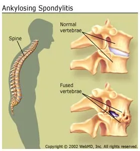
Ankylosing spondylitis is a type of arthritis that affects the spine. Ankylosing spondylitis symptoms include pain and stiffness from the neck down to the lower back. The spine’s bones (vertebrae) fuse together, resulting in a rigid spine. These changes may be mild or severe, and may lead to a stooped-over posture. Early diagnosis and treatment helps control pain and stiffness and may reduce or prevent significant deformity.

Who Is Affected by Ankylosing Spondylitis?

Ankylosing spondylitis affects about 0.1% to 0.5% of the adult population. Although it can occur at any age, spondylitis most often strikes men in their teens and 20s. It is less common and generally milder in women and more common in some Native American tribes.

What Are the Symptoms of Ankylosing Spondylitis?

The most common early symptoms of ankylosing spondylitis include:

  • Pain and stiffness. Constant pain and stiffness in the low back, buttocks, and hips that continue for more than three months. Spondylitis often starts around the sacroiliac joints, where the sacrum (the lowest major part of the spine) joins the ilium bone of the pelvis in the lower back region.
  • Bony fusion. Ankylosing spondylitis can cause an overgrowth of the bones, which may lead to abnormal joining of bones, called “bony fusion.” Fusion affecting bones of the neck, back, or hips may impair a person’s ability to perform routine activities. Fusion of the ribs to the spine or breastbone may limit a person’s ability to expand his or her chest when taking a deep breath.
  • Pain in ligaments and tendons. Spondylitis also may affect some of the ligaments and tendons that attach to bones. Tendonitis (inflammation of the tendon) may cause pain and stiffness in the area behind or beneath the heel, such as the Achilles tendon at the back of the ankle.

Ankylosing spondylitis is a systemic disease, which means symptoms may not be limited to the joints. People with the condition also may have fever, fatigue, and loss of appetite. Eye inflammation (redness and pain) occurs in some people with spondylitis. In rare cases, lung and heart problems also may develop.

What Causes Ankylosing Spondylitis?

Although the cause of ankylosing spondylitis is unknown, there is a strong genetic or family link. Most, but not all, people with spondylitis carry a gene called HLA-B27. Although people carrying this gene are more likely to develop spondylitis, it is also found in up to 10% of people who have no signs of the condition.
 

How Is Ankylosing Spondylitis Treated?

There is no cure for ankylosing spondylitis, but there are treatments that can reduce discomfort and improve function. The goals of treatment are to reduce pain and stiffness, maintain a good posture, prevent deformity, and preserve the ability to perform normal activities. When properly treated, people with ankylosing spondylitis may lead fairly normal lives. Under ideal circumstances, a team approach to treat spondylitis is recommended. Members of the treatment team typically include the patient, doctor, physical therapist, and occupational therapist. In patients with severe deformities, osteotomy and fusion can be done.

  • Physical and occupational therapy. Early intervention with physical and occupational therapy is important to maintain function and minimize deformity.
  • Exercise. A program of daily exercise helps reduce stiffness, strengthen the muscles around the joints and prevent or minimize the risk of disability. Deep breathing exercises may help keep the chest cage flexible. Swimming is an excellent form of exercise for people with ankylosing spondylitis.
  • Medications. Certain drugs help provide relief from pain and stiffness, and allow patients to perform their exercises with minimal discomfort. Nonsteroidal anti-inflammatory drugs (NSAIDs) — such as ibuprofen, naproxen, and aspirin — are the most commonly used drugs for spondylitis treatment. In moderate to severe cases, other drugs may be added to the treatment regimen. Disease-modifying antirheumatic drugs (DMARDs), such as methotrexate (Rheumatrex), can be used when NSAIDs alone are not enough to reduce the inflammation, stiffness, and pain. In addition, relatively new drugs called biologics — adalimumab (Humira), adalimumab-atto (Amjevita), a biosimilar to Humira, certolizumab pegol (Cimzia), etanercept (Enbrel), etanercept-szzs (Erelzi), a biosimilar to Enbrel, golimumab (Simponi Aria, Simponi), infliximab (Remicade), and infliximab-dyyb (Inflectra), a biosimilar to Remicade, and secukinimab (Cosentyx)– have been FDA-approved for treating ankylosing spondylitis. Also, the antidepressant Cymbalta has been approved for chronic back pain as well. Steroid injections into the joint or tendon may be helpful in some cases.
  • Surgery. Artificial joint replacement surgery may be a treatment option for some people with advanced joint disease affecting the hips or knees.


Spondyloarthropathies: Causes, Symptoms & Treatments

What are spondyloarthropathies?

Spondyloarthropathies are a family of long-term (chronic) diseases of joints. These diseases occur in children (juvenile spondyloarthropathies) and adults. They include ankylosing spondylitis, reactive arthritis, psoriatic arthritis, and joint problems linked to inflammatory bowel disease (enteropathic arthritis). Spondyloarthropathies are sometimes called spondyloarthritis.Although all spondyloarthropathies have different symptoms and outcomes, they are similar in that all of them:

  • Usually involve the attachments between your low back and the pelvis (sacroiliac joint).
  • Affect areas around the joint where your ligaments and tendons attach to bone (enthesitis), such as at the knee, foot, or hip.

It is important to recognize that the spondyloarthropathies are different from rheumatoid arthritis (RA) in adults and juvenile idiopathic arthritis (JIA) in children.

What causes spondyloarthropathies?

Experts don’t know what causes spondyloarthropathies. The presence of a particular gene, HLA-B27, is often associated with ankylosing spondylitis. Spondyloarthropathies, especially ankylosing spondylitis, are more likely to run in families than other forms of rheumatic disease, such as lupus or rheumatoid arthritis.

What are the symptoms?

Spondyloarthropathies often cause:

  • Low back pain that may spread into the buttock.
  • Morning stiffness, especially in the back or neck, that gets better during the day and after exercise.
  • Fatigue.

Although spondyloarthropathies all result in joint pain, each type also has specific symptoms.

  • Ankylosing spondylitis causes stiffness and low back pain. Over time, the pain usually moves from the lower back into the upper back. In severe cases, the affected joints in the spine fuse together, causing severe back stiffness. Other areas (such as the hips, chest wall, and heels) may also be affected. In children, symptoms usually begin in the hips, knees, heels, or big toes and later progress to the spine.
  • Reactive arthritis causes pain, swelling, and inflammation of the joints, especially in the sacroiliac joint, the attachment between the lower back and pelvis, and in the fingers, toes, and feet. The fingers and toes may swell, causing a “sausage digit.” Reactive arthritis can also cause fever, weight loss, skin rash, and inflammation. In children, the joints of the lower legs are most commonly affected.
  • Psoriatic arthritis is a form of arthritis associated with a skin condition called psoriasis. The psoriasis symptoms (scaly red patches on the skin) often precede the arthritis symptoms, sometimes by many years. The severity of the rash does not mirror the severity of the arthritis. The fingernails and toenails may show pitting or thickening and yellowing. The joint problems involve large joints, such as the hips and sacroiliac joints. Swelling of entire toes or fingers, resulting in sausage digits, also occurs.
  • Enteropathic arthritis is spinal arthritis that also involves inflammation of the intestinal wall. Symptoms can come and go. And when the abdominal pain is flaring, this arthritis may also flare. The arthritis typically affects large joints, such as the knees, hips, ankles, and elbows. In children, the arthritis may begin before the intestinal inflammation.
    A general difference between spondyloarthropathies and juvenile spondyloarthropathies is that in adults, the spine generally is affected, while in children the arms and legs are more frequently affected. Children may have 4 or fewer joints that are painful or swollen (typically the knees or ankles), inflammation of a part of the eye (iritis), and neck pain and stiffness.Spondyloarthropathies may cause inflammatory eye disease, particularly uveitis. In some cases, spondyloarthropathies can cause disabilities, particularly if bones in the spine fuse together. People who have spondyloarthropathies for a long time may develop complications in organs, such as the heart and lungs.

How are they treated?

In most cases, spondyloarthropathies are mild and may be undiagnosed for many years. Most people do not have trouble with daily activities. Treatment is focused on relieving pain and stiffness and on good posture and stretching of the affected areas to prevent stiffening and deformity. Nonsteroidal anti-inflammatory drugs (NSAIDs) are commonly used to treat pain and inflammation linked to spondyloarthropathies. Other treatment options depend on the type of spondyloarthropathy you have. For example, medicines are used to treat intestinal inflammation in enteropathic arthritis.

Wheezing: Causes, symptoms & treatments

Cause of Wheezing:

To determine the cause of your wheezing, your doctor will ask questions about your symptoms and what triggers them. For example, if you have no history of lung disease and you always wheeze after eating a certain food or at a certain time of year, the doctor may suspect that you have a food or respiratory allergy.

The doctor will listen to your lungs with a stethoscope to hear where the wheezing is and how much wheezing you have.

If this is the first time you’ve been evaluated, your doctor will probably ask you to perform a breathing test (spirometry) and may also order a chest X-ray.

Other blood tests and procedures may be necessary, depending on what the doctor learns from interviewing and examining you.

If it seems like allergies may be related to your wheezing, there are a variety of other tests your doctor may use to verify allergies, including skin testing or blood tests.

What Are the Treatments for Wheezing?

First off, see a doctor to determine the cause of your wheezing and then receive treatment for the specific cause.

If wheezing is caused by asthma, your doctor may recommend some or all of the following to reduce inflammation and open the airways:

  •  A fast-acting bronchodilator inhaler — albuterol (Proventil HFA, Ventolin HFA), levalbuterol (Xopenex ) — to dilate constricted airways when you have respiratory symptoms
  • An inhaled corticosteroid — beclomethasone (Qvar),  budesonide (Pulmicort), ciclesonide (Alvesco), flunisolide (Aerospan), fluticasone (Flovent), mometasone (Asmanex)
  • A long-acting bronchodilator/corticosteroid combination — budesonide/formoterol (Symbicort), fluticasone/salmeterol (Advair),
  • An asthma controller pill to reduce airway inflammation — montelukast (Singulair), zafirlukast (Accolate)
  • A non-sedating antihistamine pill — cetirizine (Zyrtec), fexofenadine (Allegra), loratadine (Claritin, Alavert) — or a prescription nasal spray — budesonide (Rhinocort), fluticasone propionate (Flonase), mometasone furoate (Nasonex), triamcinolone acetonide (Nasacort AQ) — if you have nasal allergies. Flonase, Nasacort Allergy 24HR and Rhinocort Allergy are also available over the counter.

If you have acute bronchitis, your doctor may recommend some or all of the following:

  • A bronchodilator — albuterol (Proventil HFA, Ventolin HFA), levalbuterol, (Xopenex) — to help ease the wheezing as the infection clears.
  • An antibiotic is usually not needed unless you have an underlying chronic lung problem or your doctor suspects a bacterial infection may be present.

Generally, any mild wheezing that accompanies acute bronchitis disappears when the infection does.


Sleeve Gastrectomy: Risk factors & Complications

As with any surgical procedure, potential risks and complications can occur. Although these problems rarely happen, it’s important to know the facts.

If you’re considering sleeve gastrectomy surgery, we encourage you to attend a bariatric and metabolic surgery informational seminar hosted by a trained surgeon.

                                                                           Image of Sleeve Gastrectomy or Stomach Sleeve

By removing a section of your stomach, leaving just a sleeve the size of a banana, you can limit food intake and feel full sooner.

Health Benefits and Weight Loss

Bariatric and metabolic surgeries, like sleeve gastrectomy, have proven to be more effective in controlling diabetes than medical management alone.

Possible post-surgery complications

One or all of the following conditions and complications are possible following all types of bariatric and metabolic surgery procedures, as well as all types of bariatric and metabolic procedures.

Potentially serious complications

Surgical:

  • Perforation of stomach/intestine or leakage, causing peritonitis or abscess
  • Internal bleeding requiring transfusion
  • Severe wound infection, opening of the wound, incisional hernia
  • Spleen injury requiring removal/other organ injury
  • Gastric outlet or bowel obstruction

Pulmonary:

  • Pneumonia, atelectasis (collapse of lung tissue), fluid in chest
  • Respiratory insufficiency, pulmonary edema (fluid in lungs)
  • Blood clots in legs/lungs (embolism)

Cardiovascular:

  • Myocardial infarction (heart attack), congestive heart failure
  • Arrhythmias (irregular heartbeats)
  • Stroke (cerebrovascular accident)

Kidney and liver:

  • Acute kidney failure
  • Liver failure
  • Hepatitis (may progress to cirrhosis)

Psychosocial:

  • Anorexia nervosa, bulimia
  • Postoperative depression, dysfunctional social problems
  • Psychosis

Death

Other complications (may become serious):

  • Minor wound or skin infection/scarring, deformity, loose skin
  • Urinary tract infection
  • Allergic reactions to drugs or medications
  • Vomiting or nausea/inability to eat certain foods/improper eating
  • Inflammation of the esophagus (esophagitis), acid reflux (heartburn)
  • Low sodium, potassium, or blood sugar; low blood pressure
  • Problems with the outlet of the stomach (narrowing or stretching)
  • Anemia, metabolic deficiency (iron, vitamins, minerals), temporary hair loss
  • Constipation, diarrhea, bloating, cramping, malodorous stool or gas
  • Development of gallstones or gallbladder disease
  • Stomach or outlet ulcers (peptic ulcer)
  • Staple-line disruption, weight gain, failure to lose satisfactory weight
  • Intolerance to refined sugars (dumping), with nausea, sweating, weakness

Evaluate all your options before considering Sleeve Gastrectomy

Be sure to discuss the procedure with your doctor. It’s important to understand that:

  • Bariatric and metabolic surgery is not cosmetic surgery. The procedures do not involve the removal of fatty tissue by cutting or suction.
  • A decision to elect surgical treatment requires an assessment of the benefits and risks to the patient and the meticulous performance of the appropriate surgical procedure.
  • The success of bariatric and metabolic surgery is dependent on long-term lifestyle changes in diet and exercise.

Safety

Metabolic and bariatric and metabolic surgery is as safe or safer than other commonly performed procedures, including gall bladder surgery. When performed at a Bariatric and Metabolic Surgery Center of Excellence, metabolic and bariatric and metabolic surgery has a mortality rate of 0.13 percent. Gall bladder removals have a mortality rate of 0.52 percent.

All surgeries present risks. These risks vary depending on weight, age, and medical history, and patients should discuss these with their doctor and bariatric and metabolic surgeon.

Pages:12