Global Treatment Services Pvt. Ltd.

Global Treatment Services

Cirrhosis: Causes, Symptoms and Treatments

Cirrhosis:

Cirrhosis is a complication of many liver diseases that is characterized by abnormal structure and function of the liver. The diseases that lead to cirrhosis do so because they injure and kill liver cells, and the inflammation and repair that is associated with the dying liver cells causes scar tissue to form. The liver cells that do not die multiply in an attempt to replace the cells that have died. This results in clusters of newly-formed liver cells (regenerative nodules) within the scar tissue. There are many causes of cirrhosis; they include chemicals (such as alcohol, fat, and certain medications), viruses, toxic metals (such as iron and copper that accumulate in the liver as a result of genetic diseases), and autoimmune liver disease in which the body’s immune system attacks the liver.
Cirrhosis is a consequence of chronic liver disease characterized by replacement of liver tissue by fibrosis, scar tissue and regenerative nodules (lumps that occur as a result of a process in which damaged tissue is regenerated), leading to loss of liver function. Cirrhosis is most commonly caused by alcoholism, hepatitis B and C, and fatty liver disease, but has many other possible causes. Some cases are idiopathic (i.e., of unknown cause). Ascites (fluid retention in the abdominal cavity) is the most common complication of cirrhosis, and is associated with a poor quality of life, increased risk of infection, and a poor long-term outcome. Other potentially life-threatening complications are hepatic encephalopathy (confusion and coma) and bleeding from esophageal varices.

Major Causes Of Liver Cirrhosis:

  1. Chronic alcohol consumption – Consuming alcohol regularly causes the development of liver cirrhosis. This is the most important and common cause of liver cirrhosis. 30 % of the individuals who drink 8 to 14 ounces of alcholol ( hard drinks) for about 10-12 years can develop liver cirrhosis. In the beginning, it is fatty liver but slowly the fat keeps on accumulating over the liver cells and hampering their functions, suffocating them and more alcohol keeps on injuring them, leading to their death and scarring.
  2. Hepatitis A,B, C, D ,E , F ,G – Although hepatitis A has not been implicated as major reason for cirrhosis of the liver but more deadly hepatitis types like B, C and G are considered devastating for the liver. The virus can remain dormant for years but can attack anytime leading to fulminant hepatic failure. Treatment of hepatitis is very important to prevent cirrhosis at later stage. Hepatitis is one of the leading causes of liver cirrhosis.
  3. Too many drugs consumed for other diseases - for example for gout, for arthritis, for skin, for allergies and so on – people keep on consuming various drugs which are metabolized in liver but ignoring the burden on liver, they keep on popping pills and putting pressure on liver, causing liver cirrhosis.
  4. Non alcoholic fatty liver disease - This disease can be caused by metabolic syndrome, diabetes, and obesity. Obesity also puts a lot of burden on the liver and slowly causes cirrhosis.
  5. Cryptogenic Cirrhosis - It is one of the major reasons for liver transplant now a days. It is called cryptogenic cirrhosis as it is not explainable. The reasons are not known to current day medical science and tht’s why it is called cryptogenic. Usually it is due to long standing obesity, diabetes and insulin resistance.
  6. Primary biliary cirrhosis is a liver disease caused by disturbance of immune system, when the immune system starts attacking our own bile ducts in the lvier. The bile ducts carries the bile From liver to the intestine. This bile helps in digestion and contains bilirubin. If these bile duct is blocked due to certain reasons, the process of liver destruction starts. Slowly as the liver cells are being destroyed, the whole liver becomes inflamed and a process of hepatic cells damage starts leading to accumulate of waste products in the liver and causing more damage.

What Are The Signs And Symptoms Of Liver Cirrhosis?

  1. Weight loss and loss of appetite
  2. Weakness and fatigue
  3. Jaundice
  4. Accumulation of water in abdominal cavity called Ascites
  5. Encephalopathy
  6. Bleeding in esophagus due to backlog/ pooling of blood. Also called esophageal varices
  7. Redness of the palm.
  8. Duputryen’s contracture.
  9. Clubbing of the fingers
  10. Enlargement of parotid and lachrymal gland
  11. Red spidery patches on the skin.
  12. Spider naevi – around naval
  13. Decreased body hair
  14. Gynaecomastia in men
  15. Hirsuitism, menstrual abnormalities in females
  16. Enlarged spleen.

Treatments:

Cirrhosis is generally irreversible, and treatment usually focuses on preventing progression and complications. In advanced stages of cirrhosis the only option is a liver transplant.

2D echo cardiogram: Procedures

An echocardiogram uses sound waves to produce images of your heart. This commonly used test allows your doctor to see how your heart is beating and pumping blood. Your doctor can use the images from an echocardiogram to identify various abnormalities in the heart muscle and valves.

Depending on what information your doctor needs, you may have one of several types of echocardiograms. Each type of echocardiogram has few risks involved.

Why it’s Done:

  • Transthoracic echocardiogram. This is a standard, noninvasive echocardiogram. A technician (sonographer) spreads gel on your chest and then presses a device known as a transducer firmly against your skin, aiming an ultrasound beam through your chest to your heart. The transducer records the sound wave echoes your heart produces. A computer converts the echoes into moving images on a monitor. If your lungs or ribs block the view, a small amount of intravenous dye may be used to improve the images.
  • Transesophageal echocardiogram. If it’s difficult to get a clear picture of your heart with a standard echocardiogram, your doctor may recommend a transesophageal echocardiogram. In this procedure, a flexible tube containing a transducer is guided down your throat and into your esophagus, which connects your mouth to your stomach. From there, the transducer can obtain more-detailed images of your heart. Your throat will be numbed, and you’ll have medications to help you relax during a transesophageal echocardiogram.
  • Doppler echocardiogram. When sound waves bounce off blood cells moving through your heart and blood vessels, they change pitch. These changes (Doppler signals) can help your doctor measure the speed and direction of the blood flow in your heart. Doppler techniques are used in most transthoracic and transesophageal echocardiograms, and they can check blood flow problems and blood pressures in the arteries of your heart that traditional ultrasound might not detect. Sometimes, the blood flow shown on the monitor is colorized to help your doctor pinpoint any problems (color flow echocardiogram).
  • Stress echocardiogram. Some heart problems — particularly those involving the coronary arteries that supply blood to your heart muscle — occur only during physical activity. For a stress echocardiogram, ultrasound images of your heart are taken before and immediately after walking on a treadmill or riding a stationary bike. If you’re unable to exercise, you may get an injection of a medication to make your heart work as hard as if you were exercising.

Results:

Your doctor will look for healthy heart valves and chambers, as well as normal heartbeats. Information from the echocardiogram may show:

  • Heart size. Weakened or damaged heart valves, high blood pressure or other diseases can cause the chambers of your heart to enlarge. Your doctor can use an echocardiogram to evaluate the need for treatment or monitor treatment effectiveness.
  • Pumping strength. An echocardiogram can help your doctor determine your heart’s pumping strength. Specific measurements may include the percentage of blood that’s pumped out of a filled ventricle with each heartbeat (ejection fraction) or the volume of blood pumped by the heart in one minute (cardiac output). If your heart isn’t pumping enough blood to meet your body’s needs, heart failure may be a concern.
  • Damage to the heart muscle. During an echocardiogram, your doctor can determine whether all parts of the heart wall are contributing normally to your heart’s pumping activity. Parts that move weakly may have been damaged during a heart attack or be receiving too little oxygen. This may indicate coronary artery disease or various other conditions.
  • Valve problems. An echocardiogram shows how your heart valves move as your heart beats. Your doctor can determine if the valves open wide enough for adequate blood flow or close fully to prevent blood leakage. Abnormal blood flow patterns and certain conditions, such as aortic valve stenosis — when the heart’s aortic valve is narrowed — can be detected as well.
  • Heart defects. Many heart defects can be detected with an echocardiogram, including problems with the heart chambers, abnormal connections between the heart and major blood vessels, and complex heart defects that are present at birth. Echocardiograms can even be used to monitor a baby’s heart development before birth.


Risks:

There are few risks involved in a standard transthoracic echocardiogram. You may feel some discomfort similar to pulling off an adhesive bandage when the technician removes the electrodes placed on your chest during the procedure.

If you have a transesophageal echocardiogram, your throat may be sore for a few hours afterward. Rarely, the tube may scrape the inside of your throat. Your oxygen level will be monitored during the exam to check for any breathing problems caused by sedation medication.

During a stress echocardiogram, exercise or medication — not the echocardiogram itself — may temporarily cause an irregular heartbeat. Serious complications, such as a heart attack, are rare.


Dyslexia: Causes, Symptoms & Treatments

Dyslexia is a specific reading disability due to a defect in the brain’s processing of graphic symbols.

It is a learning disability that alters the way the brain processes written material and is typically characterized by difficulties in word recognition, spelling, and decoding.

People with dyslexia have problems with reading comprehension.

The National Center for Learning Disabilities says that dyslexia is a neurological and often genetic condition, and not the result of poor teaching, instruction, or upbringing.

Dyslexia is not linked to intelligence.

Symptoms of dyslexia

The most common signs and symptoms associated with dyslexia are:

  • Learning to read – the child, despite having normal intelligence and receiving proper teaching and parental support, has difficulty learning to read.
  • Milestones reached later – the child learns to crawl, walk, talk, and ride a bicycle later than the majority of other kids.
  • Speech – apart from being slow to learn to speak, the child commonly mispronounces words, finds rhyming extremely challenging, and does not appear to distinguish between different word sounds.
  • Slow at learning sets of data – at school, the child takes much longer than the other children to learn the letters of the alphabet and how they are pronounced. There may also be problems remembering the days of the week, months of the year, colors, and some arithmetic tables.
  • Coordination – the child may seem clumsier than their peers. Catching a ball may be difficult.
  • Left and right – the child commonly gets “left” and “right” mixed up.
  • Reversal – numbers and letters may be reversed without realizing.
  • Spelling – might not follow a pattern of progression seen in other children. The child may learn how to spell a word today and completely forget the next day.
  • Phonology problems – phonology refers to the speech sounds in a language. If a word has more than two syllables, phonology processing becomes much more difficult. For example, with the word “unfortunately” a person with dyslexia may be able to process the sounds “un” and “ly,” but not the ones in between.
  • Concentration span – children with dyslexia commonly find it hard to concentrate. Many adults with dyslexia say this is because, after a few minutes of non-stop struggling, the child is mentally exhausted. A higher number of children with dyslexia also have ADHD (attention deficit hyperactivity disorder), compared with the rest of the population.
  • Sequencing ideas – when a person with dyslexia expresses a sequence of ideas, they may seem illogical.
  • Autoimmune conditions – people with dyslexia are more likely to develop immunological problems, such as hay fever, asthma, eczema, and other allergies.


Causes of dyslexia

A child doing homework
A child with dyslexia may have more difficulty than usual in reading, spelling, and concentrating.

Specialist doctors and researchers are not sure what causes a person to develop dyslexia.

Some evidence points to the possibility that the condition is inherited, as dyslexia often runs in families.

Genetic causes of dyslexia

A team at the Yale School of Medicine found that defects in a gene, known as DCDC2, were associated with problems in reading performance.

Treatments for dyslexia

It is important for family members and the person with dyslexia to remember that dyslexia is not a disease. We live in a society where reading and writing are integral parts of everyday life – interventions that help people with dyslexia are aimed at improving their coping skills.

There is currently no “cure” for dyslexia. There are, however, a range of specialist and well-targeted interventions that can help children and adults improve their reading and writing skills.

The sooner a child is diagnosed and receives support, the more likely he or she will achieve long-term improvements.

Psychological testing helps the teacher develop a better-targeted teaching program for the child.

A teacher who is trained in helping children with dyslexia will use a range of techniques to improve the child’s reading skills. These techniques usually involve tapping into the child’s senses, including touch, vision, and hearing.

Some children find that tracing their finger around the shape of letters helps them process data more effectively.

Parkinson’s Disease: Symptoms & Treatments

Parkinson’s disease is a movement disorder that progresses slowly. Some people will first notice a sense of weakness, difficulty walking, and stiff muscles. Others may notice a tremor of the head or hands. Parkinson’s is a progressive disorder and the symptoms gradually worsen. The general symptoms of Parkinson’s disease include:

  • Slowness of voluntary movements, especially in the initiation of such movements as walking or rolling over in bed
  • Decreased facial expression, monotonous speech, and decreased eye blinking
  • A shuffling gait with poor arm swing and stooped posture
  • Unsteady balance; difficulty rising from a sitting position
  • Continuous “pill-rolling” motion of the thumb and forefinger
  • Abnormal tone or stiffness in the trunk and extremities
  • Swallowing problems in later stages
  • Lightheadedness or fainting when standing (orthostatic hypotension).

Treatments:

What Is Pallidotomy?

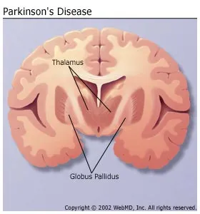
It is thought that the part of the brain called the globus pallidus becomes overactive in Parkinson’s disease. This overactivity acts like a brake and slows or diminishes bodily movement. Pallidotomy surgery permanently destroys the overactive globus pallidus to lessen the symptoms of Parkinson’s disease. This treatment can eliminate rigidity and significantly reduce tremor, bradykinesia, and balance problems. Pallidotomy can also enhance the effect of medication in people with an advanced form of the disease.

What Is Thalamotomy?

It is thought that the abnormal brain activity that causes tremor is processed through the thalamus. Thalamotomy destroys part of the thalamus to block the abnormal brain activity from reaching the muscles and causing tremor. Because thalamotomy is used only to control tremors, it is not generally recommended as a treatment for Parkinson’s disease.

parkinson


Ankle disorders: Causes, symptoms & Treatments

What causes ankle disorders?

The causes of ankle disorders vary, and can include running, jumping, and overuse. Other common causes of ankle sprains and fractures include:

  • twisting or rotating the ankle beyond the normal range of motion
  • tripping or falling
  • landing on the foot with increased force

Other injuries that can lead to tendonitis in the ankle or Achilles tendonitiscan be caused by:

  • lack of conditioning for the muscles in the leg and foot
  • excess strain on the Achilles tendon, the tendon that connects your calf muscles to your heel
  • bone spurs in the heel that rub on the Achilles tendon
  • untreated flat feet leading to additional stress on the posterior tibialis tendon

Different types of arthritis (inflammation of joints and tissues) can also affect the foot and ankle:

  • Osteoarthritis is a degenerative type of arthritis that typically begins in middle age and slowly progresses. Over time, cartilage between your bones becomes worn down. This results in pain and stiffness in your joints.
  • Rheumatoid arthritis is an autoimmune inflammatory disease. It occurs when your body mistakenly attacks its own healthy tissues. RA affects your joints and also destroys cartilage.
  • Post-traumatic arthritis occurs after an injury to your foot or ankle. Stress from the injury can cause your joints to become stiff or inflamed, even years after the injury occurred.

What are the symptoms of ankle disorders?

The symptoms of ankle disorders will vary based on the specific type of injury. Common symptoms include:
  • pain
  • swelling
  • stiffness
  • problems moving the ankle
  • inability to put any weight on the ankle
  • difficulty walking
  • bruising

Tendonitis and Achilles tendonitis may produce similar symptoms, but can be accompanied by:

  • pain along the back of the heel that gets worse throughout the day
  • thickening of the tendon
  • swelling that gets worse with physical activity
  • loud popping sounds at the back of the heel, signaling a ruptured tendon. If this occurs, seek emergency medical attention.

Treatments:

Treatment will depend on your condition and your symptoms. Treatments can include nonsurgical and surgical options. Nonsurgical options involve:

  • taking pain medications
  • taking medications to reduce swelling and inflammation, like aspirin or ibuprofen
  • resting and elevating your ankle
  • applying ice packs to reduce swelling
  • wearing compression bandages or casts to immobilize your ankle
  • getting cortisone (steroid) injections to reduce pain and swelling

Severe fractures or ruptured tendons may require surgical treatment. Surgical procedures used to treat ankle disorders include:

  • reconstruction to rebuild bones, joints, tendons, and ligaments in your ankle
  • removal of damaged tissue (debridement)
  • lengthening the calf muscles to reduce pressure on your Achilles tendon
  • fusion of the bones in your ankle to make it more stable (arthrodesis)
  • replacement of your ankle joint (arthroplasty).


Endometriosis: Causes, Symptoms & Treatments

Endometriosis is a medical condition that occurs when the lining of the uterus, called the endometrium, grows in other places, such as the fallopian tubes, ovaries or along the pelvis. When that lining breaks down, like the regular lining in the uterus that produces the menstruation, it has nowhere to go. This causes cysts, heavy periods, severe cramps and even infertility.

The endometrial tissue may also grow in the vagina, cervix, bowel or bladder, and in rare cases it may spread to other parts of the body, such as the lungs.

The cause of endometriosis is unknown, but researchers have several theories. One theory is that during menstruation, blood with endometrial cells flows back into the fallopian tubes. The cells are rooted there and grow a new lining. Another theory is that the bloodstream carries endometrial cells throughout the body.

It’s also possible that problems with the immune system contribute to the development of endometriosis, when the immune system does not properly detect and destroy endometrial tissue outside of the uterus.

Yet another theory is that abdominal cells that were present since a woman’s embryonic state retain their ability to become endometrial cells. Because endometriosis sometimes run in families, it’s theorized that a person’s genetics can contribute to the development of the condition.

The most common symptom of endometriosis is cramping during menstruation, which can get worse over time, and can be debilitating. The pain is due to internal bleeding from the lining being shed inside the body — in a place where it doesn’t belong — and can also lead to scar tissue formation, blocked fallopian tubes, and bowel problems.

Although many women experience some pain during their periods, women with endometriosis often say their menstrual pain is much worse than usual, and increases over time.

Women should see their doctor if they experience pelvic pain that persists. At first, some women with endometriosis experience pain just before the start of their period, but as the condition progresses, the pain may start in mid-cycle (during ovulation), and continue until after the woman’s period ends.

Women with the condition may also experience pain while having sex, fatigue, painful urination or bowel movements during menstruation and gastrointestinal upset. Infertility and bleeding between periods are also symptoms of endometriosis.

Some women don’t experience any symptoms at all, but realize they have the condition when they are unable to get pregnant.

Diagnosis

The only way for endometriosis to be diagnosed for certain is through laparoscopy, which is a minor surgical procedure that involves your doctor putting a thin scope into your abdomen to view your pelvic organs. If the doctor sees the extra endometrial tissue during the laparoscopy, he or she can also remove it right there to treat the condition.

However, other basic medical tests are typically performed first, before a laparoscopy. These include a pelvic exam, where the doctor manually feels for abnormalities such as cysts, or an abdominal or vaginal ultrasound, which uses sound waves to create an image of the uterus and reproductive organs. Vaginal and abdominal ultrasounds can’t definitively diagnosis endometriosis, but they can test for cysts that may be caused by the condition.

If all other causes of pelvic pain can be ruled out, the doctor can choose to treat the condition surgically or with medicine.

There are several risk factors for endometriosis none of which can really be helped. They include: never having given birth, having a mother with endometriosis, having menstrual cycles that are shorter than 27 days and bleeding that lasts longer than eight days,  having a medical condition that makes the passage of menstrual flow irregular, and having previous damage to cells that line the pelvis.

The biggest complication with endometriosis is fertility problems. About a third to a half of women with endometriosis have difficulties getting pregnant. Infertility can occur because the condition can create adhesions that trap the egg near the ovary, making it difficult for it to travel down the fallopian tube to be fertilized by sperm.

However, that doesn’t mean all women with endometriosis can’t get pregnant – it just might take them a little longer.

Treatment for endometriosis can involve pain medication (for the severe cramping), hormone therapy to slow growth of the endometrial tissue and surgery to remove the tissue. A number of factors go into determining a woman’s treatment, including her age, the severity of her symptoms, and whether the patient wants to become pregnant.

For women who are not trying to become pregnant, hormonal birth control pills are usually the first step in treatment.

For women who are trying to become pregnant, treatment may involve gonadotropin-releasing hormone agonist, which stops ovulation. This treatment is usually recommended for only six months at a time, and pregnancy is possible once treatment is stopped.

For women whose symptoms are not relieved by medication, surgery is another treatment option. Laparoscopy and laparotomy are common forms of surgery used to treat endometriosis. They require going in through the abdominal region to remove the endometrial tissue.

Most women experience relief from pain after surgery, but the pain may come back within two years. Birth-control pills and other medications may be used along with surgery to help extend the pain-free period.

However, if symptoms keep coming back after these surgical procedures, a hysterectomy, or total removal of the uterus, may be a “last resort” option. The condition is less likely to come back after this procedure.


Mini-thoracotomy: Causes, Symptoms & Treatments

What is an atrial septal defect?

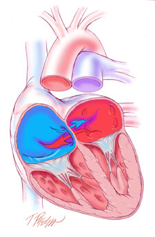
An atrial septal defect or ASD is an opening between the upper two chambers of the heart, known as the right atrium and the left atrium. A congenital heart defect, an ASD permits mixing of deoxygenated blood returning to the heart from the body (right atrium) and freshly oxygenated blood coming from the lungs (left atrium). The degree of mixing is largely related to the size of the defect and the relative pressures in each atrium. Early in life, blood from the left atrium preferentially moves into the right atrium, causing excessive blood to flow through the lungs. If this flow, otherwise known as “shunting” is significant enough, resistance to flow develops in the lung resulting in a gradual reversal of shunting of deoxygenated blood from the right atrium into the left atrium and subsequently through the body. This latter condition can lead to a condition called “cyanosis” whereby inadequately oxygenated blood is delivered to the body causing early fatigue and congestive heart failure.

Atrial Septal DefectAtrial Septal Defect

Causes:

Blood can flow between the two upper heart chambers through the ASD.

When blood flows between the two heart chambers. This is called a shunt. Pressure in the lungs may build up. Over time, there will be less oxygen in the blood that goes to the body.

Small atrial septal defects often cause very few problems, and may be discovered much later in life. Many problems can occur if the opening is large, or if there is more than one opening.  ASD is not very common.

Symptoms:

A person with no other heart defect, or a small defect (less than 5 millimeters) may not have symptoms, or the symptoms may not occur until middle age or later.

Symptoms that do occur may begin at any time after birth through childhood. They can include:

  • Difficulty breathing (dyspnea)
  • Frequent respiratory infections in children
  • Feeling the heart beat (palpitations) in adults
  • Shortness of breath with activity

How is an atrial septal defect treated?

Small atrial septal defects can often be followed conservatively without surgery, due to minimal shunting. Larger ASDs should be closed to prevent the late irreversible consequences of excessive left-to-right shunting. ASDs can be closed surgically by simply sewing them closed or, in the case of larger ASDs, placing a patch of the patient’s own tissue or synthetic material (e.g., Dacron) over it.

How is an atrial septal defect repaired?

ASD repairs require the use of cardiopulmonary bypass, otherwise known as “the heart-lung machine.” This permits the surgeon to safely open the right atrium and access the ASD in a relatively bloodless field. In some cases, the heart is also stopped for 1 to 2 hours to facilitate the repair. Repairs range from relatively simple operations to more complex procedures depending on the location, size, and characteristics of the ASD. The total duration of the operation ranges from 2 to 3 hours.

What are minimally invasive approaches to an atrial septal defect?

The most common surgical approach requires the surgeon to saw open the breastbone and spread the edges apart to gain direct access to the heart. Although this approach provides excellent access to the heart, the resulting wound requires several months to heal completely, an extended recovery period with substantial activity restrictions, and can be subject to serious complications including infection, breakdown, and even death.

Mini-ASD Repair

Minimally Invasive ApproachMinimally Invasive Approach to ASD Closure

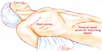
Currently at The Johns Hopkins Hospital, the most commonly employed minimally invasive approach to ASDs is a “mini-thoracotomy” which consists of a 3 inch incision made through the right side of the chest between the ribs. Heart-lung bypass is instituted with small tubes placed in the main artery and vein of the right leg through a 1 to 2 inch incision placed in the right groin crease. The heart is then stopped and the right atrium is opened to expose the ASD. At this point, specialized hand-held “chopstick” like instruments are inserted through this small incision by the surgeon to repair the defect. After the defect is repaired, the heart is then closed and restarted. Finally, heart-lung bypass is discontinued and the incisions are closed.


Systemic Sclerosis: Features and Treatments

Systemic sclerosis (SSc) is a multisystem autoimmune disease in which there is increased fibroblast activity resulting in abnormal growth of connective tissue. This causes vascular damage and fibrosis. Fibrosis occurs in skin, the gastrointestinal (GI) tract and other internal organs.

The name scleroderma is derived from the Greek for ‘hard skin’ and emphasises the dermatological component of the disease. It was described by Hippocrates. There is a localised form of scleroderma, also known as morphoea

General features

  • Fatigue.
  • Weight loss.

Skin features

  • Signs in the hand:
    • Swelling (non-pitting oedema) of fingers and toes – a common early sign; digits may look sausage-like; hand movement may be limited.
    • Skin becomes hard and thickened – this may limit joint movement or cause joint contractures; in the fingers, this is known as sclerodactyly.
    • Swelling and sclerosis reduce hand movements, so patients may be unable to make a fist, or to place the palmar surfaces together – the ‘prayer sign’.
    • Fingertips may have pitting, ulcers or loss of bulk from finger pads.
    • Raynaud’s phenomenon. This is the most common symptom and is present at some point in 90% of cases. Raynaud’s phenomenon with puffy fingers is thought to be a cardinal sign of likely SSc.
  • Calcinosis – nodules or lumps of chalky material which may break through the skin.
  • Face and mouth:
    • Tightening of facial skin.
    • Tight lips (microstomia) – can make dental hygiene difficult.
  • Telangiectasia.
  • ‘Salt and pepper’ appearance of skin, due to areas of hypopigmentation and hyperpigmentation.
  • Dry or itchy skin; reduced hair over affected skin areas.

Musculoskeletal features

  • Joint pain and swelling.
  • Myalgia (due to inflammatory myopathy).
  • Restriction of joint movement, contractures and muscle atrophy due to skin sclerosis.
  • Tendon friction rubs – palpable/audible over the flexor/extensor tendons of the hands, knees and ankles.

GI features

  • Heartburn and reflux oesophagitis.
  • Oesophageal scarring and dysphagia.
  • Delayed gastric emptying – eg, fullness after meals.
  • Reduced small bowel motility – can cause bacterial overgrowth, with bloating, malabsorption, diarrhoea and malnutrition.
  • Constipation due to reduced colonic motility.

Pulmonary features

  • Pulmonary fibrosis (interstitial lung disease):
    • Occurs in as many as 75% of scleroderma patients, but only a few develop end-stage disease.
    • Causes restrictive lung disease.
    • Symptoms and signs: exertional dyspnoea, cough, coarse basal crackles.
  • Pulmonary arterial hypertension (PAH):
    • Occurs in about 10-15% of patients with scleroderma.
    • A leading cause of death in SSc. The presence of PAH drastically reduces survival rate.
    • Symptoms and signs: exertional dyspnoea, syncope, right ventricular strain features.
    • Recent research has attempted to define predictive screening tools. These include monitoring lung function, ECG, urate levels, and N-terminal prohormone of brain natriuretic peptide (NT-proBNP); and by taking into account anti-centromere antibody (ACA) presence and history or presence of telangiectasia.

Management of GI symptoms

For upper GI symptoms:

  • Maintain upright posture after meals; raise the head of the bed; limit alcohol.
  • Proton pump inhibitors.
  • May also need H2-receptor antagonists and pro-motility agents (metoclopramide or domperidone).
  • Dilatation of oesophageal strictures if required.

For intestinal bacterial overgrowth and malabsorption:

  • Cyclical antibiotics.
  • Nutritional advice and nutritional supplements; rarely, parenteral nutrition is required.

For constipation:

  • Dietary fibre and good fluid intake.
  • Softening laxatives (such as lactulose) and/or soluble fibre (such as ispaghula).

Management of pulmonary disease

Pulmonary fibrosis (interstitial lung disease)

  • Benefits of cyclophosphamide seem clear, but must be weighed against side-effects.
  • Supportive treatment: prompt treatment of chest infections – oxygen if needed.

Pulmonary arterial hypertension (PAH)

  • Drug treatment of PAH has improved recently and includes:
    • Endothelin receptor antagonists – eg, bosentan or sitaxsentan.
    • Vasodilators – eg, sildenafil.
    • Prostaglandin derivatives – eg, iloprost (nebulised or intravenous) or epoprostenol (infusion).
  • Supportive treatment – eg, oxygen.

GI complications

See also specific GI sections under ‘Clinical features’ and ‘Management’ headings.

  • Malnutrition due to swallowing problems and other digestive issues.
  • ‘Watermelon stomach’ (gastric antral vascular ectasia):
    • May cause anaemia and GI bleeding.
    • May need endoscopic laser coagulation to prevent bleeding.
  • Obstruction and pseudo-obstruction:
    • Can occur due to reduced motility and bacterial overgrowth.
    • Can be complicated by perforation and peritonitis.
    • Pseudo-obstruction is treated initially by bowel rest and antibiotics.
    • Laparotomy may be needed.
  • Anorectal dysfunction:
    • In some cases, the rectum and anus are involved, causing faecal incontinence.
    • This may require surgery.

Scleroderma renal crisis

  • A serious complication with features of accelerated hypertension.
  • Can lead to renal failure if not treated promptly.
  • Occurs in 5-10% of patients with scleroderma and is more common in those with diffuse or rapidly progressive disease.
  • Presentation:
    • Usually presents as accelerated hypertension with oliguria, headache, fatigue, oedema, rapidly rising serum creatinine levels, proteinuria and microscopic haematuria.
    • Scleroderma renal crises can occur with apparently normal blood pressure, but the blood pressure is higher than baseline values – hence the importance of regular blood pressure monitoring.
  • Treatment is with angiotensin-converting enzyme (ACE) inhibitors, plus dialysis if necessary.
  • Testing patients with scleroderma for anti-RNA polymerase III antibodies may help identify at-risk patients.

Pulmonary complications

  • Aspiration pneumonia from severe reflux.
  • Respiratory muscle weakness if there is severe myositis or extensive skin disease involving the chest.
  • Pneumothorax

Cardiac complications

  • Many different cardiac abnormalities can be associated with SSc. These include:
    • Microvascular coronary artery disease (with resultant myocardial ischemia).
    • Myocardial fibrosis.
    • Left ventricular (LV) systolic dysfunction, LV diastolic dysfunction.
    • Pericarditis or pericardial effusion; these may cause cardiac impairment or congestive cardiac failure.
    • Arrhythmias and conduction defects (including bradyarrhythmias and tachyarrhythmias).
  • The wide variety of abnormalities makes it difficult to assess prevalence. It is likely that the subclinical cardiac involvement rates are very high.
  • Treatment is according to the clinical features.

Other complications

  • Erectile dysfunction.
  • Depression.
  • Osteoporosis – due to reduced blood flow.
  • Hypothyroidism may be associated.


Treatments:

There is no cure for SSc, and management consists of controlling symptoms and preventing complications.

Monitoring

Regular monitoring and reviews are aimed at early detection and treatment of complications. Monitoring includes:

  • Regular review of symptoms.
  • Blood pressure monitoring.
  • Renal function monitoring.
  • Lung function tests and chest CT scan.
  • ECG and echocardiography.

Non-drug treatments

  • Patient involvement and education:
    • ‘Expert patient’ programmes and the Scleroderma Society.
    • Awareness of urgent problems such as renal crisis or intestinal obstruction symptoms.
  • Physiotherapy to promote joint mobility and muscle strength.
  • Home exercises to maintain range of motion (such as gentle mouth, face and hand stretches).
  • Avoid tobacco and maintain healthy weight.
  • Nutritional advice, and supplements if needed.
  • For Raynaud’s phenomenon:
    • Prevention – avoid cold and trauma; use warm clothing or heated clothing.
    • For an attack – warm the body, hands and feet gently (the skin may be numb and unable to feel if the heat source is too hot); use gentle arm movements or gentle massage to help restore circulation.
  • Occupational therapists – for adaptations to assist in daily living.
  • Camouflage products – for cosmetic help with skin changes.


Bone Cancer: Causes, Symptoms and Treatments

Bone cancer can be distinguished into primary bone cancer or secondary bone cancer. Primary bone cancer starts in the bone, where the cancer spreads within the cells of the bone. Secondary cancer, on the other hand, starts elsewhere in the body and spreads to the bones. Examples of primary bone cancer include steosarcoma, Ewing sarcoma, malignant fibrous histiocytoma, and chondrosarcoma.

Causes:

Although bone cancer does not have a clearly defined cause, researchers have identified several factors that increase the likelihood of developing these tumours.

Osteosarcoma occurs more frequently in people who have had high-dose external radiation therapy or treatment with certain anticancer drugs; children seem to be particularly susceptible.

A small number of bone cancers are due to heredity. For example, children who have had hereditary retinoblastoma (an uncommon cancer of the eye) are at a higher risk of developing osteosarcoma, particularly if they are treated with radiation.

Additionally, people who have hereditary defects of bones and people with metal implants, which doctors sometimes use to repair fractures, are more likely to develop osteosarcoma.

The following groups of people may be at a higher risk of developing bone cancer:

  • Being a child or very young adult – most cases of bone cancer occurs in children or young adults aged up to 20
  • Patients who have received radiation therapy (radiotherapy)
  • People with a history of Paget’s disease
  • People with a close relative (parent or sibling) who has/had bone cancer
  • People with Li-Fraumeni syndrome – a rare genetic condition
  • Babies born with an umbilical hernia

Symptoms:

The most common symptom of bone tumours is pain, which gradually increases over time. A person may go weeks, months, and sometimes years before seeking help; the pain increases with the growth of the tumour.

Additional symptoms may include:

  • Fatigue
  • Fever
  • Anemia
  • Unexplained bone fractures

Many patients will not experience any symptoms, except for a painless mass. Some bone tumours may weaken the structure of the bone, causing pathologic fractures.

Treatments:

The type of treatment for bone cancer depends on several factors, including what type of bone cancer it is, where it is located, how aggressive it is, and whether it is localized or has spread. There are three approaches to bone cancer:

  • Surgery
  • Radiotherapy (radiation therapy)
  • Chemotherapy

Surgery – the aim is to remove the tumour, all of it if possible, and some of the bone tissue that surrounds it. If some of the cancer is left behind after surgically removing the tumour it may continue to grow and eventually spread. Limb sparing surgery, also known as limb salvage surgery means that surgical intervention occurs without having to amputate the limb. The surgeon may take some bone from another part of the body to replace lost bone (bone graft), or an artificial bone may be put in. In some cases, however, amputation of a limb may be necessary.

Radiation therapy is also known as radiotherapy, radiation oncology and XRT. Approximately 40% of patients of all types of cancer undergo some kind of radiotherapy. It involves the use of beams of high-energy X-rays or particles (radiation) to destroy cancer cells. Radiotherapy works by damaging the DNA inside the tumour cells, destroying their ability to reproduce. Radiotherapy can be used for different reasons:

  • Total Cure – to cure the patient by completely destroying the tumour
  • To alleviate symptoms – radiotherapy is often used to relieve pain in more advanced cancers
  • Neo-adjuvant radiotherapy (before surgery) – if a tumour is large, radiotherapy can shrink it, making it easier and less harmful to then surgically remove it
  • Adjuvant radiotherapy – given after surgery. The aim is to eliminate the cancer cells that remained behind
  • Combination therapy (radiotherapy combined with another type of therapy) – in some cases, chemo radiation – radiotherapy combined with chemotherapy – is more effective

Chemotherapy is the use of chemicals (medication) to treat disease – more specifically, it usually refers to the destruction of cancer cells. Cytotoxic medication prevents cancer cells from dividing and growing. In general, chemotherapy has 5 possible goals:

  • Total remission – to cure the patient completely. In some cases chemotherapy alone can get rid of the cancer completely
  • Combination therapy – chemotherapy can help other therapies, such as radiotherapy or surgery have more effective results
  • Delay/Prevent recurrence – chemotherapy, when used to prevent the return of a cancer, is most often used after a tumour is removed surgically
  • Slow down cancer progression – used mainly when the cancer is in its advanced stages and a cure is unlikely. Chemotherapy can slow down the advancement of the cancer
  • To relieve symptoms – also more frequently used for patients with advanced cancer

Idiopathic Thrombocytopenic Purpura (ITP): Causes, Symptoms and Treatments

What is Idiopathic Thrombocytopenic Purpura (ITP)?

Idiopathic thrombocytopenic purpura (ITP) is a platelet disorder that occurs in people who have an abnormally low number of platelets in the blood. Platelets are essential in forming blood clots, which consist of a mass of fibers and blood cells. Platelets travel to a damaged or cut area of the body and stick together to form a clot. Having fewer platelets can result in easy bruising, bleeding gums and internal bleeding.

Idiopathic means the cause is unknown.
Thrombocytopenia means a decreased number of platelets in the blood.
Purpura refers to the purple discoloring of the skin, as with a bruise.

Acute thrombocytopenic purpura is most commonly seen in young children (2 to 6 years old). The symptoms may follow a viral illness, such as chicken pox. Acute ITP usually has a very sudden onset, and the symptoms usually disappear in less than six months (often within a few weeks). The disorder usually does not recur. Acute ITP is the most common form of the disorder.
Chronic thrombocytopenic purpura can happen at any age, and the symptoms can last a minimum of six months or several years. It is more common in adults than in children, but it does affect adolescents. Two to three times more females have ITP than males. Chronic ITP can recur often and requires continual follow-up care with a hematologist.
What are the causes of ITP?

In most cases, the cause of ITP is unknown. It is not contagious, meaning a child cannot “catch it” from playing with another child with ITP. It also is important to know that nothing the parents or the child did caused the disorder.

Often, the child may have had a virus or viral infection approximately three weeks before developing ITP. It is believed that the body’s immune system, when making antibodies to fight against a virus, “accidentally” also made an antibody that can stick to the platelet cells. The body recognizes any cells with antibodies as foreign cells and destroys them. Doctors think that in people who have ITP, platelets are being destroyed because they have antibodies. That is why ITP is also referred to as immune thrombocytopenic purpura.

Although there has been research looking at whether certain medications can cause ITP, no direct link has been made with any specific medication that may cause ITP.

What are the symptoms of ITP?

A normal platelet count is 150,000 to 450,000. With ITP, the platelet count is less than 100,000. By the time significant bleeding occurs, the child may have a platelet count of less than 10,000. The lower the platelet count, the greater the risk of bleeding.

Because platelets help stop bleeding, the symptoms of ITP are related to increased bleeding. However, each child may experience symptoms differently. Symptoms can include:

  1. Purpura, which is the purple color of the skin after blood has “leaked” under it.
  2. A bruise is blood under the skin.
  3. Children with ITP can have large bruises from no known trauma. Bruises can appear at the joints of elbows Petechia, which are tiny red dots under the skin caused by very small bleeds.
  4. Nosebleeds.
  5. Bleeding in the mouth and/or in and around the gums.
  6. Blood in the vomit, urine or stool.
  7. Bleeding in the head, which is the most dangerous symptom of ITP.

Any head trauma that occurs when there are not enough platelets to stop the bleeding can be life-threatening.
The symptoms of ITP can resemble other blood disorders or medical problems. Always consult your child’s physician for a diagnosis.

How is ITP diagnosed?

In addition to a complete medical history and physical examination, diagnostic procedures for ITP can include:

Complete blood count (CBC), which is a measurement of size, number, and maturity of different blood cells in a specific volume of blood.
Additional blood and urine tests to measure bleeding time and detect possible infections.
Careful review of the child’s medications.
A bone marrow aspiration, which involves taking a small amount of bone marrow fluid, to look at the production of platelets and to rule out that any abnormal cells the marrow may be producing could lower platelet counts.
Treatment

A child’s specific treatment plan, including the frequency of visits, the length of treatment, side effects and long-term effects are determined by the:

Child’s age, overall health, and medical history
Extent of the disease
Child’s tolerance for specific medications, procedures, or therapies
Expectations for the course of the disease
The patient and family’s opinion or preference
Not all children with ITP require treatment. Close monitoring of a child’s platelets and prevention of serious bleeding complications may be the course of action chosen until the body is able to correct the disorder on its own.

When treatment is necessary, the two most common forms of treatment are:

Steroids, which can help prevent bleeding by decreasing the rate of platelet destruction. Steroids, if effective, will increase platelet counts within two to three weeks.
Intravenous gamma globulin (IVGG), a protein that contains many antibodies and also slows the destruction of platelets. It works more quickly than steroids (within 24 to 48 hours).
Other treatments for ITP can include:

Immune globulin, which is a medication that temporarily stops the spleen from destroying platelets. A child must be Rh positive and have a spleen for this medication to be effective.
Medication changes, because when a medication is the suspected cause, stopping the use of or changing the medication may be necessary.
Infection treatment to increase platelet counts, if infection is the cause for ITP.
Splenectomy to remove a child’s spleen if the spleen is the site of platelet destruction. This is considered more often in older children with chronic ITP.
Hormone therapy in teenage girls to stop their menstrual cycle when their platelets are low if excessive bleeding occurs.
Preventing bleeding

Parents of a child with ITP need to be aware of how to prevent injuries and bleeding. Consider the following:

Making the environment as safe as possible for young children by padding a crib, having the child wear a helmet, and providing protective clothing when platelet counts are low.
Restricting contact sports, riding bicycles, and rough play may be necessary.
Avoiding medications that contain aspirin, ibuprofen, naprosen, or other non-steroidal anti-inflammatory drugs because they may interfere with the body’s ability to control bleeding.
Discuss prevention measures with the child’s physician.

Pages:12