Global Treatment Services Pvt. Ltd.

Global Treatment Services

Myomas: Causes, Symptoms & Treatments

Uterine fibroids are noncancerous growths of the uterus that often appear during childbearing years. Also called leiomyomas or myomas, uterine fibroids aren’t associated with an increased risk of uterine cancer and almost never develop into cancer.

Fibroids range in size from seedlings, undetectable by the human eye, to bulky masses that can distort and enlarge the uterus. You can have a single fibroid or multiple ones. In extreme cases, multiple fibroids can expand the uterus so much that it reaches the rib cage.

fibroids


Symptoms:

  • Heavy menstrual bleeding
  • Menstrual periods lasting more than a week
  • Pelvic pressure or pain
  • Frequent urination
  • Difficulty emptying the bladder
  • Constipation
  • Backache or leg pains

Rarely, a fibroid can cause acute pain when it outgrows its blood supply, and begins to die.

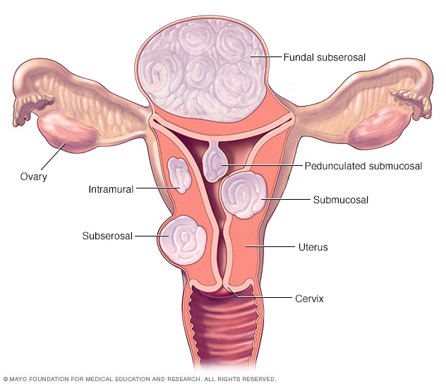
Fibroids are generally classified by their location. Intramural fibroids grow within the muscular uterine wall. Submucosal fibroids bulge into the uterine cavity. Subserosal fibroids project to the outside of the uterus.

When to see a doctor

See your doctor if you have:

  • Pelvic pain that doesn’t go away
  • Overly heavy, prolonged or painful periods
  • Spotting or bleeding between periods
  • Difficulty emptying your bladder

Seek prompt medical care if you have severe vaginal bleeding or sharp pelvic pain that comes on suddenly.

Causes

Doctors don’t know the cause of uterine fibroids, but research and clinical experience point to these factors:

  • Genetic changes. Many fibroids contain changes in genes that differ from those in normal uterine muscle cells.
  • Hormones. Estrogen and progesterone, two hormones that stimulate development of the uterine lining during each menstrual cycle in preparation for pregnancy, appear to promote the growth of fibroids. Fibroids contain more estrogen and progesterone receptors than normal uterine muscle cells do. Fibroids tend to shrink after menopause due to a decrease in hormone production.
  • Other growth factors. Substances that help the body maintain tissues, such as insulin-like growth factor, may affect fibroid growth.


Treatments:

  • Uterine artery embolization.
  • Myolysis.
  • Laparoscopic or robotic myomectomy.
  • Hysteroscopic myomectomy.
  • Endometrial ablation

Risk factors

There are few known risk factors for uterine fibroids, other than being a woman of reproductive age. Other factors that can have an impact on fibroid development include:

  • Heredity. If your mother or sister had fibroids, you’re at increased risk of developing them.
  • Race. Black women are more likely to have fibroids than women of other racial groups. In addition, black women have fibroids at younger ages, and they’re also likely to have more or larger fibroids.
  • Environmental factors. Onset of menstruation at an early age; use of birth control; obesity; a vitamin D deficiency; having a diet higher in red meat and lower in green vegetables, fruit and dairy; and drinking alcohol, including beer, appear to increase your risk of developing fibroids.

Carcinoid Tumors: Causes, Symptoms & Treatment

All carcinoid tumors, wherever they show up, affect cells that make hormones. They’re part of a group of diseases called neuroendocrine tumors (NETs). Most carcinoid tumors start in one of two areas: your lungs or your digestive system, also known as the GI tract. That includes places like your stomach, small intestine, colon, appendix, or rectum.It’s not as common, but sometimes the tumors start in your pancreas, your testicles if you’re a man, or ovaries if you’re a woman. Keep in mind that these tumors often grow slowly. And doctors often find them when they’re at an early stage, which makes them easier to treat.

Doctors don’t know for sure why people get them. But a few things may put you at a higher risk.Genetic disease. You may get carcinoid tumors if you have an illness called multiple endocrine neoplasia type 1 (MEN1). It’s a disease that’s passed down through your family. About 10% of these tumors are due to MEN1.Another condition that can raise your risk for them is neurofibromatosis type 1.Race. More African-Americans than whites get carcinoid tumors in the GI tract.Gender. Women are slightly more likely than men to have this type of cancer.

Age. Most people are diagnosed with carcinoid tumors in their 40s or 50s.

Conditions.

Symptoms:

When carcinoid tumors form on cells that make hormones, the tumors can start to make hormone-like substances of their own. This can cause a variety of symptoms, depending on where this is going on.

For instance, if you’ve got the tumors in your GI tract, you might notice things like this happening to you:

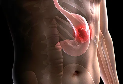
stomach cancer illustration

  • Pain in your belly
  • Diarrhea
  • Constipation
  • Feel nauseated or throw up
  • Can’t breathe right
  • Get blood in your stool
  • Lose weight
  • Feel short of breath or wheeze
  • Heartbeat starts to speed up
  • Feel weak
  • Hair starts to grow on your body and face

If you have a lung carcinoid tumor, you might get symptoms like a cough, and you sometimes might cough up some bloody mucus. You also might hear a whistling sound while you breathe, called wheezing.

When you have this type of cancer for many years, you might get a condition called carcinoid syndrome. It’s a group of symptoms that start when the tumors release certain hormones into your bloodstream.

Diagnosis:

These are diagnosis & initial investigation options for carcinoid tumors;

Biospy

Blood Urine Test

Upper endoscopy

Colonoscopy.

Capsule endoscopy.

CT, or computed tomography.

MRI, or magnetic resonance imaging

Radionuclide scanning.

Treatments options are below:

  • Surgery
  • Chemoembolization
  • Hormone therapy
  • Immunotherapy
  • Radioembolization
  • Targeted therapy


Hemangiomas: Causes, Symptoms & Treatments

Hemangiomas, or infantile hemangiomas, are noncancerous growths of blood vessels. They’re the most common growths or tumors in children. They usually grow for a period of time and then subside without treatment.

They don’t cause problems in most infants. However, some hemangiomas may open and bleed or ulcerate. This may be painful. Depending on their size and location, they may be disfiguring. Additionally, they may occur with other abnormalities of the central nervous system or spine.

The growths may also occur with other internal hemangiomas. These affect internal organs such as the liver, other parts of the gastrointestinal system, the brain, or organs of the respiratory system. The hemangiomas that affect organs usually don’t cause problems.

On the skin

Hemangiomas of the skin develop when there is an abnormal proliferation of blood vessels in one area of the body. Experts aren’t sure why blood vessels group together like this, but they believe it’s caused by certain proteins produced in the placenta during gestation (the time when you’re in the womb).

Hemangiomas of the skin can form on the top layer of skin or on the fatty layer underneath, called the subcutaneous layer. At first, a hemangioma may appear to be a red birthmark on the skin. Slowly, it will start to protrude upward from the skin.

On the liver

Hemangiomas of the liver form in and on the liver’s surface. These hemangiomas are thought to be sensitive to estrogen. During menopause, many women are prescribed replacement estrogen to minimize symptoms caused by the decline of their natural estrogen levels. This excess estrogen can spur the growth of liver hemangiomas. Similarly, pregnancy and sometimes oral contraceptive pills can increase the size of hemangiomas.

Where they occur

Besides the skin and liver, hemangiomas can grow on other areas within the body, such as the:

  • kidneys
  • lungs
  • colon
  • brain

Hemangiomas that grow in the brain cavities or other cavities within the body are called cavernous hemangiomas.

Symptoms:

Hemangiomas inside the body usually go unnoticed until they grow large or until multiple hemangiomas form. Some symptoms that indicate you might have an internal hemangioma include:

  • nausea
  • vomiting
  • abdominal discomfort
  • loss of appetite
  • unexplained weight loss
  • a feeling of fullness in the abdomen

Treatments:

A single, small hemangioma usually requires no treatment. It will likely go away on its own. However, some cases require treatment, such as skin hemangiomas that develop lesions or sores.

  1. Corticosteroid medication
  2. Beta-blockers
  3. Laser treatment
  4. Medicated gel
  5. Surgery

For hemangiomas on the organs

Hemangiomas within the body may require treatment if they grow too large or cause pain. Treatment options for these hemangiomas include:

  • surgical removal of the hemangioma
  • surgical removal of the damaged organ or damaged area
  • tying off of the main artery that supplies blood to the hemangiomas

GOUT: Causes, Symptoms & Treatments

Causes

Gout is caused initially by an excess of uric acid in the blood, or hyperuricemia. Uric acid is produced in the body during the breakdown of purines – chemical compounds that are found in high amounts in certain foods such as meat, poultry, and seafood.

Normally, uric acid is dissolved in the blood and is excreted from the body in urine via the kidneys. If too much uric acid is produced, or not enough is excreted, it can build up and form needle-like crystals that trigger inflammation and pain in the joints and surrounding tissue.

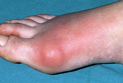
Inflamed toe joint in patient with gout

red, swollen index finger caused by acute gout

Symptoms

Gout usually becomes symptomatic suddenly without warning, often in the middle of the night.

The main symptoms are intense joint pain that subsides to discomfort, inflammation, and redness.

Gout frequently affects the large joint of the big toe, but can also affect the ankles, knees, elbows, wrists, and fingers.

TYPES

There are various stages through which gout progresses, and these are experienced as the different types of gout.

Asymptomatic hyperuricemia

It is possible for a person to have elevated uric acid levels without any outward symptoms. At this stage, treatment is not required, though urate crystals are being deposited in tissue and causing slight damage.

People with asymptomatic hyperuricemia may be advised to take steps to address any possible factors contributing to uric acid build-up.

Acute gout

This stage occurs when the urate crystals that have been deposited suddenly cause acute inflammation and intense pain. This sudden attack is referred to as a “flare” and will normally subside within 3 to 10 days. Flares can sometimes be triggered by stressful events, alcohol and drugs, as well as cold weather.

Interval or intercritical gout

This stage is the period in between attacks of acute gout. Subsequent flares may not occur for months or years, though if not treated, over time, they can last longer and occur more frequently. During this interval, further urate crystals are being deposited in tissue.

Chronic tophaceous gout

Chronic tophaceous gout is the most debilitating type of gout. Permanent damage may have occurred in the joints and the kidneys. The patient can suffer from chronic arthritis and develop tophi, big lumps of urate crystals, in cooler areas of the body such as the joints of the fingers.

It takes a long time without treatment to reach the stage of chronic tophaceous gout – around 10 years. It is very unlikely that a patient receiving proper treatment would progress to this stage.

Pseudogout

One condition that is easily confused with gout is pseudogout. The symptoms of pseudogout are very similar to those of gout.

The major difference between gout and pseudogout is that the joints are irritated by calciumphosphate crystals rather than urate crystals. Pseudogout requires different treatment to gout.

Treatment

person with gout

Gout patients often have acute inflammation around their joints.

The majority of gout cases are treated with medication. Medication can be used to treat the symptoms of gout attacks, prevent future flares, and reduce the risk of gout complications such as kidney stones

Benign Lung Nodules and Tumors : Causes, Symptoms & Treatments

A nodule is a “spot on the lung,” seen on an X-ray or computed tomography (CT) scan. In fact, a nodule shows up on about one in every 500 chest X-rays. Normal lung tissue surrounds this small round or oval solid overgrowth of tissue. It may be a single or solitary pulmonary nodule. Or, you may have multiple nodules.
Your lung nodule is more likely to be benign if:

  • You are younger than age 40.
  • You are a nonsmoker.
  • There is calcium in the nodule.
  • The nodule is small.

A benign lung tumor is an abnormal growth of tissue that serves no purpose and is found not to be cancerous. Benign lung tumors may grow from many different structures in the lung.

Causes:

Inflammation from infections such as:

  • Tuberculosis (TB)
  • A lung abscess
  • Round pneumonia (rare in adults)

Inflammation from noninfectious causes such as:

  • Rheumatoid arthritis
  • Wegener granulomatosis
  • Sarcoidosis

Symptoms: 

  • Wheezing
  • Coughing that lasts or coughing up blood
  • Shortness of breath
  • Fever, especially if pneumonia is present

Benign lung nodules and tumors usually cause no symptoms. This is why they are almost always found accidentally on a chest X-ray or CT scan. However, they may lead to symptoms like these

These are some of the more common types of benign lung tumors:

  • Hamartomas are the most common type of benign lung tumor and the third most common cause of solitary pulmonary nodules. These firm marble-like tumors are made up of tissue from the lung’s lining as well as tissue such as fat and cartilage. They are usually located in the periphery of the lung.
  • Bronchial adenomas make up about half of all benign lung tumors. They are a diverse group of tumors that arise from mucous glands and ducts of the windpipe or large airways of the lung. A mucous gland adenoma is an example of a true benign bronchial adenoma.
  • Rare neoplasms may include chondromas, fibromas, or lipomas — benign tumors made up of connective tissue or fatty tissue.

Treatments:

In many cases, your doctor may simply observe a suspicious lung nodule with multiple chest X-rays over several years. However, your doctor may suggest a biopsy or removal of an entire nodule in situations like these:

  • You are a smoker and the nodule is large.
  • You have symptoms.
  • A scan suggests the nodule might be cancerous.
  • The nodule has grown.

Surgery can often be done with small incisions and a short hospital stay. If your nodule is benign, you will not need any further treatment, except to manage any underlying problems or complications related to the nodule such as pneumonia or an obstruction.

If you need invasive surgery to remove a tumor, your doctor may recommend one or more tests beforehand to ensure your health. These might include blood tests or kidney, liver, or pulmonary (lung) function tests.

DCIS(Ductal Carcinoma In Situ ): Causes, Symptoms & Treatments

Breast cancer(DCIS) is an uncontrolled growth of breast cells. To better understand breast cancer, it helps to understand how any cancer can develop.

Cancer occurs as a result of mutations, or abnormal changes, in the genes responsible for regulating the growth of cells and keeping them healthy. The genes are in each cell’s nucleus, which acts as the “control room” of each cell. Normally, the cells in our bodies replace themselves through an orderly process of cell growth: healthy new cells take over as old ones die out. But over time, mutations can “turn on” certain genes and “turn off” others in a cell. That changed cell gains the ability to keep dividing without control or order, producing more cells just like it and forming a tumor.

Causes:

Diagnosing DCIS usually involves a combination of procedures:

  • Physical examination of the breasts: Your doctor may be able to feel a small lump in the breast during a physical examination, although a noticeable lump is rare with DCIS. In cases when DCIS cannot be felt during a physical exam, it can often be detected using mammography.
  • Mammography: DCIS is usually found by mammography. As old cancer cells die off and pile up, tiny specks of calcium (called “calcifications” or “microcalcifications”) form within the broken-down cells. The mammogram will show the cancer cells inside the ducts as a cluster of these microcalcifications, which appear either as white specks or as a shadow.
  • Biopsy: If you do have a suspicious mammogram, your doctor will probably want you to have a biopsy. There are two ways to get a biopsy done with only a little bit of surgery (more invasive biopsies are rarely needed for DCIS):
    • Fine needle aspiration biopsy: A very small, hollow needle is inserted into the breast. A sample of cells is removed and examined under the microscope. This method leaves no scars.
    • Core needle biopsy: A larger needle is inserted to remove several bigger samples of tissue from the area that looks suspicious. In order to get the core needle through the skin, the surgeon must make a tiny incision. This leaves a very tiny scar that is barely visible after a few weeks.

    If a needle biopsy is not able to remove cells or tissue, or it does not give definite results (inconclusive), a more involved biopsy may be necessary. These biopsies are more like regular surgery than needle biopsies:

    • Incisional biopsy: Incisional biopsy removes a small piece of tissue for examination.
    • Excisional biopsy: Excisional biopsy attempts to remove the entire suspicious lump of tissue from the breast.

Biopsies are done only to make the diagnosis. If DCIS is diagnosed, more surgery is needed to ensure all of the cancer is removed along with “clear margins,” which means that a border of healthy tissue around the cancer is also removed. Usually this means having lumpectomy, or in some cases (a large area of DCIS, for example), mastectomy.

After the biopsy, the pathologist analyzes the piece of breast tissue and reports back on the:

  • type and grade of the DCIS: how abnormal the cells look when compared with normal breast cells, and how fast they are growing
  • hormone-receptor status: Whether or not the cancer cells have receptors (proteins in a cell that receive messages from hormones) for the hormones estrogen and/or progesterone. If estrogen and/or progesterone receptors are present, this means that the cancer cells’ growth is fueled by these hormones.

Type and grade of DCIS

All DCIS is considered stage 0 breast cancer — the earliest stage possible. “Stage” describes how far the cancer has spread beyond the site of the original tumor. Even though DCIS is always considered stage 0, it can be any size and be located in any number of areas inside the breast.

Knowing the type and grade of DCIS can help you and your doctor decide on the best treatment for you.

When a pathologist looks at the tissue removed during the biopsy, he or she determines whether or not any abnormal cells are present. If abnormal cells are present, the pathologist will note how different the cells look compared with normal, healthy breast cells. The image shows the range of possible findings, from normal cells all the way to invasive ductal cancer.

Range of DCIS growth

Breast cancer: Stages

  • Normal cells
  • Ductal hyperplasia or “overgrowth” means that too many cells are present.
  • Atypical ductal hyperplasia means that there are too many cells (hyperplasia) and they are starting to take on an abnormal appearance (atypical or “not typical”).
  • Ductal carcinoma in situ (DCIS) means that there are too many cells and they have the features of cancer, but they are still confined to the inside of the duct. (DCIS is stage 0 breast cancer.)
  • DCIS-MI (DCIS with microinvasion) means that a few of the cancer cells have started to break through the wall of the duct. DCIS-MI is stage I breast cancer.
  • Invasive ductal cancer (IDC) means that the cancer cells have broken beyond the breast duct. The breast cancer is no longer a DCIS but an invasive ductal carcinoma, the most common type of breast cancer. IDC can be diagnosed at any stage from I-IV.

Symptoms:

DCIS generally has no signs or symptoms. A small number of people may have a lump in the breast or some discharge coming out of the nipple. About 80% of DCIS cases are found by mammography.

Treatments:

  • Lumpectomy followed by radiation therapy: This is the most common treatment for DCIS. Lumpectomy is sometimes called breast-conserving treatment because most of the breast is saved.
  • Mastectomy: Mastectomy, or removal of the breast, is recommended in some cases.
  • Lumpectomy alone(radiation therapy)
  • Hormonal therapy after surgery: These treatments, which block or lower the amount of estrogen in the body, are typically used if the DCIS tests positive for hormone receptors.

Chemotherapy, a form of treatment that sends anti-cancer medications throughout the body, is generally not needed for DCIS. DCIS is non-invasive and remains within the breast duct, so there is no need to treat cancer cells that might have traveled to other areas of the body.

Orchiectomy : Procedure details

If you’ve been diagnosed with testicular cancer, your doctor has probably talked to you about orchiectomy, surgery to remove one or both testicles. Testicles, or testes, are the male sex organs that make sperm and the hormone testosterone.

Why Get Orchiectomy?

It’s usually needed to treat testicular cancer. But it can also help if a testicle is damaged by infection or injury. Sometimes it’s part of prostate or breast cancer treatment.

When doctors suspect you have cancer, they usually take off a piece of a tumor and look for telltale cells under a microscope. They can’t really do that with testicular cancer because there’s the risk the cancer will spread. Instead, they almost always do what’s called a radical inguinal orchiectomy.

It’s called “radical” because it removes the spermatic cord along with the testicle and tumor. The cord has blood and lymph vessels that could let the cancer spread to other parts of the body.

In a simple orchiectomy, the doctor only removes one or both testicles. This surgery can ease symptoms, prevent problems from prostate cancer, and treat male breast cancer.

Precautions before Surgery:

If you still have one testicle, you should still be able to get an erection and have sex. If both are removed, your body won’t be able to make sperm. If you want to have children, you may want to store sperm before the procedure. Talk to your doctor to plan ahead.

Epilepsy (apasmara) : Ayurvedic Treatments

Epilepsy

The major sign in this condition is the occurrence of seizures which happen when our brain cells that communicate through electric impulses start sending out the wrong signals. These seizures can last anywhere from several seconds to a few minutes. Having just one seizure does not mean you have epilepsy. Generally, the occurrence of several seizures are a clear indication of epilepsy.

Epilepsy and Ayurveda

Epilepsy is called Apasmara (Apa = loss, smara = memory, intelligence and/or consciousness). Akshepaka is a disease that is characterized by convulsions. In Ayurveda, the convulsions are caused due to an imbalance of the Vata dosha. The loss of consciousness in terms of doshic influence can be attributed to Pitta dosha. When one loses consciousness soon after seeing the colors red, blue and black and also recovers quickly, this is considered as a sign of Vata disturbance. Similarly, Pitta disturbances happen due to seeing the colors light or dark red or yellow and recovering with heavy sweating.

Convulsions in Apasmara are followed by losing consciousness and frothing at the mouth. Due to its cardinal sign of loss of consciousness or memory, epilepsy or epileptic attacks are commonly known as the ‘falling disease’ or ‘fits.’ It is a serious disorder of the central nervous system that affects both children and adults alike.

Depending on the dominance of the three doshas and their combined effect, it is classified into four types.

A fifth type, called Yoshapasmara, which is more prevalent among women. Yoshapasmara is another type of epileptic seizure elaborated in our texts and finds relevance in the condition of hysteria. Many a times, it is ignored as a tendency or hereditary factor. This has grave impact on the growth and well-being of the female suffering and the people associated with her. But on a positive note, it is treatable and can bring about a great change in the person afflicted by this.

Types of Epileptic Seizures

Seizures are classified in two main categories:
1. Partial seizures involve a part of the brain. They can be:

  • Simple partial seizures. Symptoms may include involuntary twitching of the muscles or arms and legs; changes in vision; vertigo; and having unusual tastes or smells.
    The person does not lose consciousness.
  • Complex partial seizures. Symptoms may be like those of partial seizures, but the person does lose awareness for a time. The person may do things over and over, like walking in a circle, rubbing the hands together, or staring into space.

2. Generalized seizures involve much more or all of the brain. They can be:

  • Absence seizures (petit mal). Symptoms may include staring and brief loss of consciousness.
  • Myoclonic seizures. Symptoms may include jerking or twitching of the limbs on both sides of the body.
  • Tonic-clonic seizures (grand mal). Symptoms may include loss of consciousness, shaking or jerking of the body, and loss of bladder control. The person may experience an aura or an unusual feeling before the seizure starts. These seizures can last from 5 to 20 minutes.

Causes of Epilepsy

According to Ayurveda, the causes of epilepsy could be kama (passion), krodha (anger), lobha (greed), moha (temptation), harsha (ectasy), soka (grief) chinta (worry), udvega (anxiety), etc.
Today, we can largely group the causes as:

  • Hereditary influences
  • Serious shock or injury to brain or nervous system
  • Diseases like meningitis and typhoid
  • Allergic reaction to certain food substances
  • Circulatory disorders like a stroke or a heart attack
  • Fevers
  • Drug abuse or overuse: Chronic alcoholism, Lead poisoning, Use of hallucinogens and stimulants like cocaine, Antidepressants or sleep aids like benzodiazepines and barbiturates
  • Mental conflict: Long term grief, passion or anger, Subconscious fear or hatred
  • Deficient mineral assimilation like inadequate hemoglobin or essential minerals like magnesium
  • Consumption of junk/processed foods and absence of a regularized lifestyle habit for those with epileptic tendencies

Symptoms of Epilepsy

Presymptomatic phase

  • Involuntary twitching of eyebrows and rapid deviating movements of the eyes
  • Drooling
  • Rigidity of the muscles
  • Fatigue
  • Feeling of spasms or congestion in the heart
  • Disinterest in food
  • Hallucination or hearing sounds or voices
  • Body ache

Symptomatic phase

  • In case of petit mal, there is momentary loss of consciousness with no convulsions except a slight rigidity. In this case, the attack stops within a few seconds.
  • Grand mal, on the other hand, has a more pronounced effect. Violent convulsions accompanied by a sudden loss of consciousness, twitching of the muscles, biting of the tongue, distorted fixation of limbs, rotation of the head and deviation of the eyes continues for much longer.
  • Mild or vigorous shaking of arms and legs
  • Head retraction to one side
  • Constriction of fingers
  • Accidental urination
  • Making sounds like whimpering or crying
  • Vomiting which could be frothing around the mouth or emesis of stomach contents
  • Loss of consciousness or blacking out

Seizure First Aid: What to Do During

  • Firstly, it is necessary to understand that thinking someone may swallow their tongue during a seizure is nothing but a common myth.
  • One must avoid putting anything in the mouth of the patient during an attack.
  • Do not try to restrain the person in any way. If possible, remove the individual’s eyeglasses, tie, and/or scarf.
  • Turn the person to their side. This can help saliva flow out of the mouth by clearing the airways.
  • Do not try to feed food or drink to the person at the time or soon after. This could cause choking.

Effects of Epilepsy on Everyday Life

Living with epilepsy can be tough and cause the sufferer many social problems. A person suffering from such a condition usually also gets attacks of depression, withdrawal from society, loss of health due to abnormal eating habits among other things.

Those with well-controlled seizures have a different set of issues as those experienced by people with poorly controlled seizures. It is a fact that having epileptic seizures can indeed affect the lifestyle of the sufferer. For example, young people striving towards academic goals, getting a driver’s license, seeking employment and travelling can all become challenging.

Top among epilepsy management tips can be to practice meditation and other relaxation techniques. Yoga and pranayama along with meditation can have a healing effect on brain chemistry by countering the effects of stress which often disturb electrical activities in the brain.

It is important to develop a seizure management plan which needs to be shared with friends, family and other pertinent people so they know what to do. Learning about your condition can give you confidence. Read about it as much as possible, join support groups and maintain a journal of personal triggers so you can safety proof yourself.

Seizure Precautions List

If you have epilepsy, the risk for accidents and sustaining injuries tends to become higher. During an epileptic seizure, it is quite possible that you can’t control where you fall or have jerky muscle movements where you hit against something. This situation is potentially dangerous as you can get physically injured in the form of cuts, bruises, burns, etc.
Here are a few things you can do to stay safe at home.

  • Cushion your falls with padding on the floor like soft carpeting, cork and rubber flooring.
  • Use home linen like carpets, covers and curtains with natural wool or other natural fibers as synthetic material can cause friction burns.
  • Cover sharp edges, keep obstacles out of the way and use wire organizers to avoid tripping over them.
  • Use toughened safety glass or double glazing as far as possible.
  • Electric cookers can be safer than gas burners as there will be no open flame. Also, cooking on the back burner is safer as you are less likely to lean into the direct flame.
  • Use cordless electrical devices as far as possible.
  • Opt for a low bed so that you don’t fall from height, if at all.

Some tips for staying safe outside

  • When outside your home, it is a good idea to carry a card or wear medical jewelry that signifies your condition so it is easier to get help.
  • States do have restrictions on driving if you suffer from epilepsy. Know the laws where you live, It’s better to be safe than sorry.
  • If you like to go swimming or indulge in other sports, use a buddy system. It is ideal that this person is aware of how to handle a seizure situation if it arises.
  • If it’s possible, sit down in the shower rather than stand to avoid injuries.

Ayurvedic Treatment of Epilepsy

A doctor consultation is a must for diagnosing and treating epilepsy effectively. Medication, counseling and lifestyle go hand in hand to have a complete therapeutic experience. The good news is that there is a cure for epilepsy in Ayurveda – at least, it be managed to a large extent of being able to lead a completely normal life.

  • Counselling: Assurance and guidance helps overcome the depression and anxiety and may also enhance the effects of the medications.
  • Panchakarma: Strong detoxification procedures are highly recommended in such cases. The type of purification method chosen to restore the brain’s activity to normal would depend on the specific dosha that is vitiated.
    Vata dosha: In this case, the body is cleansed using enema (basti) to balance Vata dosha. This dosha gets aggravated due to stress, lack of sleep and acute mental exertion. Constipation and problems of the GI tract can also bring about this type of epilepsy. Vata is calmed with the help of medicated oil massage (abhyanga) as well as by pouring a stream of oil on the head (shirodhara) which can calm the mind. Herbs like shankhapushpi, ashwagandha and brahmi are used to normalize brain activity.
    Pitta dosha: The aggravation of this dosha is corrected by purgation (virechana). This dosha is usually disturbed due to high heat. The possible causes of epilepsy due to Pitta dosha could be diseases like encephalitis and inflammation of the head.
    Kapha dosha: Toxins are removed from the system by inducing vomiting (vamana). This is often due to a blockage of the nervous system. One of the primary signs of Kaphic epilepsy can be the excessive secretion of saliva. It could be set off by a sedentary or inactive lifestyle. Tulsi and calamus root are commonly used to treat Kapha type epilepsy.
  • Rejuvenation and revitalization through Rasayana therapy reduces the recurrence of attacks and promotes complete mind and body coordination for a fruitful life. The Ayurvedic epilepsy natural treatment are Panchayagavya Ghrita, Mahapanchagavya Ghrita, Vachadhya Ghrita along with the various herbs given below.
 

Pages:12