Global Treatment Services Pvt. Ltd.

Global Treatment Services

  • Home
  • Profile
  • Our Services
  • Contact Us
Browsing posts in: Orthopaedic

Frozen Shoulder in Diabetic patients

Sep 29, 2026 Posted by admin Orthopaedic 0 comments

Overview

Frozen shoulder is often thought of as a local shoulder problem. But for people living with diabetes, the risk can be substantially higher.

An earlier meta-analysis found that people with diabetes were about five times more likely to develop frozen shoulder than people without diabetes. More recent evidence continues to confirm a strong association, with a 2026 systematic review and meta-analysis estimating that people with diabetes had approximately 3.7 times higher odds of developing adhesive capsulitis.

So why does diabetes make the shoulder more vulnerable?

The answer appears to involve a combination of chronic blood sugar exposure, changes in collagen, inflammation and fibrosis within the shoulder capsule.

Frozen shoulder, or adhesive capsulitis, occurs when the capsule surrounding the shoulder joint becomes inflamed, thickened and progressively stiff.

The shoulder is normally one of the body’s most mobile joints. It relies on a flexible joint capsule to allow movements such as:

  • Raising the arm overhead
  • Reaching behind the back
  • Putting on a shirt
  • Combing the hair
  • Reaching for objects on a high shelf

With frozen shoulder, these movements gradually become painful and restricted.

Unlike many other causes of shoulder pain, frozen shoulder typically causes restriction of both active and passive movement. In other words, even when someone else tries to move the arm, the shoulder remains stiff.

Frozen shoulder generally progresses through stages:

1. Freezing stage

Pain gradually increases and movement starts becoming restricted. Night pain can be particularly troublesome.

2. Frozen stage

Pain may begin to settle, but stiffness becomes more pronounced. Everyday activities can become difficult.

3. Thawing stage

Shoulder movement gradually improves, although recovery can take months and sometimes considerably longer.

The underlying biology involves inflammation followed by fibrosis and abnormal collagen deposition within the joint capsule.

Treatment and management

Treatment for a frozen shoulder focuses on easing pain and recovering normal movement using pain relief, physical therapy, and gentle home exercises.

Pain Relief and Medications
  • Oral pain relievers: Over-the-counter options like ibuprofen or naproxen help lower swelling and discomfort.

  • Steroid injections: Cortisone shots directly into the joint reduce severe inflammation, especially during the early painful stage.

  • Hydrodilatation: A doctor injects sterile fluid into the joint capsule to stretch the tissue and make movement easier
Physical Therapy and Stretches

  • Supervised therapy: A physical therapist guides you through safe stretches to loosen the shoulder capsule.

  • Heat and cold therapy: Apply an ice pack to numb sharp pain or use a warm towel/heating pad to relax tight muscles before doing stretches.

  • Gentle movement: Do easy home movements like pendulum swings or table slides, avoiding any sharp or forced pain
Advanced Procedures

  • Manipulation under anesthesia: A doctor gently moves your arm while you sleep under general anesthesia to break up tight scar tissue.

  • Surgery: Arthroscopic surgery to remove scar tissue is rare and only used if other treatments fail.
  • The appropriate treatment depends on the stage of frozen shoulder, severity of symptoms, medical history and individual response to treatment.

    People with diabetes should also discuss any treatment that could affect blood glucose levels with their healthcare professional. This is particularly relevant when corticosteroids are being considered.

Learn More About Us

Above article is for  reading purpose only however,If you have joint pain or any other health related issues please connect us via email : query@gtsmeditour.com and you can visit our website: www.gtsmeditour.com and book an appointment , assistance will arrive to you ..!



Compartment Syndrome – Fasciotomy

Sep 28, 2026 Posted by admin Orthopaedic 0 comments

Overview

If you or someone else is experiencing signs of compartment syndrome after an injury or surgery, this is a critical medical emergency requiring immediate evaluation at an emergency department. Is this related to a recent injury, surgery, or specific symptoms you are currently observing. A fasciotomy is an emergency surgical procedure that cuts open the tight connective tissue surrounding muscles to relieve dangerous internal pressure. You might need this surgery if you have muscle compartment syndrome. Cutting through the fascia that encloses your muscle compartment releases the severe pressure inside, relieving pain and restoring blood flow. Fascia is a tough connective tissue that wraps around your muscles and other body parts. A group of muscles, nerves and blood vessels enclosed within a layer of fascia is called a muscle compartment. fasciotomy also treats chronic compartment syndrome. This is less often an emergency. Chronic compartment syndrome develops more gradually, but sometimes, it gradually becomes severe.

  • Bone fractures: Especially breakages in the lower leg (tibia) or forearm.
  • Crush injuries: Damage from car accidents or heavy falling objects.
  • Tight casts or bandages: External wraps that squeeze the limb too tightly as swelling starts.
  • Severe burns:
    Deep skin and tissue damage that leads to heavy swelling.
  • Reperfusion injury: A sudden return of blood flow after it was blocked for a long time.
  • Blood vessel damage or bleeding disorders: Internal pooling of blood from trauma or anticoagulant medicines.
  • Animal bites: Venous or toxic reactions from venomous creatures.

Symptoms

The five classic signs of compartment syndrome are pain, pallor, paresthesia, paralysis, and pulselessness

The 5 Signs (The 5 Ps)
  • Pain: Severe pain that feels much worse than expected for the injury. It does not get better with pain medicine or elevating the limb.
  • Pallor: The skin over the affected area looks pale, shiny, or dusky because blood flow is blocked.
  • Paresthesia: A tingling, burning, or “pins and needles” feeling caused by pressure on the nerves.
  • Paralysis: Muscle weakness or the total inability to move the limb. This is a dangerous late sign.
  • Pulselessness: A weak or missing pulse in the affected arm or leg. This is an advanced and severe sign

Signs and symptoms by chronic exertional compartment syndrome  can include:

  • Aching, burning or cramping pain in a compartment of the affected limb
  • Tightness in the affected limb
  • Numbness or tingling in the affected limb
  • Weakness of the affected limb
  • Foot drop, in severe cases, if legs are affected
  • Occasionally, swelling or bulging as a result of a muscle hernia

Pain caused by chronic exertional compartment syndrome typically follows this pattern:

  • Begins consistently after a certain time, distance or intensity of exertion after you start exercising the affected limb
  • Progressively worsens as you exercise
  • Becomes less intense or stops completely within 15 minutes of stopping the activity
  • Over time, recovery time after exercise may increase

Taking a complete break from exercise or performing only low-impact activity might relieve your symptoms, but relief is usually only temporary. Once you take up running again, for instance, those familiar symptoms usually come back.

Diagnostic Testing and Measurements

  • Delta pressure (Diastolic minus compartment pressure): Calculated by subtracting the measured compartment pressure from the patient’s diastolic blood pressure. A difference (delta pressure) of 30 mmHg or less strongly indicates acute compartment syndrome.
  • Absolute pressure threshold: Compartment pressure readings above 30 to 40 mmHg in a high-risk setting often mandate surgical release.
  • Continuous monitoring: Used in unconscious or unreliable patients where clinical exams are difficult.
  • Treatment

    Treatment for compartment syndrome depends entirely on whether the condition is acute or chronic.

    Acute Compartment Syndrome
    Acute compartment syndrome is a dangerous medical emergency that needs immediate treatment to prevent permanent muscle damage, kidney failure, or amputation.
    Remove Constriction: Doctors immediately cut off any tight casts, splints, or bandages wrapped around the limb
    Limb Position: Medical staff keep the affected arm or leg level with the heart to maintain blood flow. (Elevating the limb above the heart is strictly avoided because it lowers blood flow)
    Surgical Decompression (Fasciotomy): If pressure stays high, a surgeon performs an emergency operation called a fasciotomy. The surgeon cuts open the skin and the tight connective tissue (fascia) covering the muscle to let the pressure escape. The wound is left open and closed days later or covered with a skin graft.
    Chronic (Exertional) Compartment Syndrome
    Chronic compartment syndrome is linked to exercise and is rarely an emergency.
    Non-Surgical Care: Rest, stopping the painful activity, changing exercise habits, physical therapy, and using orthotic shoe inserts can help mild cases. Pain medications or anti-inflammatory drugs may also reduce discomfort.
    Surgery: If conservative options fail and pain stops you from exercising, a surgical fasciotomy to release the tight fascia provides a permanent fix.
    Although surgery is effective for most people, it’s not without risk and, in some cases, it may not completely alleviate symptoms associated with chronic exertional compartment syndrome. Complications of the surgery can include infection, permanent nerve damage, numbness, weakness, bruising and scarring.
    This article is for information purpose only, and not medical advise, for assistance relevant to any surgical procedures you can connect us via email : query@gtsmeditour.com  and get the complimentary opinion and treatment plan abroad.

    Plantar fasciitis

    Jun 2, 2024 Posted by admin Orthopaedic 0 comments

    Plantar fasciitis is one of the most common causes of heel pain. It involves inflammation of a thick band of tissue that runs across the bottom of each foot and connects the heel bone to the toes, known as the plantar fascia.

    Plantar fasciitis commonly causes stabbing pain that often occurs with your first steps in the morning. As you get up and move, the pain normally decreases, but it might return after long periods of standing or when you stand up after sitting.

    The cause of plantar fasciitis is poorly understood. It is more common in runners and in people who are overweight.

    plantar

    Causes

    The plantar fascia is a band of tissue, called fascia,that connects your heel bone to the base of your toes. It supports the arch of the foot and absorbs shock when walking.

    Tension and stress on the fascia can cause small tears. Repeated stretching and tearing of the facia can irritate or inflame it, although the cause remains unclear in many cases of plantar fasciitis.

    Symptoms

    Plantar fasciitis typically causes a stabbing pain in the bottom of your foot near the heel. The pain is usually the worst with the first few steps after awakening, although it also can be triggered by long periods of standing or when you get up from sitting.

    Diagnosis

    Plantar fasciitis is diagnosed based on your medical history and physical exam. During the exam, your health care professional will check for areas of tenderness in your foot. The location of your pain can help determine its cause.

    Imaging tests

    Usually no tests are needed. Your health care professional might suggest an X-ray or MRI to make sure another problem, such as a stress fracture, is not causing your pain.

    Sometimes an X-ray shows a piece of bone sticking out from the heel bone. This is called a bone spur. In the past, these bone spurs were often blamed for heel pain and removed surgically. But many people who have bone spurs on their heels have no heel pain.

    Risk factors

    Even though plantar fasciitis can develop without an obvious cause, some factors can increase your risk of developing this condition. They include:

    • Plantar fasciitis is most common in people between the ages of 40 and 60.
    • Certain types of exercise.Activities that place a lot of stress on your heel and attached tissue — such as long-distance running, ballet dancing and aerobic dance — can contribute to the onset of plantar fasciitis.
    • Foot mechanics.Flat feet, a high arch or even an atypical pattern of walking can affect the way weight is distributed when you’re standing and can put added stress on the plantar fascia.
    • Excess pounds put extra stress on your plantar fascia.
    • Occupations that keep you on your feet.Factory workers, teachers and others who spend most of their work hours walking or standing on hard surfaces can be at increased risk of plantar fasciitis.

    Treatment

    Most people who have plantar fasciitis recover in several months with conservative treatment, such as icing the painful area, stretching, and modifying or staying away from activities that cause pain.

    Medicines

    Pain relievers you can buy without a prescription such as ibuprofen (Advil, Motrin IB, others) and naproxen sodium (Aleve) can ease the pain and inflammation of plantar fasciitis.

    Therapies

    Physical therapy or using special devices might relieve symptoms. Treatment may include:

    • Physical therapy.A physical therapist can show you exercises to stretch the plantar fascia and Achilles tendon and to strengthen lower leg muscles. A therapist also might teach you to apply athletic taping to support the bottom of your foot.
    • Night splints.Your care team might recommend that you wear a splint that holds the plantar fascia and Achilles tendon in a lengthened position overnight to promote stretching while you sleep.
    • Your health care professional might prescribe off-the-shelf or custom-fitted arch supports, called orthotics, to distribute the pressure on your feet more evenly.
    • Walking boot, canes or crutches.Your health care professional might suggest one of these for a brief period either to keep you from moving your foot or to keep you from placing your full weight on your foot.

    Surgical or other procedures

    If more-conservative measures aren’t working after several months, your health care professional might recommend:

    • Injecting steroid medicine into the tender area can provide temporary pain relief. Multiple shots aren’t recommended because they can weaken your plantar fascia and possibly cause it to rupture. Platelet-rich plasma obtained from your own blood can be injected into the tender area to promote tissue healing. Ultrasound imaging during injections can assist in precise needle placement.
    • Extracorporeal shock wave therapy.Sound waves are directed at the area of heel pain to stimulate healing. This is for chronic plantar fasciitis that hasn’t responded to more-conservative treatments. Some studies show promising results, though this therapy hasn’t been shown to be consistently effective.
    • Ultrasonic tissue repair.This minimally invasive technology uses ultrasound imaging to guide a needlelike probe into the damaged plantar fascia tissue. The probe tip then vibrates rapidly to break up the damaged tissue, which is suctioned out.
    • Few people need surgery to detach the plantar fascia from the heel bone. It is generally an option only when the pain is serious and other treatments have failed. It can be done as an open procedure or through a small incision with local anesthesia

    Complications

    Ignoring plantar fasciitis can result in chronic heel pain that hinders your regular activities. You’re likely to change your walk to try to avoid plantar fasciitis pain, which might lead to foot, knee, hip or back problems.


    All about Laminectomy

    Dec 29, 2020 Posted by admin Orthopaedic 0 comments

    istockphoto-1185251495-170667a

    What is Laminectomy?

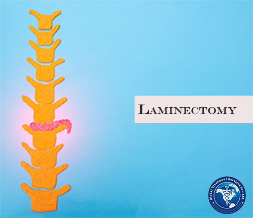
    Laminectomy also known as decompression/Lumbar surgery, that creates space by removing the lamina (the back part of a vertebra that covers your spinal canal.) laminectomy enlarges your spinal canal to relieve pressure on the spinal cord or nerves.This pressure is most commonly caused by bony overgrowths within the spinal canal, which can occur in people who have arthritis in their spines. These overgrowths are sometimes referred to as bone spurs, but they’re a normal side effect of the aging process in some people.

    Causes:

    • mild to severe back pain
    • numbness or weakness in the legs
    • difficulty walking
    • difficulty controlling bladder or bowel movements

    A laminectomy is only used if your symptoms interfere with daily life. It’s performed when less invasive treatments have failed.

    Why it’s done

    Bony overgrowths within the spinal canal can narrow the space available for your spinal cord and nerves. This pressure can cause pain, weakness or numbness that can radiate down your arms or legs.

    Because the laminectomy restores spinal canal space but does not cure you of arthritis, it more reliably relieves radiating symptoms from compressed nerves than it does back pain from spinal joints.

    Your doctor may recommend laminectomy if:

    • Conservative treatment, such as medication or physical therapy, fails to improve your symptoms
    • You have muscle weakness or numbness that makes standing or walking difficult
    • You experience loss of bowel or bladder control

    In some situations, laminectomy may be necessary as part of surgery to treat a herniated spinal disk. Your surgeon may need to remove part of the lamina to gain access to the damaged disk.

    Risks

    Laminectomy is generally a safe procedure. But as with any surgery, complications may occur. Potential complications include:

    • Bleeding
    • Infection
    • Blood clots
    • Nerve injury
    • Spinal fluid leak

    During laminectomy

    Surgeons usually perform laminectomy using general anesthesia, so you’re unconscious during the procedure.

    The surgical team monitors your heart rate, blood pressure and blood oxygen levels throughout the procedure. After you’re unconscious and can’t feel any pain:

    • The surgeon makes an incision in your back over the affected vertebrae and moves the muscles away from your spine as needed. Small instruments are used to remove the appropriate lamina. The size of the incision may vary depending on your condition and body size. Minimally invasive surgeries typically use smaller incisions than those used for open procedures.
    • If laminectomy is being performed as part of surgical treatment for a herniated disk, the surgeon also removes the herniated portion of the disk and any pieces that have broken loose (diskectomy).
    • If one of your vertebrae has slipped over another or if you have curvature of the spine, spinal fusion may be necessary to stabilize your spine. During spinal fusion, the surgeon permanently connects two or more of your vertebrae together using bone grafts and, if necessary, metal rods and screws.
    • Depending on your condition and individual needs, the surgeon may use a smaller (minimally invasive) incision and a special surgical microscope to perform the operation.

    After laminectomy

    After surgery, you’re moved to a recovery room where the health care team watches for complications from the surgery and anesthesia. You may also be asked to move your arms and legs. Your doctor may prescribe medication to relieve pain at the incision site.

    You might go home the same day as the surgery, although some people may need a short hospital stay. Your doctor may recommend physical therapy after a laminectomy to improve your strength and flexibility.

    Depending on the amount of lifting, walking and sitting your job involves, you may be able to return to work within a few weeks. If you also have spinal fusion, your recovery time will be longer.

    Takeaway

    A laminectomy is only used if your symptoms interfere with daily life. It’s performed when less invasive treatments have failed.

    Most people report measurable improvement in their symptoms after laminectomy, particularly a decrease in pain that radiates down the leg or arm. But this benefit may lessen over time if you have a particularly aggressive form of arthritis. Laminectomy is less likely to improve pain in the back itself. A laminectomy will often relieve many symptoms of spinal stenosis. However, it can’t prevent spine problems in the future and it may not completely relieve pain in everyone.People who also have a spinal fusion are more likely to have spinal problems in the future.

    if you are looking for treatment or second opinion for the same from the best doctors  share your latest reports  email us : info@gtsmeditour.com


    Tennis elbow

    Feb 14, 2019 Posted by admin Orthopaedic 0 comments

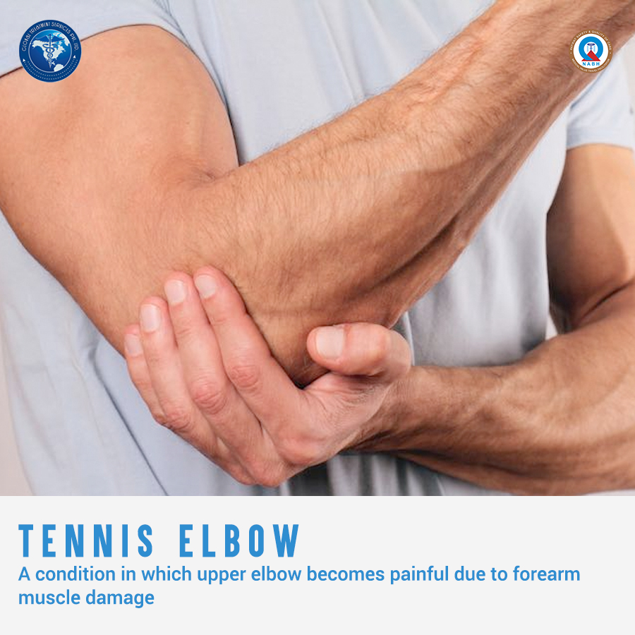
    Tennis elbow or ‘Lateral epicondylitis’ is a condition of persistent pain on the outer side of the elbow.It is caused by an overuse of forearm muscles resulting in repetitive injury to the tendons attached to the elbow.It can become responsible for substantial pain and loss of function of the affected limb.As the name may confuse,it is not only associated with the sport ‘Tennis’.It can be due to any other sport or work related activities.10.feb14tenniselbow

    Condition explained

    The extensor muscle called  the extensor carpi radialis brevis, helps to straighten and stabilize the wrist.This muscle attaches to a part of the elbow bone called the lateral epicondyle (thus the medical name ‘lateral epicondylitis’). It is the tendons that connect and transmit a muscle’s force to the bone. In lateral epicondylitis, tendon’s attachment to the bone is degenerated due to repetitive use, weakening and placing greater stress on the  forearm muscles. This can lead to pain associated with activities in which this muscle is active.

    Causes

    Here are some potential causes of this condition:

    • Overuse: This can be both non-work and work-related. Overuse can happen from “repetitive” gripping and grasping activities such as cutting, plumbing,gardening,carpentry, painting,  etc.
    • Trauma: Although less common, a direct blow to the elbow may result in swelling of the tendon that can lead to degeneration. This can make the elbow more susceptible to an overuse injury.

    Who is affected?

    Tennis elbow is most commonly seen in following groups of people:

    • Manual Laborers
      People who work with their hands like plumbers or bricklayers are at greater risk of developing tennis elbow.
    • Sports Participants
      Sports participants, especially racquet sport players and throwing sports persons(discus and javelin),are prone to developing tennis elbow.
    • People doing activities that involve fine, repetitive hand and wrist movements – such as using scissors or typing

    It is present in 40% of all racquet sport players and 15% of people working in repetitive manual trades like . It can occur at any age, however, sufferers are generally between the ages of 35 and 50.Predictably, the side affected is usually associated with handedness, but it can occur in the non-dominant arm. Males and Females are affected equally.

    Symptoms

    The typical symptom is pain in outer side of elbow,the area also becomes tender.But the pain can radiate into forearm and wrist also.

    • Pain when performing gripping tasks or resisted wrist/finger extension
    • Pain when the muscles are stretched.
    • Tenderness directly over the bony epicondyle

    There may be rigger points in the wrist muscles.In severe cases, there may be local swelling .Also some activities, such as making a fist, shaking hands, carrying bags like a briefcase, turning on taps may be painful.Some sufferers will also have neck stiffness and tenderness, as well as signs of nerve irritation.

    Diagnosis and treatment

    Usually, doctors are able to make the diagnosis based on the history and a physical examination.Additional tests such as X-rays or ultrasound may be needed to exclude other causes and/or assess the severity of tendon damage.
    Initial treatment involves relative rest and avoiding activities that aggravate the pain.If you are diagnosed with tennis elbow, activities that strain affected muscles and tendons should be immediately stopped.Taking analgesics may help ease mild pain and inflammation caused by tennis elbow.. Wearing a tennis elbow brace can help to protect the tendon.
    Physiotherapy has been shown to be effective in the short and long-term management of tennis elbow.Physiotherapy treatment can include gentle mobilisation of your neck and elbow joints, electrotherapy, elbow kinesio taping, muscle stretches, neural mobilisations, massage and strengthening.
    A small percentage of patients diagnosed with tennis elbow may finally require surgical treatment. Patients may consider surgery if conservative treatments are not effective after a period of 6 – 12 months.

    Untreated Tennis Elbows can last anywhere from 6 months to 2 years. You are also prone to recurrence.Aside from pain, the major complication of tennis elbow is that it impedes your ability to perform physical activities, such as certain sports or types of work.

    For any queries regarding the procedure and treatment facilities,email us at query@gtsmeditour.com .

    Read about:Endometriosis

    Recent Posts

    • Frozen Shoulder in Diabetic patients
    • Compartment Syndrome – Fasciotomy
    • Collodion baby syndrome
    • Infertility
    • Dysbiosis

    Recent Comments

    • Mr. Green on Lower back pain: Treatments
    • admin on Hepatitis A : Treatments
    • elizabeth on Hepatitis A : Treatments
    • admin on Kidney Transplantation: Procedure, Images
    • nakiberu josephine on Kidney Transplantation: Procedure, Images

    Archives

    • September 2026
    • July 2026
    • June 2026
    • May 2026
    • April 2026
    • March 2026
    • February 2026
    • January 2026
    • December 2025
    • September 2025
    • August 2025
    • July 2025
    • June 2025
    • May 2025
    • April 2025
    • March 2025
    • February 2025
    • January 2025
    • December 2024
    • November 2024
    • October 2024
    • September 2024
    • August 2024
    • July 2024
    • June 2024
    • May 2024
    • December 2021
    • August 2021
    • June 2021
    • May 2021
    • April 2021
    • March 2021
    • February 2021
    • December 2020
    • November 2020
    • October 2020
    • March 2020
    • February 2020
    • January 2020
    • November 2019
    • September 2019
    • August 2019
    • July 2019
    • June 2019
    • May 2019
    • April 2019
    • March 2019
    • February 2019
    • January 2019
    • December 2018
    • November 2018
    • October 2018
    • September 2018
    • August 2018
    • July 2018
    • June 2018
    • May 2018
    • April 2018
    • March 2018
    • February 2018
    • January 2018
    • December 2017
    • November 2017
    • October 2017
    • September 2017
    • August 2017
    • July 2017
    • June 2017
    • May 2017
    • April 2017
    • March 2017
    • February 2017
    • January 2017
    • December 2016
    • November 2016
    • October 2016
    • September 2016
    • August 2016
    • July 2016
    • June 2016
    • May 2016
    • April 2016
    • March 2016
    • February 2016
    • January 2016
    • December 2015
    • November 2015
    • October 2015
    • September 2015
    • August 2015
    • July 2015
    • June 2015
    • May 2015
    • April 2015

    Categories

    • Activity
    • Andrology
    • Ayurveda
    • Bariatrics
    • Cardiology
    • Child health
    • Cosmetic Surgeries
    • Dentistry
    • Dermatology
    • Digestive Disorders
    • Endocrinologist
    • Endocrinology
    • Event
    • Gastroenterology
    • General
    • General Health
    • geriatric psychiatrist
    • Gynaecology
    • Health & Nutrition
    • Infertility
    • Mental health
    • Nephrology
    • Neurology
    • Neuropathy
    • neuropsychiatrist
    • OBG
    • Oncology
    • Ophthalmology
    • Orthopaedic
    • Otorhinolaryngologist.
    • Paediatrics
    • Plastic surgery
    • podiatrist
    • Psychiatry
    • Pulmonology
    • Rheumatologist
    • Robotic Assisted Surgeries
    • Senior care
    • skin cancer
    • Speciality
    • Therapies
    • Therapists
    • Uncategorized
    • Urology
    • vascular
    • Women Healthcare

    Meta

    • Log in
    • Entries RSS
    • Comments RSS
    • WordPress.org
    © 2015 Global Treatment Services Pvt Ltd.