Global Treatment Services Pvt. Ltd.

Global Treatment Services

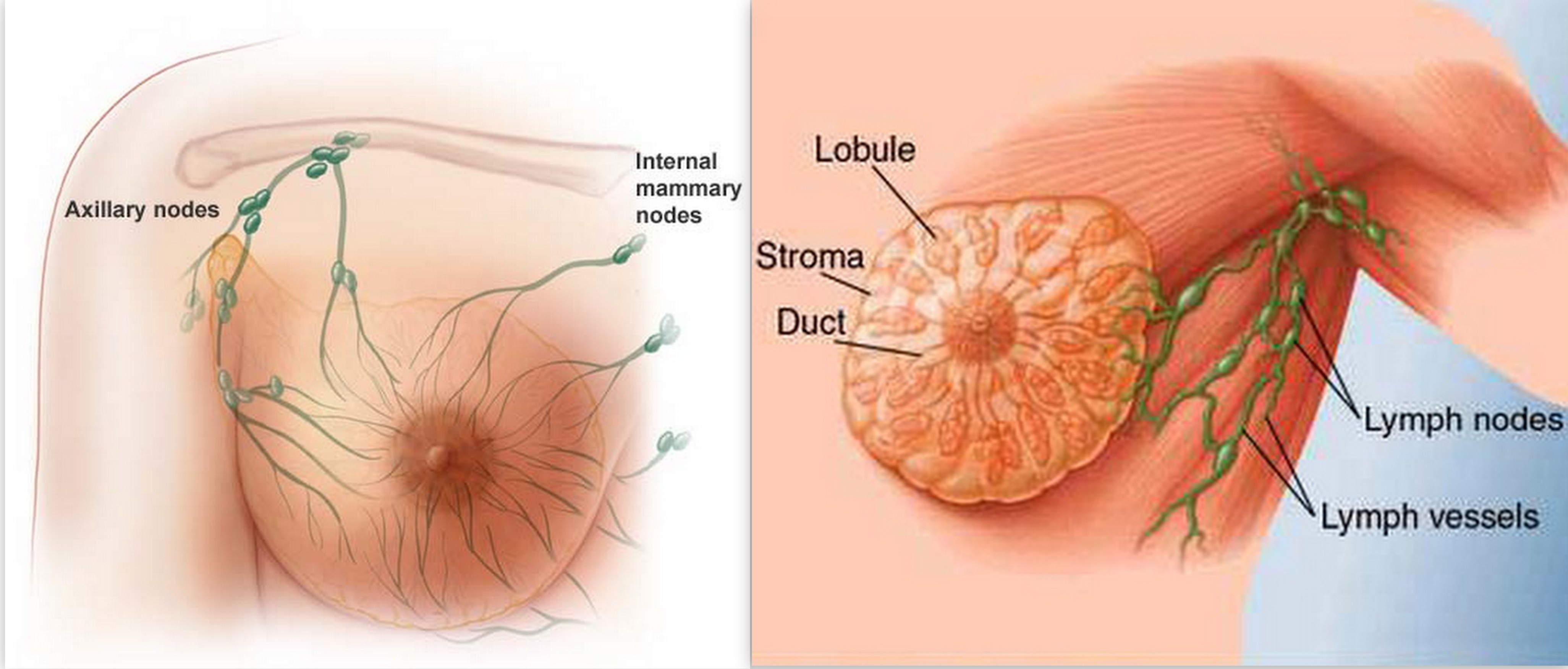
Breast Cancer : Causes, Types and Treatments

Breast cancer definition

Breast cancer is a malignant tumor (a collection of cancer cells) arising from the cells of the breast. Although breast cancer predominantly occurs in women, it can also affect men. This article deals with breast cancer in women.

What are the statistics on male breast cancer?

Breast cancer is rare in men but typically has a significantly worse outcome. This is partially related to the often late diagnosis of male breast cancer, when the cancer has already spread.

Symptoms are similar to the symptoms in women, with the most common symptom being a lump or change in skin of the breast tissue or nipple discharge. Although it can occur at any age, male breast cancer usually occurs in men over 60 years of age.

What are the different types of breast cancer? Where does breast cancer come from?

There are many types of breast cancer. Some are more common than others, and there are also combinations of cancers. Some of the most common types of cancer are as follows:

  • Ductal carcinoma in situ: The most common type of noninvasive breast cancer is ductal carcinoma in situ (DCIS). This type of cancer has not spread and therefore usually has a very high cure rate.
  • Invasive ductal carcinoma: This cancer starts in a duct of the breast and grows into the surrounding tissue. It is the most common form of breast cancer. About 80% of invasive breast cancers are invasive ductal carcinoma.
  • Invasive lobular carcinoma: This breast cancer starts in the glands of the breast that produce milk. Approximately 10% of invasive breast cancers are invasive lobular carcinoma.
  • The remainder of breast cancers are much less common and include the following:
  • Mucinous carcinoma are formed from mucus-producing cancer cells. Mixed tumors contain a variety of cell types.
  • Medullary carcinoma is an infiltrating breast cancer that presents with well-defined boundaries between the cancerous and noncancerous tissue.
  • Inflammatory breast cancer: This cancer makes the skin of the breast appear red and feel warm (giving it the appearance of an infection). These changes are due to the blockage of lymph vessels by cancer cells.
  • Triple-negative breast cancers: This is a subtype of invasive cancer with cells that lack estrogen and progesterone receptors and have no excess of a specific protein (HER2) on their surface. It tends to appear more often in younger women and African-American women.
  • Paget’s disease of the nipple: This cancer starts in the ducts of the breast and spreads to the nipple and the area surrounding the nipple. It usually presents with crusting and redness around the nipple.
  • Adenoid cystic carcinoma: These cancers have both glandular and cystic features. They tend not to spread aggressively and have a good prognosis.
  • Lobular carcinoma in situ: This is not a cancer but an area of abnormal cell growth that can lead to invasive breast cancer later in life.


Related image

What causes breast cancer?

There are many risk factors that increase the chance of developing breast cancer. Although we know some of these risk factors, we don’t know the cause of breast cancer or how these factors cause the development of a cancer cell.

We know that normal breast cells become cancerous because of mutations in the DNA, and although some of these are inherited, most DNA changes related to breast cells are acquired during one’s life.

Proto-oncogenes help cells grow. If these cells mutate, they can increase growth of cells without any control. Such mutations are referred to as oncogenes. Such uncontrolled cell growth can lead to cancer.

What about antiperspirants or deodorants as causes of breast cancer?

Research has shown that parabens (a preservative used in deodorants) can build up in breast tissues. However, this study did not show that parabens cause breast cancer or that the parabens (which are found in many other products) were linked to the use of deodorants.

A 2002 study did not show any increased risk for breast cancer in women using an underarm deodorant or antiperspirant. A 2003 study showed an earlier age for breast cancer diagnosis in women who shaved their underarms more frequently and used underarm deodorants.

Treatments:

Patients with breast cancer have many treatment options. Most treatments are adjusted specifically to the type of cancer and the staging group. Treatment options are being adjusted frequently and your health care provider will have the information on the current standard of care available. Treatment options should be discussed with a health care team. The following are the basic treatment modalities used in the treatment of breast cancer.

Surgery

Most women with breast cancer will require surgery. Broadly, the surgical therapies for breast cancer can be divided into breast-conserving surgery and mastectomy.

Breast-conserving surgery

This surgery will only remove part of the breast (sometimes referred to as partial mastectomy). The extent of the surgery is determined by the size and location of the tumor.

In a lumpectomy, only the breast lump and some surrounding tissue is removed. The surrounding tissue (surgical margins) are inspected for cancer cells. If no cancer cells are found, this is called “negative” or “clear margins.” Frequently, radiation therapy is given after lumpectomies.

Mastectomy

During a mastectomy (sometimes also referred to as a simple mastectomy), all the breast tissue is removed. If immediate reconstruction is considered, a skin-sparing mastectomy is sometimes performed. In this surgery, all the breast tissue is removed as well, but the overlying skin is preserved.

Radical mastectomy

During this surgery, the surgeon removes the axillary lymph nodes as well as the chest wall muscle in addition to the breast. This procedure is done much less frequently than in the past, as in most cases, a modified radical mastectomy is as effective.

Modified radical mastectomy

This surgery removes the axillary lymph nodes in addition to the breast tissue. Depending on the stage of the cancer, a health care team might give someone a choice between a lumpectomy and a mastectomy. Lumpectomy allows sparing of the breast but usually requires radiation therapy afterward. If lumpectomy is indicated, long-term follow-up shows no advantage of a mastectomy over the lumpectomy.

Preventive surgery

For a small group of patients who have a very high risk of breast cancer, surgery to remove the breasts may be an option. Although this reduces the risk significantly, a small chance of developing cancer remains.

Double mastectomy is a surgical option to prevent breast cancer. This prophylactic (preventive) surgery can decrease the risk of breast cancer by about 90% for women at moderate to high risk for breast cancer.

Such an approach should be carefully discussed with a health care team.

The discussion about whether to undergo any preventive surgery should include

  • genetic testing for BRCA1 or BRCA2 gene mutations,
  • full review of risk factors,
  • family history of cancer and specifically breast cancer, and
  • other preventive options such as medications.

Radiation therapy

Radiation therapy destroys cancer cells with high energy rays. There are two ways to administer radiation therapy.

External beam radiation

This is the usual way radiation therapy is given for breast cancer. A beam of radiation is focused onto the affected area by an external machine. The extent of the treatment is determined by a health care team and is based on the surgical procedure performed and whether lymph nodes were affected or not.

The local area will usually be marked after the radiation team has determined the exact location for the treatments. Usually, the treatment is given five days a week for five to six weeks.

Brachytherapy

This form of delivering radiation uses radioactive seeds or pellets. Instead of a beam from the outside delivering the radiation, these seeds are implanted into the breast next to the cancer.

Chemotherapy

Chemotherapy is treatment of cancers with medications that travel through the bloodstream to the cancer cells. These medications are given either by intravenous injection or by mouth.

Chemotherapy can have different indications and may be performed in different settings as follows:

  • Adjuvant chemotherapy: If surgery has removed all the visible cancer, there is still the possibility that cancer cells have broken off or are left behind. If chemotherapy is given to assure that these small amounts of cells are killed as well, it is called adjunct chemotherapy. Chemotherapy is not given in all cases, since some women have a very low risk of recurrence even without chemotherapy, depending upon the tumor type and characteristics.
  • Neoadjuvant chemotherapy: If chemotherapy is given before surgery, it is referred to as neoadjuvant chemotherapy. Although there seems to be no advantage to long-term survival whether the therapy is given before or after surgery, there are advantages to see if the cancer responds to the therapy and by shrinking the cancer before surgical removal.
  • Chemotherapy for advanced cancer: If the cancer has metastasized to distant sites in the body, chemotherapy can be used for treatment. With cases of metastatic breast cancer, the health-care team will need to determine the most appropriate length of treatment.

There are many different chemotherapeutic agents that are either given alone or in combination. Usually, these drugs are given in cycles with certain treatment intervals followed by a rest period. The cycle length and rest intervals differ from drug to drug.

Hormone therapy

This therapy is often used to help reduce the risk of cancer reoccurrence after surgery, but it can also be used as adjunct treatment.

Estrogen (a hormone produced by the ovaries) promotes the growth of a few breast cancers, specifically those containing receptors for estrogen (ER positive) or progesterone (PR positive). The following drugs are used in hormone therapy:

  • Tamoxifen (Nolvadex): This drug prevents estrogen from binding to estrogen receptors on breast cells.
  • Toremifene (Fareston) works similar to Tamoxifen and is only indicated in metastatic breast cancer.
  • Fulvestrant (Faslodex): This drug eliminates the estrogen receptor and can be used even if tamoxifen is no longer useful.
  • Aromatase inhibitors: They stop estrogen production in postmenopausal women. Examples are letrozole (Femara), anastrozole (Arimidex), and exemestane (Aromasin).

Targeted therapy

As we are learning more about gene changes and their involvement in causing cancer, drugs are being developed that specifically target the cancer cells. They tend to have fewer side effects than chemotherapy (as they target only the cancer cells) but usually are still used in adjunct with chemotherapy.

Alternative treatments

Whenever a disease has the potential for much harm and death, physicians search for alternative treatments. As a patient or the loved one of a patient, there may be an inclination to try everything and leave no option unexplored. The danger in this approach is usually found in the fact that the patient might not avail themselves of existing, proven therapies. One should discuss any interest in alternative treatments with a health care team and together explore the different options.

Lung cancer: causes symptoms and prevention

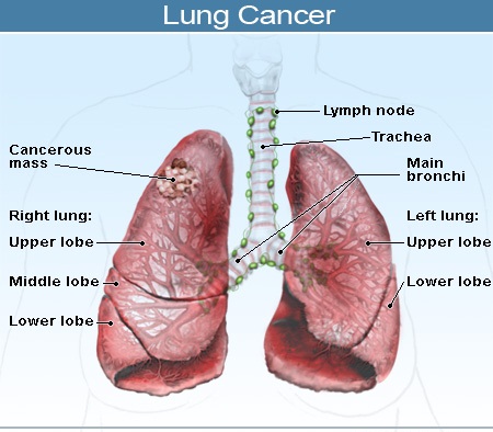
Lung cancer is a malignant lung disease primarily caused by cigarette smoking. It often has no obvious symptoms until the disease is quite advanced, and has a low rate of survival. Treatment for lung cancer mainly involves surgery and chemotherapy, while radiation therapy and targeted drug therapies may also be used.

Related image

What is lung cancer?

Lung cancer results from abnormal growth of cells in the lining of the lungs, leading to the growth of a malignant tumour.

There are two different types of lung cancer – small cell lung cancer (SCLC) and non-small cell lung cancer (NSCLC) – which differ in terms of how they grow and spread to other parts of the body and how they’re treated.

Causes

The development of lung cancer is strongly associated with cigarette smoking – approximately 90% of lung cancers are attributable to tobacco use. Pipe and cigar smoking can also cause lung cancer, but the risk is not as high as with cigarette smoking. Tobacco smoke contains more than 4,000 chemical compounds, many of which are cancer-causing (carcinogens).  Passive smoking, i.e. the inhalation of tobacco smoke by non-smokers who live or work with smokers, is also an established risk factor for the development of lung cancer.

Although the majority of lung cancers are linked to tobacco smoking, not all smokers go on to develop lung cancer suggesting that genetic susceptibility (i.e. family history) may play a role in the development of lung cancer.  Other causes of lung cancer include air pollution (from vehicles, industry, and power generation) and inhalation of asbestos fibres (usually in the workplace).

Image result for lung cancer causes symptoms and prevention

Signs, symptoms, and diagnosis

Symptoms of lung cancer are varied and warning signs are not always obvious. Up to 25% of people who get lung cancer display no symptoms. In people who do display symptoms, they include the following:

  • Persistent cough and hoarseness
  • Shortness of breath, wheezing, and chest pain
  • Blood-streaked sputum
  • Chest pain
  • Frequent episodes of bronchitis or pneumonia
  • Weight loss, weakness, and fatigue.

A wide range of diagnostic tests and procedures are used to diagnose lung cancer, including taking a patient history, physical examination, blood tests, chest x-rays, computerized tomography (CT) and positron emission tomography (PET) scans, bone scans, bronchoscopy, cytological studies of sputum, bronchial washings, and fine needle biopsy. Many of these procedures are also used to determine the stage of the cancer.

Staging of lung cancer

The stage of a lung cancer refers to the extent to which the cancer has spread to other parts of the body. Staging helps to determine how the cancer should be treated. Lung cancer can spread to any organ in the body but the liver, brain, and bones are the most common sites. The two types of lung cancer are staged differently. A simplified overview of staging is as follows:

NSCLC
Stage I: cancer that is confined to the lung
Stage II: cancer that is confined to the chest
Stage III: cancer that is confined to the chest but with larger and more aggressive tumours than at stage II
Stage VI: cancer that has spread to other parts of the body.

SCLC
Limited-stage: cancer confined to the area of the chest
Extensive-stage: cancer that has spread to other parts of the body.

Treatment

Treatment for cancer involves a combination of surgery to remove cancer cells, and chemotherapy and radiation therapy to kill cancer cells. Lung cancer is incurable unless complete surgical removal of the tumour cells can be achieved.

Surgery is the most effective treatment for lung cancer but only about 20% of lung cancers are suitable for surgery i.e. Stage I and II NSCLC and cancer that has not spread beyond the lung.

Radiation therapy may be used for both NSCLC and SCLC and is a good option for people who are not suitable for surgery or who refuse surgery.

Chemotherapy is used for both NSCLC and SCLC. Chemotherapy drugs may be given alone or in combination with surgery or radiation therapy. Chemotherapy is the treatment of first choice for SCLC since it has usually spread extensively in the body by the time it has been diagnosed.

Also used in the treatment of lung cancer are targeted therapies. These are drugs (gefitinib and erlotinib) or antibodies (cetuximab, bevacizumab) that block the growth and spread of cancer by interfering with specific molecules involved in tumour growth and progression. They are used in some patients with NSCLC that does not respond to standard chemotherapy.

Prevention

The most effective measure that can be taken to prevent the development of lung cancer is to stop smoking. Reducing exposure to passive smoking is also an effective method of prevention.

Bone Cancer: Causes, Symptoms & Treatments

What Is Bone Cancer?

Bone cancer occurs when a tumor, or abnormal mass of tissue, forms in a bone. A tumor may be malignant, which means it’s growing aggressively and spreading to other parts of the body. A malignant tumor is often referred to as cancerous. Cancer that begins in the bones is rare.

Types of Bone Cancer

Primary bone cancers are the most serious of all bone cancers. They form directly in the bones or surrounding tissue, such as cartilage.

Cancer can also spread, or metastasize, from another part of your body to your bones. This is known as secondary bone cancer, and this type is more common than primary bone cancer.

Common types of primary bone cancers include:

Multiple Myeloma (MM)

Multiple myeloma is the most common type of bone cancer. It occurs when cancer cells grow in the bone marrow and cause tumors in various bones. MM usually affects older adults. Among bone cancers, MM has one of the best prognoses, and many people who have it don’t require treatment.

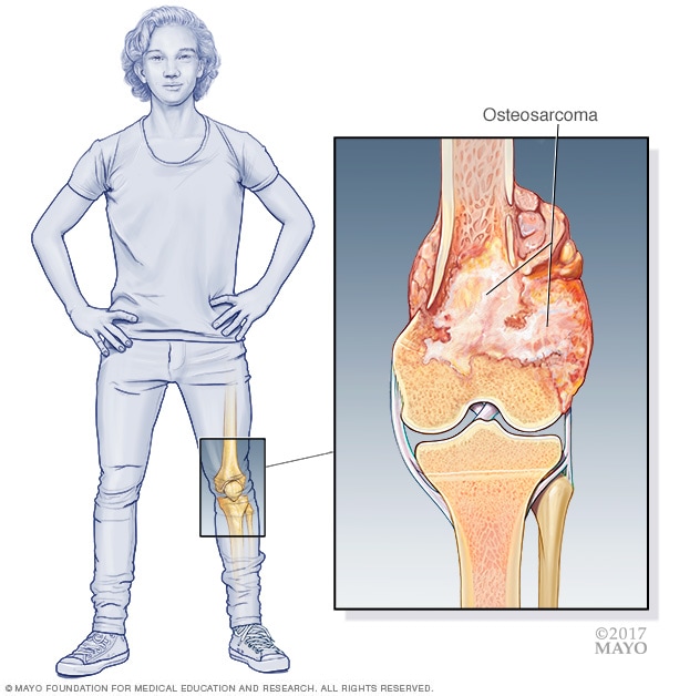
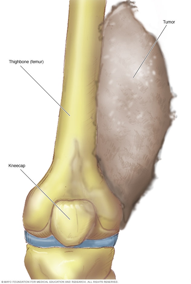
Osteosarcoma (Osteogenic Sarcoma)

Illustration showing osteosarcoma<br /><br /><br /><br />

Osteosarcoma, or osteogenic sarcoma, generally affects children and adolescents, but it can also occur in adults. It has a tendency to originate at the tips of the long bones in the arms and legs. Osteosarcoma may also start in the hips, shoulders, or other locations. It affects the hard tissue that provides the outer layer of your bones.

Chondrosarcoma

Chondrosarcoma may occur in the pelvis, thigh areas, and shoulders of older adults. It forms in the subchondral tissue, which is the tough connective tissue between your bones. This is the second most common primary cancer involving the bones.

Ewing’s Sarcoma

Illustration showing Ewing's sarcoma<br /><br /><br /><br />

Ewing’s sarcoma is a rare cancer that either begins in the soft tissues surrounding the bones or directly in the bones of children and young adults. The long bones of the body, such as the arms and legs, and the pelvis are commonly affected.

What Are the Symptoms of Bone Cancer?

  • pain and swelling in the affected bones
  • palpable hard mass in the long bones of the limbs
  • feeling tired or fatigued

Less common symptoms include:

  • easily broken bones
  • weight loss

What Causes Bone Cancer?

The cause of bone cancer isn’t exactly known, but there are certain factors that may contribute to or increase a person’s chances of forming abnormal growths in the bone. These include:

Abnormal Cellular Growth

Healthy cells continually divide and replace older cells. After completing this process they die. Abnormal cells, however, continue living. They start forming masses of tissue that turn into tumors.

Radiation Therapy

Radiation therapy, which kills dangerous cancer cells, can be used to treat bone cancer. However, osteosarcoma may form in some people who receive the treatment. The use of high dosages of radiation may be a factor in this development.

 Who Is at Risk for Bone Cancer?

The following may be risk factors for bone cancer:

  • having a family history of cancer, especially bone cancer
  • having received radiation treatment or therapy in the past
  • having Paget’s disease, which is a condition that causes the bones to break down and then grow back abnormally
  • currently or previously having had multiple tumors in the cartilage, which is the connective tissue in the bone

GIST Cancer : Causes & Symptoms

About the gastrointestinal tract

The gastrointestinal (GI or digestive) tract includes the:

  • Esophagus
  • Stomach
  • Gallbladder and bile ducts
  • Liver
  • Pancreas
  • Small intestine
  • Colon
  • Rectum
  • Anus
  • Lining of the gut

The GI tract plays a central role in digesting food and liquid and in processing waste.  When you swallow food, it is pushed down a muscular tube called the esophagus and enters the stomach. The muscles in the stomach mix the food and release gastric juices that help break down and digest the food.

The food then moves into the small intestine, or small bowel, for further digestion before entering the large intestine. The large intestine helps remove waste from the body. The colon makes up the first 5 to 6 feet of the large intestine. The rectum makes up the last 6 inches, ending at the anus.

About gastrointestinal stromal tumor (GIST)

A tumor begins when healthy cells change and grow out of control, forming a mass called a tumor. A tumor can be cancerous or benign. A cancerous tumor is malignant, meaning it can grow and spread to other parts of the body. A benign tumor means the tumor can grow but will not spread. A tumor can start in any part of the GI tract. There are several different types of GI tumors, including gastrointestinal stromal tumor (GIST).

GISTs are different from more common types of GI tumors because of the type of tissue in which they start. GISTs belong to a group of cancers called soft-tissue sarcomas. Soft-tissue sarcomas develop in the tissues that support and connect the body. The sarcoma cells resemble the cells that hold the body together, including fat cells, muscles, nerves, tendons, joints, blood vessels, and lymph vessels.

Doctors used to think that GISTs were muscle or nerve tumors. However, research shows that GIST begins in “pacemaker” cells found in the walls of the GI tract. These pacemaker cells are called interstitial cells of Cajal (ICCs), and they send signals to the GI tract to help move food and liquid through the digestive system.


Symptoms :

People with GIST often do not experience any specific symptoms or signs. When symptoms do occur, they may be vague. Or, the cause of a symptom may be a different medical condition that is not a tumor.

  • Pain or discomfort in the abdomen
  • A mass in the abdomen that you can feel with your hand
  • Bowel obstruction
  • Nausea and vomiting
  • Vomiting blood
  • Blood in the stool
  • Fatigue due to anemia, which is a low red blood cell count and is often caused by bleeding in the GI tract.

Stages:

For other types of GI tumors, the most common staging tool used is called the TNM system. TNM is an abbreviation for tumor (T), node (N), and metastasis (M). However, the use of the TNM system for GIST is not required. Instead, doctors look at different factors to determine a patient’s prognosis and the specific risk of how aggressive an individual tumor will be. Specifically, doctors want to figure out how quickly the GIST may grow and the chance the tumor will come back, or recur, after surgery.

Doctors commonly use the factors listed below to determine how aggressive a GIST that has been removed could be:

  • The size of the tumor
  • The mitotic count, which describes the number of dividing cells
  • The location where the tumor started

A GIST that has spread to distant areas of the body is considered higher risk because it has a greater likelihood of coming back, called recurrent GIST.


Salivary Gland cancer : Causes, Symptoms & Teatments

The major salivary glands are on each side of the face and below the tongue. Several important nerves and other structures run through or near salivary glands and can be affected by salivary tumors.

Possible signs and symptoms of salivary gland cancer include:

  • A lump or swelling in your mouth, cheek, jaw, or neck
  • Pain in your mouth, cheek, jaw, ear, or neck that does not go away
  • A difference between the size and/or shape of the left and right sides of your face or neck
  • Numbness in part of your face
  • Weakness of the muscles on one side of your face
  • Trouble opening your mouth widely
  • Fluid draining from an ear
  • Trouble swallowing

A risk factor is anything that affects your chance of getting a disease such as cancer. Different cancers have different risk factors. Some risk factors, like smoking, can be changed. Others, like a person’s age or family history, can’t be changed.

But having a risk factor, or even many risk factors, does not mean that you will get the disease. And many people who get the disease may have few or no known risk factors.

A few risk factors are known to make a person more likely to develop salivary gland cancer.

Older age

The risk of salivary gland goes up as people get older.

Male gender

Salivary gland cancers are more common in men than in women.

Radiation exposure

Radiation treatment to the head and neck area for other medical reasons increases your risk of salivary gland cancer.

Workplace exposure to certain radioactive substances may also increase the risk of salivary gland cancer.

Family history

Very rarely, members of some families seem to have a higher than usual risk of developing salivary gland cancers. But most people who get salivary gland cancer do not have a family history of this disease.

Other possible risk factors

Certain workplace exposures

Some studies have suggested that people who work with certain metals (nickel alloy dust) or minerals (silica dust), and people who work in asbestos mining, plumbing, rubber products manufacturing, and some types of woodworking may be at increased risk for salivary gland cancer, but these links are not certain. The rarity of these cancers makes this hard to study.

Tobacco and alcohol use

Tobacco and alcohol can increase the risk for several cancers of the head and neck area, but they have not been strongly linked to salivary gland cancers in most studies.

Diet

Some studies have found that a diet low in vegetables and high in animal fat may increase the risk of salivary gland cancer, but more research is needed to confirm this possible link.

Cell phone use

One study has suggested an increased risk of parotid gland tumors among heavy cell phone users. In this study, most of the tumors seen were benign (not cancer). Other studies looking at this issue have not found such a link. Research in this area is still in progress.

If you’ve been diagnosed with salivary gland cancer, your cancer care team will discuss your treatment options with you. It’s important to weigh the benefits of each treatment option against the possible risks and side effects.

Which treatments are used for salivary gland cancer?

Common treatment options for salivary gland cancer include:

  • Surgery
  • Radiation therapy
  • Chemotherapy

Vulvodynia: Causes, Symptoms & Treatments

Vulvodynia is a burning, stinging but often unexplained pain affecting the skin around the vagina, or the vulva.

Although the condition is persistent and without an obvious cause, there are steps which can be taken to help relieve the discomfort.

Vulvodynia that is always present or comes on without a trigger is called unprovoked vulvodynia. If the pain starts when the vulva is touched, such as during sex or when changing a tampon, doctors call this provoked vulvodynia.

Vulvodynia cannot be transmitted during sex and is not a sign of a more serious condition, such as cancer.

Vestibulodynia is a related condition where pain affects the vestibule, which is the entrance to the vagina.

Types of vulvodynia

Vulvodynia affects the vulva which is the external female genital organs. This includes the labia, clitoris and vaginal opening.

The two main types of vulvodynia are:

  • Generalised vulvodynia which is pain in different areas of the vulva which occurs at different times. Vulvar pain may be constant or occasional. It may or may not be prompted by touch or pressure but this may make the pain worse.
  • Vestibulodynia is pain in the vestibule which is the entrance to the vagina. Often a burning sensation, this type of vulvar pain comes on during or after touch or pressure such as during sexual intercourse.

Possible causes of vulvodynia

Doctors do not know what causes vulvodynia. There is no evidence that infections, such as sexually transmitted infections, cause it.

Researchers are trying to find the causes of vulvodynia. These may include:

  • Nerve injury or irritation
  • Abnormal response in vulvar cells to an infection or trauma
  • Genetic factors that make the vulva respond poorly to chronic inflammation
  • Hypersensitivity to yeast infections
  • Muscle spasms
  • Allergies or irritation to chemicals or other substances
  • Hormonal changes
  • History of sexual abuse
  • Frequent antibiotic use

Carpel Tunnel Syndrome: Causes, Symptoms & Treatments

What is carpal tunnel syndrome?

Carpal tunnel syndrome is numbness, tingling, weakness, and other problems in your hand because of pressure on the median nerve in your wrist.

The median nerve and several tendons run from your forearm to your hand through a small space in your wrist called the carpal tunnel . The median nerve controls movement and feeling in your thumb and first three fingers (not your little finger).

What causes carpal tunnel syndrome?

Pressure on the median nerve causes carpal tunnel syndrome. This pressure can come from swelling or anything that makes the carpal tunnel smaller. Many things can cause this swelling, including:

  • Illnesses such as hypothyroidism, rheumatoid arthritis, and diabetes.
  • Making the same hand movements over and over, especially if the wrist is bent down (your hands lower than your wrists), or making the same wrist movements over and over.
  • Pregnancy.

Causes

Carpal tunnel syndrome is caused by pressure on the median nerve.

The median nerve runs from your forearm through a passageway in your wrist (carpal tunnel) to your hand. It provides sensation to the palm side of your thumb and fingers, except the little finger. It also provides nerve signals to move the muscles around the base of your thumb (motor function).

Anything that squeezes or irritates the median nerve in the carpal tunnel space may lead to carpal tunnel syndrome. A wrist fracture can narrow the carpal tunnel and irritate the nerve, as can the swelling and inflammation resulting from rheumatoid arthritis.

There is no single cause in many cases. It may be that a combination of risk factors contributes to the development of the condition.

Risk factors

A number of factors have been associated with carpal tunnel syndrome. Although they may not directly cause carpal tunnel syndrome, they may increase your chances of developing or aggravating median nerve damage. These include:

  • Anatomic factors. A wrist fracture or dislocation, or arthritis that deforms the small bones in the wrist, can alter the space within the carpal tunnel and put pressure on the median nerve.People with smaller carpal tunnels may be more likely to have carpal tunnel syndrome.
  • Sex. Carpal tunnel syndrome is generally more common in women. This may be because the carpal tunnel area is relatively smaller in women than in men.Women who have carpal tunnel syndrome may also have smaller carpal tunnels than women who don’t have the condition.
  • Nerve-damaging conditions. Some chronic illnesses, such as diabetes, increase your risk of nerve damage, including damage to your median nerve.
  • Inflammatory conditions. Illnesses that are characterized by inflammation, such as rheumatoid arthritis, can affect the lining around the tendons in your wrist and put pressure on your median nerve.
  • Obesity. Being obese is a significant risk factor for carpal tunnel syndrome.
  • Alterations in the balance of body fluids. Fluid retention may increase the pressure within your carpal tunnel, irritating the median nerve. This is common during pregnancy and menopause. Carpal tunnel syndrome associated with pregnancy generally resolves on its own after pregnancy.
  • Other medical conditions. Certain conditions, such as menopause, obesity, thyroid disorders and kidney failure, may increase your chances of carpal tunnel syndrome.
  • Workplace factors. It’s possible that working with vibrating tools or on an assembly line that requires prolonged or repetitive flexing of the wrist may create harmful pressure on the median nerve or worsen existing nerve damage.However, the scientific evidence is conflicting and these factors haven’t been established as direct causes of carpal tunnel syndrome.Several studies have evaluated whether there is an association between computer use and carpal tunnel syndrome. However, there has not been enough quality and consistent evidence to support extensive computer use as a risk factor for carpal tunnel syndrome, although it may cause a different form of hand pain.

Prevention

There are no proven strategies to prevent carpal tunnel syndrome, but you can minimize stress on your hands and wrists with these methods:

  • Reduce your force and relax your grip. If your work involves a cash register or keyboard, for instance, hit the keys softly. For prolonged handwriting, use a big pen with an oversized, soft grip adapter and free-flowing ink.
  • Take frequent breaks. Gently stretch and bend hands and wrists periodically. Alternate tasks when possible. This is especially important if you use equipment that vibrates or that requires you to exert a great amount of force.
  • Watch your form. Avoid bending your wrist all the way up or down. A relaxed middle position is best. Keep your keyboard at elbow height or slightly lower.
  • Improve your posture. Incorrect posture rolls shoulders forward, shortening your neck and shoulder muscles and compressing nerves in your neck. This can affect your wrists, fingers and hands.
  • Change your computer mouse. Make sure that your computer mouse is comfortable and doesn’t strain your wrist.
  • Keep your hands warm. You’re more likely to develop hand pain and stiffness if you work in a cold environment. If you can’t control the temperature at work, put on fingerless gloves that keep your hands and wrists warm.

Lumbar Radiculopathy: Causes, Symptoms & Treatments

Within the body, several sensitive nerves branch away from the spinal column through foraminal canals before travelling to peripheral regions of the body. These nerves relay sensory information to and from the central nervous system and enable us to interact with the world around us. Sometimes, however, nerves can begin to operate ineffectively or improperly relay information because they are being compressed and damaged as they exit the spine. The medical term for this painful condition is known as “radiculopathy.”

When radiculopathy is experienced, one or more nerves becomes diseased or aggravated as the result of abnormal pressure, which generally develops in or near the foraminal canals. Nerves can become irritated or pinched in any section of the spine, but radiculopathy is most frequently seen in the lumbar (lower back) and cervical (neck) regions of the back. These two regions are most susceptible to injury because the lower back bears much of the upper body’s weight and because the upper spine is sensitive and almost constantly in motion. Radiculopathy can usually be treated through a combination of conservative methods, although surgery may be recommended in extreme cases.

Radiculopathy Causes

There is a diverse array of causes for radiculopathy, usually discovered in conjunction with an injury or other spinal condition. Some of the most common factors that may contribute to nerve compression include:

  • Frequent, improper movements, including those that might occur in heavy lifting, highly physical or manual labor and contact sports
  • Degenerative, age-related diseases
  • Genetic predisposition, congenital spine difficulties or a family history of radiculopathy
  • Overly thickened ligaments
  • Other spinal conditions, such as scoliosis, bone spurs and disc herniation
  • Other health issues, including diabetes, tumors and infection
  • Inflammation or displaced materials that may result from a sudden injury like a traumatic fall, motor vehicle accident or sports incident

All of these elements may also narrow the foraminal canals, leading to restricted nerves and debilitating symptoms. To care for your spine and the nerves that surround it, avoid activities that overly strain the backbone and seek prompt, professional medical attention if an issue is suspected.

Radiculopathy Symptoms

Symptoms of this condition are often shared with the other, underlying issues causing the radiculopathy to occur in the first place.

  • Pain, numbness and tingling in the back, arms or legs
  • Sciatica (discomfort that radiates through the lower back and limbs)
  • A hypersensitivity to light touch and tenderness in the region in question
  • Weakness in the affected area and associated extremities
  • An inability to perform everyday activities or movements

Because any of the nerves leaving the spinal canal may suffer from radiculopathy, symptoms vary widely depending on the location of the affected region. If you are unsure where your pain is stemming from, we offer advanced pain-mapping procedures to pinpoint the precise site of your hurting nerve.

If you or a loved one encounter incontinence of the bladder or bowels in tandem with back pain, These symptoms are indicators of a life-threatening ailment known as “cauda equina syndrome”.

Treatments:

Taking the first step toward relief is as simple as scheduling a consultation at our state-of-the-art-facilities. During this appointment, one of our spine experts will carefully and compassionately evaluate your medical history, physical state and symptoms. To confirm a diagnosis of radiculopathy, imaging tests like a C.T. scan, MRI or X-ray may be requested, as well as a pain-mapping procedure. Once the location and severity of your compressed nerve has been ascertained, we will work with you to design a treatment plan that meets your unique needs and goals.

Whenever possible, we encourage patients to consider conservative, non-invasive treatment options before resorting to surgical intervention. Some of the non-surgical methods of care commonly implemented to treat radiculopathy include:

  • Physical Therapy and Specialized Exercises: Oftentimes, physical therapists are able to strengthen the spine and reduce pressure on certain nerves by guiding the body through therapeutic movements.
  • Pain-Relieving and Anti-Inflammatory Medication: When used temporarily, certain medications can reduce the amount of pain experienced by a patient and allow them to take up other treatment modalities, such as physical therapy, without being extremely uncomfortable.
  • Injections: Localized injections can sometimes eliminate a patient’s pain for as long as 6 months at a time, often allowing compressed nerves to recover and thus reducing the need for surgery.
  • Limited activity for a set period of time
  • Heating and cooling treatments to maximize blood flow and the transmission of nutrients
  • Spinal decompression therapy

Typically, radiculopathy will clear up when conservative treatment methods are dedicatedly employed. In severe cases, however, minimally invasive surgery may be needed to obtain full pain relief. Our expert spine surgeons can oftentimes treat radiculopathy with a posterolateral lumbar fusion or other advanced minimally invasive technique.

Because we offer some of the most state-of-the-art surgical options available, patients realize many benefits when compared to traditional open surgery. Some of these benefits include:

  • Tiny incisions
  • Less blood loss and scarring
  • Minimal trauma to surrounding muscles and tissue
  • Quicker recovery times that allow patients to return to work, school and other daily activities sooner
  • Faster pain relief

Boil Infection : Causes, Symptoms & Treatments

A boil is a skin infection that starts in a hair follicle or oil gland. At first, the skin turns red in the area of the infection, and a tender lump develops. After four to seven days, the lump starts turning white as pus collects under the skin.

  • The most common places for boils to appear are on the face, neck, armpits, shoulders, and buttocks. When one forms on the eyelid, it is called a sty.
  • If several boils appear in a group, this is a more serious type of infection called a carbuncle.

Image result for boil nose causes, symptoms & treatment

Causes of Boils

Most boils are caused by a germ (staphylococcal bacteria). This germ enters the body through tiny nicks or cuts in the skin or can travel down the hair to the follicle.

These health problems make people more susceptible to skin infections:

  • Diabetes
  • Problems with the immune system
  • Poor nutrition
  • Poor hygiene
  • Exposure to harsh chemicals that irritate the skin

Symptoms of Boils

A boil starts as a hard, red, painful lump usually about half an inch in size. Over the next few days, the lump becomes softer, larger, and more painful. Soon a pocket of pus forms on the top of the boil. These are the signs of a severe infection:

  • The skin around the boil becomes infected. It turns red, painful, warm, and swollen.
  • More boils may appear around the original one.
  • A fever may develop.
  • Lymph nodes may become swollen.

When to Seek Medical Care

  • You start running a fever.
  • You have swollen lymph nodes.
  • The skin around the boil turns red or red streaks appear.
  • The pain becomes severe.
  • The boil does not drain.
  • A second boil appears.
  • You have a heart murmur, diabetes, any problem with your immune system, or use immune suppressing drugs (for example, corticosteroids or chemotherapy) and you develop a boil.
  • Boils usually do not need immediate emergency attention. However, if you are in poor health and you develop high fever and chills along with the infection, a trip to a hospital’s emergency room is needed.

Medical Treatment for Boils

If there are concerns about the seriousness of the infection, additional bloodtests will be performed. The doctor may prescribe antibiotics if the infection is severe. If the boil is drained, a culture may be done to determine the type of bacteria causing the infection and to assess if an appropriate antibiotic was given.

Next Steps — Follow-up

Whether the boil is drained at home or is lanced by a doctor, you will need to clean the infected area two to three times a day until the wound is healed. Apply an antibiotic ointment after washing and cover with a bandage. If the area turns red or looks as if it is getting infected again, contact your doctor.

Hip related diseases : Causes, Symptoms & Treatments

The hip joint can withstand repeated motion and a fair amount of wear and tear. This ball-and-socket joint — the body’s largest — fits together in a way that allows for fluid movement.

Whenever you use the hip (for example, by going for a run), a cushion of cartilage helps prevent friction as the hip bone moves in its socket.

Despite its durability, the hip joint isn’t indestructible. With age and use, the cartilage can wear down or become damaged. Muscles and tendons in the hip can get overused. Bones in the hip can break during a fall or other injury. Any of these conditions can lead to hip pain.

If your hips are sore, here is a rundown of what might be causing your discomfort and how to get hip pain relief.

Image result for hip diseases causes, symptoms & treatments

Arthritis. Osteoarthritis and rheumatoid arthritis are among the most common causes of hip pain, especially in older adults. Arthritis leads to inflammation of the hip joint and the breakdown of the cartilage that cushions your hip bones. The pain gradually gets worse. People with arthritis also feel stiffness and have reduced range of motion in the hip.

Hip fractures. With age, the bones can become weak and brittle. Weakened bones are more likely to break during a fall.

Bursitis. Bursae are sacs of liquid found between tissues such as bone, muscles, and tendons. They ease the friction from these tissues rubbing together. When bursae get inflamed, they can cause pain. Inflammation of bursae is usually due to repetitive activities that overwork or irritate the hip joint.

Tendinitis. Tendons are the thick bands of tissue that attach bones to muscles. Tendinitis is inflammation or irritation of the tendons. It’s usually caused by repetitive stress from overuse.

Muscle or tendon strain. Repeated activities can put strain on the muscles, tendons, and ligaments that support the hips. When they become inflamed due to overuse, they can cause pain and prevent the hip from working normally.

Hip labral tear. This is a rip in the ring of cartilage (called the labrum) that follows the outside rim of the socket of your hip joint. Along with cushioning your hip joint, your labrum acts like a rubber seal or gasket to help hold the ball at the top of your thighbone securely within your hip socket. Athletes and people who perform repetitive twisting movements are at higher risk of developing this problem.

Cancers. Tumors that start in the bone or that spread to the bone can cause pain in the hips, as well as in other bones of the body.

Avascular necrosis (also called osteonecrosis). This condition happens when blood flow to the hip bone slows and the bone tissue dies. Although it can affect other bones, avascular necrosis most often happens in the hip. It can be caused by a hip fracture or dislocation, or from the long-term use of high-dose steroids (such as prednisone), among other causes.

Symptoms of Hip Pain

Depending on the condition that’s causing your hip pain, you might feel the discomfort in your:

  • Thigh
  • Inside of the hip joint
  • Groin
  • Outside of the hip joint
  • Buttocks

Sometimes pain from other areas of the body, such as the back or groin (from a hernia), can radiate to the hip.

You might notice that your pain gets worse with activity, especially if it’s caused by arthritis. Along with the pain, you might have reduced range of motion. Some people develop a limp from persistent hip pain.

Hip Pain Relief :

If your hip pain is caused by a muscle or tendon strain, osteoarthritis, or tendinitis, you can usually relieve it with an over-the-counter pain medication such as Tylenol or a nonsteroidal anti-inflammatory drug such as Motrin or Aleve.

Rheumatoid arthritis treatments also include prescription anti-inflammatory medications such as corticosteroids, disease-modifying anti-rheumatic drugs (DMARDs) like methotrexate and sulfasalazine, and biologics, which target the immune system.

Another way to relieve hip pain is by holding ice to the area for about 15 minutes a few times a day. Try to rest the affected joint as much as possible until you feel better. You may also try heating the area. A warm bath or shower can help ready your muscle for stretching exercises that can lessen pain.

If you have arthritis, exercising the hip joint with low-impact exercises, stretching, and resistance training can reduce pain and improve joint mobility. For example, swimming is a good non-impact exercise for arthritis. Physical therapy can also help increase your range of motion.

When osteoarthritis becomes so severe that the pain is intense or the hip joint becomes deformed, a total hip replacement(arthroplasty) may be a consideration. People who fracture their hip sometimes need surgery to fix the fracture or replace the hip.

Get medical help right away if:

  • The hip pain came on suddenly.
  • A fall or other injury triggered the hip pain.
  • Your joint looks deformed or is bleeding.
  • You heard a popping noise in the joint when you injured it.
  • The pain is intense.
  • You can’t put any weight on your hip.
  • You can’t move your leg or hip.
Pages:1...30313233343536...81