Global Treatment Services Pvt. Ltd.

Global Treatment Services

Bilopancreatic Diversion: Causes and details of procedure

A biliopancreatic diversion changes the normal process of digestion by making the stomach smaller. It allows food to bypass part of the small intestine so that you absorb fewer calories. Because of the risks, this surgery is for people who are more than severely obese and who haven’t been able to lose weight any other way. Super obesity means that you have a BMI(body mass index) of 50 or higher.

After surgery, you will feel full more quickly than when your stomach was its original size. This reduces the amount of food you will want to eat. Bypassing part of the intestine also means that you will absorb fewer calories. This leads to weight loss. But your best chance of keeping weight off after surgery is by adopting healthy habits, such as healthy eating and regular physical activity.

There are two biliopancreatic diversion surgeries: a biliopancreatic diversion and a biliopancreatic diversion with duodenal switch. Most surgeons will not perform duodenal switch surgery unless you are super obese (BMI of 50 or higher) and your weight is causing serious health problems.

  • In a biliopancreatic diversion , part of the stomach is removed. The remaining part of the stomach is connected to the lower portion of the small intestine. This is a high-risk surgery that can cause long-term health problems, because your body has a harder time absorbing food and nutrients. People who have this surgery must take vitamin and mineral supplements for the rest of their lives, which can be expensive.
  • In a biliopancreatic diversion with duodenal switch, a different part of the stomach is removed and the surgeon leaves the pylorus intact. The pylorus is the valve that controls food drainage from the stomach. This surgery is high-risk and can cause long-term health problems, because your body has a harder time absorbing food and nutrients. People who have this surgery must take vitamin and mineral supplements for the rest of their lives, which can be expensive. Another name for this surgery is duodenal switch.

These procedures can be done by making a large cut in the belly (an open procedure) or by making a small cut and using small tools and a camera to guide the surgery (laparoscopy).

What To Expect After Surgery

You will have some belly pain and may need pain medicine for the first week or so after surgery. The cut that the doctor makes (incision) may be tender and sore. Because the surgery makes your stomach smaller, you will get full more quickly when you eat. Food also may empty into the small intestine too quickly. This is called dumping syndrome. It can cause diarrhea and make you feel faint, shaky, and nauseated. It also can make it hard for your body to get enough nutrition. Having a duodenal switch reduces the risk of dumping syndrome.

Depending on how the surgery was done (open or laparoscopic), you’ll have to watch your activity during recovery. If you had open surgery, avoid heavy lifting or strenuous exercise while you are recovering so that your belly can heal. In this case, you will probably be able to return to work or your normal routine in 4 to 6 weeks.

Eating after surgery

Your doctor will give you specific instructions about what to eat after the surgery. For about the first month after surgery, your stomach can only handle small amounts of soft foods and liquids while you are healing. It is important to try to sip water throughout the day to avoid becoming dehydrated. You may notice that your bowel movements are not regular right after your surgery. This is common. Try to avoid constipation and straining with bowel movements.

Bit by bit, you will be able to add solid foods back into your diet. You must be careful to chew food well and to stop eating when you feel full. This can take some getting used to, because you will feel full after eating much less food than you are used to eating. If you do not chew your food well or do not stop eating soon enough, you may feel discomfort or nausea and may sometimes vomit. If you drink a lot of high calorie liquid such as soda or fruit juice, you may not lose weight. If you continually overeat, your stomach may stretch. If your stomach stretches, you will not benefit from your surgery.

This surgery removes the part of the intestine where many minerals and vitamins are most easily absorbed. Because of this, you may have a deficiency in iron, calcium, magnesium, or vitamins. It’s important to make sure you get enough nutrients in your daily meals to prevent vitamin and mineral deficiencies. You may need to work with a dietitian to plan meals. And you may need to take extra vitamin B12.

Why It Is Done

Weight loss surgery is suitable for people who are severely overweight and who have not been able to lose weight with diet, exercise, or medicine.

Most surgeons will not perform duodenal switch surgery unless you are super obese (body mass index (BMI) of 50 or higher) and your weight is causing serious health problem.

It is important to think of this surgery as a tool to help you lose weight. It is not an instant fix. You will still need to eat a healthy diet and get regular exercise. This will help you reach your weight goal and avoid regaining the weight you lose.

How Well It Works

Biliopancreatic diversion surgeries are effective. Most people lose 75% to 80% of their excess weight (the weight above what is considered healthy) and stay at their new weight. Ten years after weight loss surgery, many people have gained back 20% to 25% of the weight they lost. The long-term success is highest in people who are realistic about how much weight will be lost, keep appointments with the medical team, follow the recommended eating plan, and are physically active.

Risks

Risks common to all surgeries for weight loss include an infection in the incision, a leak from the stomach into the abdominal cavity or where the intestine is connected (resulting in an infection called peritonitis), and a blood clot in the legs (deep vein thrombosis, or DVT) or lung (pulmonary embolism). Some people develop gallstones or a nutritional deficiency condition such as anemia or osteoporosis.

Biliopancreatic diversion surgery has short-term and long-term risks, including:

  • Dumping syndrome. This causes nausea, weakness, sweating, faintness, and possibly diarrhea soon after eating. These symptoms get worse if you eat highly refined, high-calorie foods (like sweets). Sometimes you may become so weak that you have to lie down until the symptoms pass.
  • A higher risk of osteoporosis. This happens because your body can’t absorb nutrients as well as it used to.
  • Bad smelling stools and diarrhea. This can occur because of poor absorption of protein, fat, calcium, iron, and vitamins B12, A, D, E, and K.
  • Poor nutrition. Eating less and less absorption may mean that you are not getting enough nutrients, which can cause health problems. You will have to take vitamin supplements for the rest of your life.

What To Think About

Weight loss surgery does not remove fatty tissue. It is not cosmetic surgery.

Some studies show that people who have weight-loss surgery are less likely to die from heart problems, diabetes, or cancer compared to obese people who did not have the surgery.

Artificial Heart Valve Surgeries: Types & Procedures

When treatment for heart valve disease includes surgery, it can be traditional or minimally invasive balloon valvuloplasty.

What Happens During Traditional Heart Valve Surgery?

You’ll get medicine to help you sleep, and a surgeon will make a cut down the center of your sternum (also called your breastbone) so he can reach your heart. He’ll then repair or replace the heart valves that need it.

What Happens During Minimally Invasive Heart Valve Surgery?

It’s done through smaller cuts. This type of surgery lowers:

  • Blood loss
  • Trauma
  • How much time you’ll spend in the hospital

Your surgeon will review your test results first to see if you’re a candidate for this procedure.

Often, the surgeon and cardiologist will use something called a transesophageal echo before and after the surgery to see how the valve is working. It uses sound waves to give your doctor a real-time look at your heart and blood vessels.

What Is Heart Valve Repair Surgery?

The mitral valve is the one repaired most often. But the aortic, pulmonic, and tricuspid valves may be helped this way, too.

If your valve can be repaired, you’ll probably have one of the procedures below:

Commissurotomy: The surgeon takes away calcium deposits and other scar tissue from the valve’s leaflets (sometimes called flaps). This is usually done for people who have severely narrowed valves and shouldn’t have balloon valvotomy.

Decalcification: Calcium deposits are removed to allow the leaflets to be more flexible and close properly.

Reshape leaflets: If one of the leaflets is floppy, a part of it may be cut out, and then the flap will be sewn back together. This lets the valve close more tightly. This is also called quadrangular resection.

Chordal transfer: If the anterior leaflet of your mitral valve is floppy (your doctor may say it has prolapse), the tendons that connect your valves — called the chordae — are moved from your posterior leaflet to your anterior leaflet. Then, the posterior leaflet is fixed by the reshape leaflets procedure.

Annulus support: Your doctor may reshape or tighten the ring of tissue that supports your valve (called the valve annulus) if it’s too wide. He’ll sew a ring structure around it. The ring may be made of tissue or synthetic material.

Patched leaflets: Your surgeon may use tissue patches to repair any leaflets that have tears or holes.

The advantages of heart valve repair surgery include:

  • You’ll have less need for life-long blood thinner medication.
  • Your heart muscle will stay strong longer.

What If My Heart Valve Cannot Be Repaired?

When you have aortic or pulmonic valve disease, valve replacement surgery is usually done.

In rare cases, the aortic valve can be repaired.

If your heart valves cannot be repaired, your doctor will replace them.

During the surgery, the damaged valve is removed and a new valve is sewn to the tissue that supported the original. The new valve can be:

Mechanical: It’s made totally of mechanical parts that your body will accept. Something called a bi-leaflet valve is used most often. It’s two carbon leaflets in a ring covered by polyester knit fabric.

Their advantage is they’re designed to last many years.

There are also potential drawbacks. People who get these have to take blood thinner medication for the rest of their life to prevent clots from forming in it. These clots can raise your chance of having a stroke. Also, some people hear a ticking sound. It’s the valve leaflets opening and closing.

Biological: Tissue valves (your doctor may call them biologic or bioprosthetic valves) are made of human or animal tissue. It may come from pigs or cows. The valves may have some artificial parts to help give it support and help its placement.

With these, most people don’t need to take life-long blood thinners, unless they have other conditions (like atrial fibrillation) that make them needed. Some of these valves can last as long as 17 years.

Homograft: Also called allograft, it’s a valve removed from a donated human heart that’s preserved and frozen under sterile conditions. One may replace a diseased valve.

Are There Nonsurgical Options for Heart Valve Disease?

Balloon valvotomy can make the opening of a narrowed (stenotic) valve wider. It’s sometimes used for folks who have:

  • Mitral valve stenosis (narrowing of the mitral valve) with symptoms
  • Aortic stenosis (narrowing of the aortic valve), but aren’t able to have surgery
  • Pulmonic stenosis (narrowing of the pulmonic valve)

In this procedure, a catheter is put into a blood vessel in the groin and guided to the heart. The tip is steered inside the narrowed valve. Once there, a tiny balloon is inflated and deflated several times to widen the opening.

Once the cardiologist sees the valve has been made wide enough, the balloon is taken out.

During this, the cardiologist may do an echocardiogram (ultrasound of your heart) to get a better look at the valve.

Hypersomnolence: causes, symptoms & treatments

Hypersomnolence is characterized by recurrent episodes of excessive daytime sleepiness or prolonged nighttime sleep. It has previously been referred to as “hypersomnia,” but this name does not capture both components of its definition.

Rather than feeling tired due to lack of or interrupted sleep at night, persons with hypersomnolence are compelled to nap repeatedly during the day, often at inappropriate times such as during work, during a meal, or in the middle of a conversation. These daytime naps usually provide no relief from symptoms.

Patients often have difficulty waking from a long sleep and may feel disoriented. Other symptoms include:

  • anxiety
  • increased irritation
  • decreased energy
  • restlessness
  • slow thinking
  • slow speech
  • loss of appetite
  • hallucinations
  • memory difficulty

Some patients lose the ability to function in family, social, occupational, or other settings.

Some may have a genetic predisposition to hypersomnolence; in others, there is no known cause.

Hypersomnolence typically affects adolescents and young adults.

Specific Diagnostic Criteria for Hypersomnolence

The predominant feature is excessive sleepiness for at least 1 month (in acute conditions) or at least 3 months (in persistent conditions) as evidenced by either prolonged sleep episodes or daytime sleep episodes that occur at least 3 times per week.

  • The excessive sleepiness causes clinically significant distress or impairment in social, occupational, or other important areas of functioning.
  • The excessive sleepiness is not better accounted for by insomnia and does not occur exclusively during the course of another sleep disorder (e.g., narcolepsy, breathing-related sleep disorder, circadian rhythm sleep disorder, or a parasomnia)
  • It cannot be accounted for by an inadequate amount of sleep.
  • The disturbance is not due to the direct physiological effects of a substance (e.g., a drug of abuse, a medication) or a general medical condition.

Hypersomnolence can co-occur with another mental or medical disorders, though this condition cannot adequately explain the predominant complaint of hypersomnolence. In other words, the hypersomnolence is significant enough to warrant its own clinical attention and treatment.

It can result from a physical problem, such as a tumor, head trauma, or injury to the central nervous system. Medical conditions including multiple sclerosis, depression, encephalitis, epilepsy, or obesity may also contribute to the disorder.

Treatments:

Hypersomnolence, also known as hypersomnia, involves repeated or prolonged bouts of sleep or sleepiness at inappropriate times, such as during the daytime or morning hours when the person is required to be awake.

The ideal treatment for hypersomnolence is based upon the symptoms experienced. Stimulant medications, such as dose-controlled amphetamines, most often prescribed for ADHD, can be used to sustain alertness in individuals with hypersomnolence. Several examples include d-amphetamine, methylphenidate (an ingredient in brand names, Ritalin and Concerta) and modafinil. Other drugs used to treat hypersomnolence include clonidine, levodopa, bromocriptine, activating antidepressants, and monoamine oxidase inhibitors.

Behavioral techniques can also be helpful for regulating one’s sleep schedule in ways that promote optimal day-to-day functioning. For example, avoiding late-night work and social activities may avoid delayed bedtime (one cause of excessive daytime sleepiness). Patients should also avoid ingesting alcohol and caffeine in the hours close to bedtime.

Astigmatism of eye: Causes, Symptoms & Treatments

What Is Astigmatism?

It’s a big word, but it simply means your eye isn’t completely round. Almost all of us have it to some degree.

A normal eyeball is shaped like a perfectly round ball. Light comes into it and bends evenly, which gives you a clear view. But if your eye is shaped more like a football or the back of a spoon, light gets bent more in one direction than another. That means only part of an object is in focus. Objects at a distance may look blurry and wavy. It’s fairly easy for an eye doctor to fix with glasses, contacts, or surgery.

What Causes It?

It’s totally natural and most people are born with it. We don’t know the exact cause. You can also get it after an eye injury, eye disease, or surgery. There’s a myth that you can get it if you read in low light or sit too close to the TV, but that isn’t true.

What Are the Symptoms?

Blurry vision. You might chalk it up to fatigue or eyestrain, but it’s the major sign of astigmatism. If you can’t see clearly, schedule an eye exam to find the source of your problems.

How Is It Diagnosed?

You’ll need a thorough eye exam. Your doctor may also find another problem — you could be nearsighted or farsighted. Because astigmatismsymptoms come on slowly, you should go to an eye doctor if you notice changes in your vision.

How Is Astigmatism Treated?

Almost all cases can be corrected with glasses or contacts. But if you only have a slight astigmatism — your doctor may refer to it as a degree — and you don’t have another vision problem, you may not need them.

Irregular astigmatism is far less common and is linked to problems with your cornea, the front part of the eye. A common one is keratoconus, in which your normally round cornea becomes cone-shaped.

There are two treatments for the common levels of astigmatism:

Corrective lenses. That means glasses or contacts. If you have astigmatism, your doctor will probably prescribe a special type of soft contact lensescalled toric. They can be made to bend light more in one direction than the other. If your case is more severe, you might go with a gas-permeable rigid contact lens. Your eye doctor will figure out which one is best for you.

Refractive surgery. This laser surgery changes the shape of your cornea. There’s more than one type, so your doctor will help you pick the one that’s right for you. You’ll need to have healthy eyes with no retina problems or corneal scars.

Major depression: Causes, symptoms & treatments

A constant sense of hopelessness and despair is a sign you may have major depression, also known as clinical depression.

With major depression, it may be difficult to work, study, sleep, eat, and enjoy friends and activities. Some people have clinical depression only once in their life, while others have it several times in a lifetime.

Major depression can sometimes occur from one generation to the next in families, but may affect people with no family history of the illness.

What Is Major or Clinical Depression?

Most people feel sad or low at some point in their lives. But clinical depression is marked by a depressed mood most of the day, sometimes particularly in the morning, and a loss of interest in normal activities and relationships — symptoms that are present every day for at least 2 weeks. In addition, according to the DSM-5 – a manual used to diagnose mental health conditions — you may have other symptoms with major depression. Those symptoms might include:

  • Fatigue or loss of energy almost every day
  • Feelings of worthlessness or guilt almost every day
  • Impaired concentration, indecisiveness
  • Insomnia or hypersomnia (excessive sleeping) almost every day
  • Markedly diminished interest or pleasure in almost all activities nearly every day (called anhedonia, this symptom can be indicated by reports from significant others)
  • Restlessness or feeling slowed down
  • Recurring thoughts of death or suicide
  • Significant weight loss or gain (a change of more than 5% of body weight in a month)

Are Women at Higher Risk for Major Depression?

Almost twice as many women as men have major or clinical depression; hormonal changes during puberty, menstruation, pregnancy, miscarriage, and menopause, may increase the risk.

Other factors that boost the risk of clinical depression in women who are biologically vulnerable to it include increased stress at home or at work, balancing family life with career, and caring for an aging parent. Raising a child alone will also increase the risk.

What Are the Signs of Major Depression in Men?

Depression in men is significantly underreported. Men who suffer from clinical depression are less likely to seek help or even talk about their experience.

Signs of depression in men may include irritability, anger, or drug and alcohol abuse (substance abuse can also be a cause of depression rather than the result of it). Suppressing negative feelings can result in violent behavior directed both inwardly and outwardly. It can also result in an increase in illness, suicide, and homicide.

What Triggers Major Depression?

Some common triggers or causes of major depression include:

  • Loss of a loved one through death, divorce, or separation
  • Social isolation or feelings of being deprived
  • Major life changes — moving, graduation, job change, retirement
  • Personal conflicts in relationships, either with a significant other or a superior
  • Physical, sexual, or emotional abuse

How Is Major Depression Diagnosed?

A health professional — such as your primary care doctor or a psychiatrist — will perform a thorough medical evaluation. You might receive a screening for depression at a regular doctor’s visit. The professional will ask about your personal and family psychiatric history and ask you questions that screen for the symptoms of major depression.

There is no blood test, X-ray, or other laboratory test that can be used to diagnose major depression. However, your doctor may run blood tests to help detect any other medical problems that have symptoms similar to those of depression. For example, hypothyroidism can cause some of the same symptoms as depression, as can alcohol or drug use and abuse, some medications, and stroke.

How Is Major Depression Treated?

Major or clinical depression is a serious but treatable illness. Depending on the severity of symptoms, your primary care doctor or a psychiatrist may recommend treatment with an antidepressant medication. He or she may also suggest psychotherapy, or talk therapy, in which you address your emotional state.

Sometimes, other medications are added to the antidepressant to boost its effectiveness. Certain medicines work better for some people. It may be necessary for your doctor to try different drugs at different doses to determine which medicine works best for you.

There are other treatment options for clinical depression — such as electroconvulsive therapy, also called ECT or shock therapy — that can be used if drugs prove ineffective or symptoms are severe.

Can Major Depression Be Prevented?

Once you have had an episode of major depression, you are at high risk of having another. The best way to prevent another episode of depression is to be aware of the triggers or causes of major depression (see above) and to continue taking the prescribed medication to avoid relapse. It is also important to know what the symptoms of major depression are and to talk with your doctor early if you have any of these symptoms.

Oculoplasty: Types of procedure

WHAT IS OCULOPLASTY?

Oculoplasty is the art and science of plastic surgery around the eye. The eyeball is a delicate structure, protected from harm by eyelids in front and the bony cup (socket) behind. Behind the eyeball run the nerves , arteries and muscles, which carry messages to the brain, move the eyeball, and provide nutrition to the eye. Oculoplasty deals with all these structures surrounding the eyeball.

WHY SHOULD AN EYE SURGEON DO PLASTIC SURGERY?

Oculoplastic surgeons first train as ophthalmologists, when they learn in details about the eye. During further higher training, they start to handle plastic surgery. They have the best knowledge about the eye and its surrounding; as eye surgeons, they are also ready to handle the most delicate of structures. This combination of skills makes the oculoplastic surgeons the best to do plastic surgery around the eye.

Types of Surgeries:

  • Ptosis:  Ptosis is drooping of the upper lid. It may be present by birth, or appear later in life. Ptosis is usually corrected by surgery.
  • Entropion– Entropion is in-turning of the eyelid. The eyelashes rub on the eye and cause pain and watering; treatment is by surgery, Ectropion– Ectropion is turning outwards of the eyelid margin, causing redness and watering (Link to gallery)Lagophthalmos- where the patient cannot close his eye; this can cause danger to the eye, pain and infection.
  • Eyelid tumors: Lid tearrepairs with grafts and flaps
  • ORBIT: Orbital fractures– A fracture in the bones around the eye can cause double vision, and a sunken small appearance of the eye. The fracture is repaired with an implant or plate. The best results are obtained in surgery within 2 weeks of injury, but surgery can also be done later.


Schizophrenia : Causes, symptoms & treatments

What is Schizoaffective Disorder?

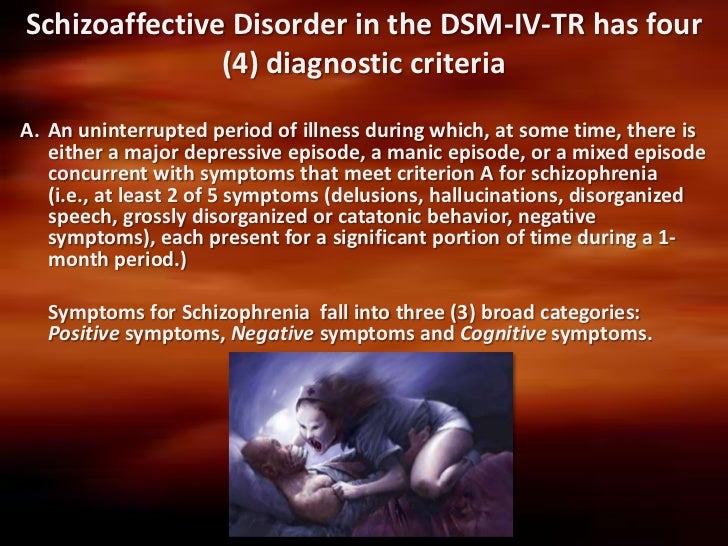
Schizoaffective disorder is a mental disorder in which the individual reflects symptoms that occur both in schizophrenia and mood disorder (major depressive or bipolar disorder). However, studies suggest that schizoaffective disorder resembles schizophrenia more than bipolar disorder. According to a report by National Alliance on Mental Illness, schizoaffective disorder is observed in about 0.3% of the population. Men and women experience schizoaffective disorder equally, but men are often reported to develop it at an earlier age.

Schizophrenia

Schizophrenia is a severe mental disorder that affects about 21 million people worldwide. It is characterized by distortions in thinking, perception, emotions, language, sense of self and behavior.

Bipolar and major depressive disorder

Bipolar disorder has been estimated to affect about 60 million people worldwide. It typically includes both manic and depressive episodes separated by periods of normal mood. The main difference between bipolar disorder and depressive disorder are the mania symptoms. When mood swings between manic and depressed states occur, it is referred to as bipolar disorder and when depression occurs alone it is a unipolar in nature, as it does not involve mania.

Mixed type- In this type of schizoaffective disorder the person has symptoms of schizophrenia, depression and mania.

What are the Causes of Schizoaffective Disorder?

The exact cause of schizoaffective disorder is unknown. But the following causes are the main factors. They include:

  • Genetics: Schizoaffective disorder tends to run in patients with a family history of the condition. It is not necessary that a person with a family history will continue to have the illness but there is a greater chance of them developing the illness. Studies reveal that it shows substantial familial overlap with both schizophrenia and bipolar disorder.
  • Brain chemistry and structure: Brain function and structure play an important role in the etiology of schizoaffective disorder. It may be induced by a neurotransmitter imbalance in a feedback-regulated system. It may involve imbalances in neurotransmitters such as dopamine, serotonin, norepinephrine, and glutamate that help regulate mood.
  • Stress: Stressful events or trauma such as early parental loss, any family conflict, or physical or sexual abuse can trigger symptoms or an onset of the illness. This is more likely to be a cause if one had experienced any such triggers when he or she was too young to know how to cope with them.
  • Environmental factors: Some factors like malnutrition, maternal illness or prenatal exposure to toxins may also cause schizoaffective disorder.
  • Drug or substance use: Psychoactive drugs such as LSD (Lysergic Acid) have been linked to the development of schizoaffective disorder. Also, cannabis use especially before the age of 15 years has also been reported to be a causative agent for this disorder.

What are the Signs and Symptoms of Schizoaffective Disorder?

The specific symptoms themselves may vary from person to person but the main symptoms include:

  • Hallucinations- Imagining, seeing, hearing, or even smelling things that do not actually exist.

  • Delusions- Having fixed, false beliefs inspite of having a strong evidence against them. It also includes delusions of persecution like believing that they are threatened, spied or attacked by someone.

  • Disorganized thinking- A person may switch very quickly from one topic to another unrelated topic and might have trouble organizing his thoughts and connecting them with a logical base.
  • Catatonic behavior- It may include bizarre behaviors and overall disability in performing daily activities such as bathing, dressing properly or even eating regularly.
  • Depressed mood- If a person has been diagnosed with schizoaffective disorder depressive type then they may have loss of interest in things they used to like before. Insomnia or hypersomnia is also observed.
  • Manic behavior- The person may also experience feelings of euphoria, inflated self-esteem, racing thoughts, increased risky behavior and other symptoms of mania.

Some physical symptoms are also observed; they include:

  • Changes in physical appearance
  • Poor hygiene
  • Significant weight loss
  • Psychomotor agitation
  • Fatigue or loss of energy

What are the Complications of Schizoaffective Disorder?

Individuals who have schizoaffective disorder that remains untreated or improperly diagnosed may run the risk of developing numerous complications. Examples may include:

  • Substance use and abuse
  • Onset of self-harming behaviors
  • Co-occurring Disorders

The most commonly occurring disorders alongside schizoaffective disorder are:

  • Post traumatic stress disorder (PTSD)
  • Generalized anxiety disorder
  • Obsessive compulsive disorder
  • Obesity
  • Diabetes

How do you Treat Schizoaffective Disorder?

Schizoaffective disorder can be managed effectively with medication and therapy. It belongs to the category of recurrent disorders and therefore, needs prophylactic treatment. National Institute for Health and Care Excellence (NICE) recommends that one should be treated with a combination of medication and talking therapies.

Medications-Certain medications are offered first, especially if an individual is first diagnosed during a psychotic episode.


It includes:

  • An antipsychotic drug- such as olanzapine or quetiapine.
  • mood stabilizer- such as lithium or valproate – especially if you have manic episodes rather than depression. Lamotrigine, is prescribed in case of bipolar disorder.
  • An anti-depressant- is used in case of symptoms that switch between mania and depression. Combination of anti-depressants with anti-psychotics are also used; for example, sertraline or fluoxetine plus haloperidol or risperidone.

Cognitive behavior therapy (CBT) – It acts as an add-on to medication and can help a person cope with the illness in a better way. It focuses on a person’s thoughts, beliefs, and how these affect their mood and actions. It also helps to identify and change any negative thoughts or behavior and replace them with adaptive thoughts.

Back Acne: Causes, prevention & Treatment

Causes

There are two causes of back acne: an increase in oil production or irritated skin. When your skin glands produce too much oil due to stress, genetics, hormonal imbalance or a poor diet, the increase in oil overproduction can clog up your skin follicles, preventing dead skin from escaping. This allows bacteria to breed and acne breakouts to occur. When clothing is warm and tight against the skin, acne may thrive because tight clothing can also trap dead skin cells. Tight clothing or a wearing a backpack may aggravate back acne by spreading the bacteria, causing fresh outbreaks.

Prevention

Wearing loose, cotton clothing may help prevent the spread of back acne. Instead of wearing a backpack, carry a handheld bag to decrease skin irritations on your back. Wash the skin on your back, regularly and immediately after exercise — but do not shower more than twice a day. Excessive bathing or showering may dry and irritate the skin on your back. To reduce the bacteria production on your skin’s surface, use an antibacterial soap daily. Exfoliate your back with a sugar-based scrub a few times a week to remove dead skin cells that may clog your follicles. Do not wear shirts more than once and keep bed sheets clean. If you sweat a lot, wipe your back with a soft towel regularly.

Treatment

Because the skin on your back is tougher and denser than the skin on your face, treatments differ depending on the severity of your back acne. If you have mild back acne and washing the skin regularly is not enough to control it, topical treatments sold in drugstores may kill bacteria, dry the oil and remove dead skin cells. If over-the-counter treatments are too weak, a doctor may prescribe a topical cream for moderate cases of acne. In severe cases, Accutane may be prescribed. This medication comes in pill form and is derived from Vitamin A. Accutane has the power to shrink oil glands, but comes with serious side effects, such as dry eyes and lips, aching joints, itching and blurred vision. If your back acne leads to scars or cysts, you may be a candidate for laser treatment or surgery, but consult your doctor before scheduling any procedures.

Considerations

If you get red marks or irritations on your back, check with your doctor to ensure you are suffering from acne and not eczema, as this requires a completely different treatment.

Hair Transplant surgery: Types & Steps

Hair transplant surgery is surgery that is performed to restore hair to areas of the scalp that are bald or that have thinning hair. There are multiple types of hair replacement surgery. Most commonly, these involve hair transplantation, but flap surgery, tissue expansion of the scalp and scalp reduction surgery, are also methods used for hair replacement. Each of these types of surgeries can be used alone, or in combination, to provide the patient with the best possible outcome for hair replacement.

Hair transplantation involves removing small pieces of hair-bearing scalp from a donor site and using them as grafts to be relocated to a bald or thinning area of the scalp.

Flaps surgeries involve moving hair bearing scalp tissue into bald areas of the scalp.

Tissue expansion allows the hair bearing scalp to be expanded to help cover areas of the scalp that have no hair.

Scalp reduction surgery involves surgically removing bald areas of the scalp and advancing, or bringing together, the hair bearing areas of the scalp.

If you and your surgeon have determined that hair transplant surgery is the best option for you, you can feel comfortable knowing that board-certified plastic surgeons have been successfully performing these types of procedures for more than thirty years.

The truth about hair loss

Baldness is often blamed on poor circulation to the scalp, vitamin deficiencies, dandruff and even excessive hat wearing. All of these theories have been disproved. It’s also untrue that hair loss can be determined by looking at your maternal grandfather, or that 40-year-old men who haven’t lost their hair will never lose it.

Hair loss is primarily caused by a combination of:

  • Aging
  • A change in hormones
  • A family history of baldness

Hair transplantation steps:

Hair transplantation involves removing small punch grafts from the hair bearing scalp or a larger piece of this scalp from a donor area and cutting this into smaller pieces to use as grafts. These grafts are then relocating to a bald or thinning area of the scalp. The grafts created in this manner differ in size and shape. Round-shaped punch grafts usually contain about 10-15 hairs. The much smaller mini-graft contains about two to four hairs; and the micro-graft, one to two hairs. Slit grafts, which are inserted into slits created in the scalp, contain about four to 10 hairs each; strip grafts are long and thin and contain 30-40 hairs.

Generally, several surgical sessions may be needed to achieve satisfactory fullness, and a healing interval of several months is usually recommended between each session. It may take up to two years before you see the final result with a full transplant series. The amount of coverage you’ll need is partly dependent upon the color and texture of your hair. Coarse, gray or light-colored hair affords better coverage than fine, dark-colored hair. The number of large plugs transplanted in the first session varies with each individual, but the average is about 50. For mini-grafts or micro-grafts, the number can be up to 700 per session.

Just before surgery, the “donor area” will be trimmed short so that the grafts can be easily accessed and removed. For punch grafts, your doctor may use a special tube-like instrument made of sharp carbon steel that punches the round graft out of the donor site so it can be replaced in the area to be covered—generally the frontal hairline. For other types of grafts, your doctor will use a scalpel to remove small sections of hair-bearing scalp, which will be divided into tiny sections and transplanted into tiny holes or slits within the scalp. When grafts are taken, your doctor may periodically inject small amounts of saline solution into the scalp to maintain proper skin strength. The donor site holes may be closed with stitches—for punch grafts, a single stitch may close each punch site; for other types of grafts, a small, straight-line scar will result. The stitches are usually concealed with the surrounding hair.

To maintain healthy circulation in the scalp, the grafts are placed about one-eighth of an inch apart. In later sessions, the spaces between the plugs will be filled in with additional grafts. Your doctor will take great care in removing and placement of grafts to ensure that the transplanted hair will grow in a natural direction and that hair growth at the donor site is not adversely affected.

After the grafting session is complete, the scalp will be cleansed and covered with gauze. You may have to wear a pressure bandage for a day or two. Some doctors allow their patients to recover bandage-free.

Tissue Expansion

Another technique used in the treatment of baldness is called tissue expansion. Plastic surgeons are the leaders in tissue expansion, a procedure commonly used in reconstructive surgery to repair burn wounds and injuries with significant skin loss. Its application in hair transplant surgery has yielded dramatic results-significant coverage in a relatively short amount of time.

In this technique, a balloon-like device called a tissue expander is inserted beneath hair-bearing scalp that lies next to a bald area. The device is gradually inflated with salt water over a period of weeks, causing the skin to expand and grow new skin cells. This causes a bulge beneath the hair-bearing scalp, especially after several weeks.

When the skin beneath the hair has stretched enough-usually about two months after the first operation-another procedure is performed to bring the expanded skin over to cover the adjacent bald area. For more information about tissue expansion, ask your plastic surgeon for the American Society of Plastic Surgeons, Inc. brochure entitled, Tissue Expansion: Creating New Skin from Old.

Flap Surgery

Flap surgery on the scalp has been performed successfully for more than 20 years. This procedure is capable of quickly covering large areas of baldness and is customized for each individual patient. The size of the flap and its placement are largely dependent upon the patient’s goals and needs. One flap can do the work of 350 or more punch grafts.

A section of bald scalp is cut out and a flap of hair-bearing skin is lifted off the surface while still attached at one end. The hair-bearing flap is brought into its new position and sewn into place, while remaining “tethered” to its original blood supply. As you heal, you’ll notice that the scar is camouflaged—or at least obscured—by relocated hair, which grows to the very edge of the incision.

In recent years, plastic surgeons have made significant advances in flap techniques, combining flap surgery and scalp reduction for better coverage of the crown; or with tissue expansion, to provide better frontal coverage and a more natural hairline.

Scalp Reduction

This technique is sometimes referred to as advancement flap surgery because sections of hair-bearing scalp are pulled forward or “advanced” to fill in a bald crown.

Scalp reduction is for coverage of bald areas at the top and back of the head. It’s not beneficial for coverage of the frontal hairline. After the scalp is injected with a local anesthetic, a segment of bald scalp is removed. The pattern of the section of removed scalp varies widely, depending on the patient’s goals. If a large amount of coverage is needed, doctors commonly remove a segment of scalp in an inverted Y-shape. Excisions may also be shaped like a U, a pointed oval or some other figure.

The skin surrounding the cut-out area is loosened and pulled, so that the sections of hair-bearing scalp can be brought together and closed with stitches. It’s likely that you’ll feel a strong tugging at this point, and occasional pain.

Procedure Steps

Before: Hair transplant candidates should have some noticeable hair loss with healthy hair growth at the back and sides of the head to serve as donor areas.
hair transplant

Step 1: A tube-like instrument punches round grafts from the donor site to be placed in the area where hair replacement is desired.
hair transplant

Step 2: A tube-like instrument punches round grafts from the donor site to be placed in the area where hair replacement is desired.
hair transplant

Step 3: When the skin beneath the hair has stretched enough, it is surgically placed over the bald area.
hair transplant

Step 4: During flap surgery, a section of bald scalp is cut out and a flap of hair-bearing skin is sewn into its place.
hair transplant

Step 5: The patterns used in scalp reduction vary widely, yet all meet the goal of bringing hair and scalp together to cover bald areas.
hair transplant

After: The results of hair transplant surgery can enhance your appearance and self-confidence.

Umbilical Hernia: Causes, symptoms & treatments

Umbilical Hernia

  • It is a common type of Hernia and constitutes around 25% of all Hernia cases.
  • Umbilical Hernia is often noted at birth as a protrusion at the bellybutton (the umbilicus).
  • Umbilical Hernia is caused due to weakened opening in the abdominal wall, which should normally close before birth but fails to close completely.


Umbilical Hernia

Causes of umbilical hernia in adults

Umblical hernia that is acquired happens due to intra-abdominal pressure from lifting heavy objects, multiple pregnancies, long history of coughing and obesity.

During pregnancy, the umbilical cord traverses through a little opening or belly button in the baby’s abdominal muscles. This slit normally closes just prior to the birth. If the muscles do not join properly in the middle line of the abdomen, the weakness of the abdominal wall can result in umbilical hernia either at that time or later in life.

Complications of umbilical hernia in adults

Adults who have umbilical hernia are likely to experience intestinal obstruction. In such a case, emergency surgery may be needed. In case of children, the complications are not so common, and they happen only when the abdominal tissue is incarcerated and cannot be retracted into the abdominal cavity. This results in reduced blood supply to the trapped intestine leading to pain in the umbilical area and damage.


Umbilical hernia in adults diagnosis

In the physical exam, the doctor will ask you to do blood tests and imaging studies like an X-ray or abdominal ultrasound may be done to find out complications.

Importantly this type of hernia must be distinguished from a paraumbilical hernia, which occurs in adults and involves a defect in the midline near to the umbilicus, and from omphalocele.

Umblical Hernia in adults Treatment

When the hole is small, say less than 1 or 2 centimeters, it is believed that 90 percent of umbilical hernia cases close in three years. If hernias do not enlarge or are asymptomatic, then there may or may not be a need for surgery.

When is surgery required for umbilical hernia?

In adults, surgery is required as a part of umbilical hernia treatment, epscially if the said hernia becomes bigger or causes pain. During surgery, a little incision is made below the navel. The herniated tissue is pushed back to the abdominal cavity, and the gap in the abdominal wall is stitched. People can go to work within a few hours of surgery and normal activities can be resumed in two to four weeks.

In children,umbilical hernia surgery is needed only if the hernia is large and cause pain. The doctor suggest operation when the hernia gets bigger after the age of 1 or 2 of it is seen to be blocking the intestines.


Pages:1...26272829303132...81