Global Treatment Services Pvt. Ltd.

Global Treatment Services

Blastomycosis: Causes, Symptoms & Treatments

Blastomycosis is an infection caused by a fungus called Blastomyces. The fungus lives in the environment, particularly in moist soil and in decomposing matter such as wood and leaves. People can get blastomycosis after breathing in the microscopic fungal spores from the air. Although most people who breathe in the spores don’t get sick, some of those who do may have flu-like symptoms, and the infection can sometimes become serious if it is not treated.

Symptoms of Blastomycosis:

Man with thermometer in his mouth while holding his head.

Fever is a common symptom of blastomycosis.

Approximately half of people who are infected with the fungus Blastomyces will show symptoms. The symptoms of blastomycosis are often similar to the symptoms of flu or other lung infections, and can include:

  • Fever
  • Cough
  • Night sweats
  • Muscle aches or joint pain
  • Weight loss
  • Chest pain
  • Fatigue (extreme tiredness)

How soon do the symptoms of blastomycosis appear?

Symptoms of blastomycosis usually appear between 3 weeks and 3 months after a person breathes in the fungal spores.

Severe blastomycosis

In some people, particularly those who have weakened immune systems, blastomycosis can spread from the lungs to other parts of the body, such as the skin, bones and joints, and the central nervous system (the brain and spinal cord).

How is blastomycosis treated?

Most people with blastomycosis will need treatment with prescription antifungal medication. Itraconazole is a type of antifungal medication that is typically used to treat mild to moderate blastomycosis. Amphotericin B is usually recommended for severe blastomycosis in the lungs or infections that have spread to other parts of the body. Depending on the severity of the infection and the person’s immune status, the course of treatment can range from six months to one year.

Blastomycosis Risk & Prevention:

Who gets blastomycosis?

Anyone can get blastomycosis if they’ve been in an area where Blastomyces lives in the environment. People who participate in outdoor activities that expose them to wooded areas (such as forestry work, hunting, and camping) in these areas may be at higher risk for getting blastomycosis.People who have weakened immune systems may be more likely to develop severe blastomycosis than people who are otherwise healthy.

Is blastomycosis contagious?

No. Blastomycosis can’t spread between people or between people and animals.

Can my pets get blastomycosis?

Yes. Pets, particularly dogs, can get blastomycosis, but it is not contagious between animals and people.The symptoms of blastomycosis in animals are similar to the symptoms in humans. If you are concerned about your pet’s risk of getting blastomycosis or if you think that your pet has blastomycosis, please talk to a veterinarian.

How can I prevent blastomycosis?

There is no vaccine to prevent blastomycosis, and it may not be possible to completely avoid being exposed to the fungus that causes blastomycosis in areas where it is common in the environment. People who have weakened immune systems may want to consider avoiding activities that involve disrupting soil in these areas.

Reproductive Genetics: Details of different methods

Reproductive Genetics

It is estimated that genetics contribute up to 10 percent of infertility issues or recurrent pregnancy losses in couples. The risk of finding chromosomal abnormalities grows as the age of the mother increases. Most embryos with an incorrect number of chromosomes fail to implant or miscarry during the first trimester of pregnancy. Genetic testing protocols have the potential to help many of those couples in their quest to have a family. Our comprehensive list of genetic testing services can greatly increase your chances of becoming a mother.

Pre-implantation Genetic Screening (PGS)

PGS is a last generation genetic test that allows us to screen for chromosomal abnormalities on embryos during an IVF cycle. The procedure requires to make a small biopsy on the embryos on which the analysis is performed. PGS analyse all 24 chromosome types using array Comparative Genomic Hybridisation (aCGH), in order to detect aneuploidies (alterations in the number of chromosomes) and select chromosomally normal embryos for transfer which are the most likely to develop to term and to be born as a healthy baby. The most common syndromes caused by chromosome aneuploidies are Down syndrome, Edwards syndrome and Patau syndrome.

Who is this for

Most of spontaneous miscarriages and some birth defects are caused by chromosomal abnormalities and the risks of having a child with a chromosome abnormality increases with maternal age. For this reason, PGS is especially recommended for:

i. Women over 35 years of age
ii. Couples who have experienced several spontaneous miscarriages of unknown cause
iii. Couples with several cycles of IVF that have not achieved pregnancy
iv. Men with low sperm concentration
v. Couples with a previous pregnancy with chromosomal abnormality

Pre-implantation Genetic Diagnosis (PGD)

The Concept

While PGD also analyzes a small piece of the embryos during an IVF cycle to detect genetic alterations, it is focussed on single gene diseases or mutations. Common examples of monogenic disorders are cystic fibrosis, fragile X syndrome, spinal muscular dystrophies (SMA), Duchenne muscular dystrophy (DMD), Huntington disease. PGD helps couples who have an increased risk for a genetic disease to conceive a healthy pregnancy.

Who is this for

  • Fertile couples who have a known history of a genetic condition in their families.
  • Any condition for which the couple’s offspring are at risk.


Endometrial Receptivity Array [ERA ]

The Concept

ERA is a personalized genetic test to diagnose the state of endometrial receptivity in the window of implantation. ERA determines the exact time period during which an embryo needs to be transferred into the woman’s uterus to achieve a successful pregnancy.

Who is this for

This test is advised

  • for patients who have had implantation failure with embryos of good morphological quality (at least 3 failed embryo transfers for younger women or two in patients 37 years or more).
  • for patients with apparently normal uterus and with normal endometrial thickness (≤6mm), in which no problems are apparent.

Hiatal Hernia: Causes, Symptoms & Treatments

The food that we eat has a long journey through a convoluted tube that we refer to as the digestive tract. Each section of the digestive tract has specific functions. When one part is affected, it can have a perpetuating effect on other parts of the tract. The digestive tract like any part of the body is prone to a host of conditions. If any of these conditions impede movement of food through the tract then it can become backed up and give rise to unusual sensations like a stuck stomach.

What does stuck stomach mean?

We often explain symptoms that we do understand in lay terms that have little to no medical significance. One of these colloquial terms is a ‘stuck stomach’ or as if somethings feels like it is ‘stuck in the stomach’. The latter is often associated with a meal in that there is the sensation that the food is stuck in the stomach. These sensations may not be caused by any underlying medical condition. This does not mean that it is imaginary but rather than the perception of something being stuck in the stomach is incorrect.

However, there are times where food and fluid can become obstructed in the stomach or even the stomach itself can become strangulated. In the early stages there may only be some vague symptoms but with time it can become severe. The two most notable conditions in this regard is a hiatal hernia and gastroparesis leading to delayed gastric emptying. Understanding what causes these conditions will help in making sense of strange sensations like there is something stuck in the stomach.

Causes

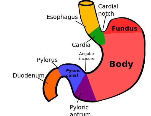
The esophagus runs down from the throat, across the chest cavity and into the abdominal cavity where it joins the stomach. The diaphragm separates the chest and abdominal cavities and the esophagus passes through a small opening in the diaphragm known as the esophageal hiatus. Food is churned in the stomach where it is mechanically broken down and chemically digested by the strong gastric acid and stomach enzymes. Eventually a semi-liquid mixture known as gastric chyme is pumped out through the pylorus of the stomach into the duodenum of the small intestine. This ensures that food which enters the stomach is processed and expelled in an orderly manner to facilitate digestion.

stomach

Hiatal Hernia

A hiatal hernia is a condition where the upper portion of the stomach become stuck in the esophageal hiatus. Normally this hiatus in the diaphragm is small and tight to ensure that only the esophagus can pass through. However, in a hiatal hernia the opening allows for the upper portion of the stomach to push through. The affected portion of the stomach becomes stuck and squeezed in the hiatus for short periods of time until it releases and slips back down into the abdominal cavity.

Causes

  • Enlarged esophageal hiatus from birth.
  • Injury to the diaphragm.
  • Repeated pressure with coughing, vomiting, when lifting heavy objects or straining during bowel movements.

Symptoms

  • Difficulty swallowing
  • Heartburn
  • Belching
  • Feeling full after small meals
  • Vomiting (sometimes bloody vomit)
  • Abdominal or chest pain

Gastroparesis

Gastroparesis is a condition where the stomach does not empty properly because the muscles that control movement through the stomach do not function as it should. Often the problem lies with the nerves that control these muscles. It is the vagus nerve that controls these muscles and it can malfunction for several reasons. Food may remain in the stomach for longer than normal. It can even become backed up and food can push up into the esophagus.

Causes

The cause of gastroparesis is not always known as is therefore referred to as idiopathic gastroparesis. Some of the known causes include:

  • Damage during surgery
  • Diabetes mellitus (diabetic neuropathy)
  • Medication like narcotic painkillers and certain antidepressants
  • Parkinson’s disease
  • Rare conditions like scleroderma and amyloidosis

Symptoms

  • Nausea and vomiting
  • Feeling of fullness
  • Changes in appetite
  • Abdominal bloating
  • Abdominal pain

Foreign Body

A foreign body in the stomach is any non-edible object in the stomach that was swallowed either intentionally or accidentally. It most often seen in young children. Sometimes the object can become lodged in the esophagus and not enter the stomach. However, it is possible that the object enters the stomach but cannot exit because it becomes lodged in the pylorus of the stomach. The symptoms are largely the same as gastroparesis. Another condition that is mainly seen in infants is pyloric stenosis which also presents in a similar way to gastroparesis.

Surgery:

Surgical procedures to treat a stomach hernia are reserved for severe cases that are unresponsive to lifestyle changes and medication. It is also indicated for cases where there are complications of the stomach hernia and in particular the acid reflux associated with it. These surgical procedures include Nissen fundoplication and laparoscopic paraesophageal hernia repair.

Nissen fundoplication where a portion of the upper stomach (fundus) is wrapped around the lower end of the esophagus. It is the procedure of choice in most stomach hernia cases and can be done as a laparoscopic procedure. This procedure is also used for gastroesophageal reflux disease (GERD).

Laparoscopic paraesophageal hernia repair is done by the means of a synthetic mesh to secure the area after removing the hernia sac from the thoracic cavity

Parenchymal Liver diseases: Causes, Symptoms & Treatments

The liver is the largest solid organ in the body, and it performs many complex functions. It produces important proteins, aids in digestion, helps blood to clot, and metabolizes cholesterol and glucose. Additionally, it aids in the removal of toxins from the digestive tract and the blood. Accordingly, when the liver becomes diseased from any cause, a number of symptoms result. These include nausea, vomiting, weight loss, fatigue, jaundice and pain in the right upper quadrant of the abdomen. Usually, about 75 percent of liver function must be lost before liver function is affected and symptoms appear.

Alcohol abuse is the number one cause of liver disease in the world. Alcohol is toxic to liver cells, and chronic alcohol abuse causes inflammation, or alcoholic hepatitis, and eventual scarring of the liver, or cirrhosis, which causes the liver to fail. However, drugs such as acetaminophen and cholesterol-controlling drugs known as statins may also cause liver disease, especially in people who drink alcohol. Additionally, viral hepatitis causes inflammation of liver cells and may also lead to liver failure.

Parenchymal liver disease affects the actual liver cells directly instead of those of supporting tissue, such as the bile ducts. According to Dictionary.com, the word “parenchymal” is an anatomical term that refers to the “specific tissue of an animal organ.” Many conditions cause this type of liver damage, including cirrhosis, hepatitis and non-alcoholic fatty liver disease.

Causes:

The term liver disease is used to describe several conditions that affect the liver’s ability to function normally. It can be caused by chronic inflammation of the liver, which can result from a number of conditions. Liver disease resulting from fat build-up in the liver, called non-alcoholic fatty liver disease, is associated with obesity.

Some other possible causes of liver disease are autoimmune diseases such as HIV and hepatitis, a variety of viruses and parasites, and cancers of the liver or bile ducts. Cancers that spread to the liver from elsewhere in the body can also lead to liver disease. It warns against eating mushrooms in the wild, as certain varieties can cause acute liver failure.

Certain medications can cause inflammation that could lead to liver disease as a result of prolonged use. Certain antibiotics and birth control drugs can lead to liver inflammation, so people who take these kinds of medications should talk to their doctors about liver health.

Treatments:

Supportive care is reserved for those with hepatitis A in order to make sure the patient remains hydrated as the body fights infection. Gallstones are often treated with gallbladder removal surgery. Long-term medical care is a treatment option for controlling and lowering the effects of liver disease.

In order to control how much protein is absorbed in patients with cirrhosis and final-stage liver disease, medications are often used. High blood ammonia levels, coma, disorientation and fatigue are common in a liver with cirrhosis, since the organ is unable to properly metabolize waste. Diuretics and a low-sodium diet may be recommended to treat water retention.

To remove fluid built up in the abdominal cavity, a needle and syringe can be used as a drainage method. If the fluid isn’t removed in time, it can result in an infection. If a patient’s liver completely fails, a liver transplant may be the only viable option. Complications of liver failure include weight loss, infection and bleeding.

Edema: Types, causes, symptoms & Treatments

Symptoms of Edema

Your symptoms will depend on the amount of swelling you have and where you have it.

Edema in a small area from an infection or inflammation (like a mosquito bite) may cause no symptoms. On the other hand, a large allergic reaction (such as from a bee sting) may cause edema on your entire arm that can bring pain and limit your arm’s movement.

Food allergies and allergic reactions to medicine may cause tongue or throat edema. This can be life-threatening if it interferes with your breathing.

Leg edema can make the legs feel heavy. This can affect walking. In edema and heart disease, for example, the legs may easily weigh an extra 5 or 10 pounds each. Severe leg edema can interfere with blood flow, leading to ulcers on the skin.

Pulmonary edema causes shortness of breath and sometimes low oxygen levels in the blood. Some people with pulmonary edema may have a cough.

There may be an indent or a “pit” that remains for a while after you push on the skin in some types of edema. This is called pitting edema. If the tissue springs back to its normal shape, it’s called non-pitting edema. It’s a symptom that may help your doctor figure out the cause of your edema.

Treatment of Edema

To treat edema, you often must treat its underlying cause. For example, you might take allergy medications to treat swelling from allergies.

Edema from a block in fluid drainage can sometimes be treated by getting the drainage flowing again. A blood clot in the leg is treated with blood thinners. They break down the clot and get drainage back to normal. A tumor that blocks blood or lymph can sometimes be shrunk or removed with surgery, chemotherapy, or radiation.

Leg edema related to congestive heart failure or liver disease can be treated with a diuretic (sometimes called a ”water pill”) like furosemide (Lasix). When you can pee more, fluid from the legs can flow back into the blood. Limiting how much sodium you eat can also help.

Colon Polyps: Causes, Symptoms & Treatments

Colon polyp is a small clump of cells that forms on the lining of the colon. Most colon polyps are harmless. But over time, some colon polyps can develop into colon cancer, which is often fatal when found in its later stages.

There are two main categories of polyps, non-neoplastic and neoplastic. Non-neoplastic polyps include hyperplastic polyps, inflammatory polyps and hamartomatous polyps. These types of polyps typically do not become cancerous. Neoplastic polyps include adenomas and serrated types. In general, the larger a polyp, the greater the risk of cancer, especially with neoplastic polyps.

Anyone can develop colon polyps. You’re at higher risk if you’re 50 or older, are overweight or a smoker, or have a personal or family history of colon polyps or colon cancer.

Colon polyps often don’t cause symptoms. It’s important to have regular screening tests, such as a colonoscopy, because colon polyps found in the early stages can usually be removed safely and completely. The best prevention for colon cancer is regular screening for polyps.

Symptoms

Colon polyps often cause no symptoms. You might not know you have a polyp until your doctor finds it during an examination of your bowel.

But some people with colon polyps experience:

  • Rectal bleeding. This can be a sign of colon polyps or cancer or other conditions, such as hemorrhoids or minor tears in your anus.
  • Change in stool color. Blood can show up as red streaks in your stool or make stool appear black. A change in color may also be caused by foods, medications and supplements.
  • Change in bowel habits. Constipation or diarrhea that lasts longer than a week may indicate the presence of a large colon polyp. But a number of other conditions can also cause changes in bowel habits.
  • Pain. A large colon polyp can partially obstruct your bowel, leading to crampy abdominal pain.
  • Iron deficiency anemia. Bleeding from polyps can occur slowly over time, without visible blood in your stool. Chronic bleeding robs your body of the iron needed to produce the substance that allows red blood cells to carry oxygen to your body (hemoglobin). The result is iron deficiency anemia, which can make you feel tired and short of breath.

When to see a doctor

See your doctor if you experience:

  • Abdominal pain
  • Blood in your stool
  • A change in your bowel habits that lasts longer than a week

You should be screened regularly for polyps if:

  • You’re age 50 or older.
  • You have risk factors, such as a family history of colon cancer. Some high-risk individuals should begin regular screening much earlier than age 50.

Causes:

There are two main categories of polyps, non-neoplastic and neoplastic. Non-neoplastic polyps include hyperplastic polyps, inflammatory polyps and hamartomatous polyps. Non-neoplastic polyps typically do not become cancerous.

Inflammatory polyps may be seen with ulcerative colitis or Crohn’s disease of the colon. Although the polyps themselves are not a significant threat, having ulcerative colitis or Crohn’s disease of the colon increases your overall risk of colon cancer.

Neoplastic polyps include adenomas and serrated types. Most colon polyps are adenomas. Serrated polyps may become cancerous, depending on their size and location in the colon. In general, the larger a polyp, the greater the risk of cancer, especially with neoplastic polyps.

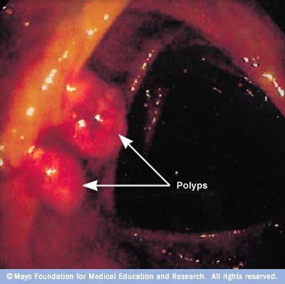
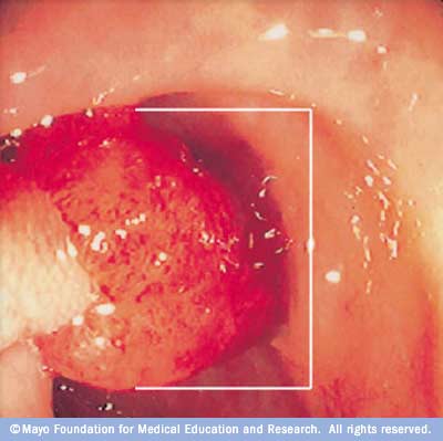
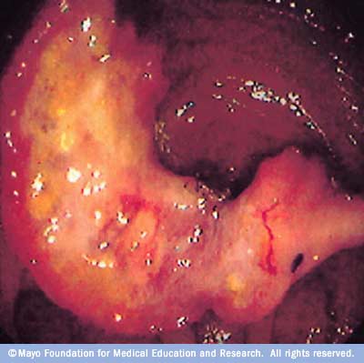
Image showing small polyps

Treatment

Your doctor is likely to remove all polyps discovered during a bowel examination. The options for removal include:

  • Removal with forceps or a wire loop (polypectomy). If a polyp is larger than 0.4 inches (about 1 centimeter), a liquid may be injected under it to lift and isolate the polyp from surrounding tissue so that it can be removed.
  • Minimally invasive surgery. Polyps that are too large or that can’t be removed safely during screening are usually removed laparoscopically, which is performed by inserting an instrument called a laparoscope into the bowel.
  • Colon and rectum removal. If you have a rare inherited syndrome, such as FAP, you may need surgery to remove your colon and rectum (total proctocolectomy).

Some types of colon polyp are far likelier to become malignant than are others. But a doctor who specializes in analyzing tissue samples (pathologist) usually must examine polyp tissue under a microscope to determine whether it’s potentially cancerous.

Follow-up care

If you have had an adenomatous polyp or a serrated polyp, you are at increased risk of colon cancer. The level of risk depends on the size, number and characteristics of the adenomatous polyps that were removed.

You’ll need follow-up screenings for polyps. Your doctor is likely to recommend a colonoscopy:

  • In five to 10 years if you had only one or two small adenomas
  • In three years if you had more than two adenomas, adenomas measuring 0.4 inches (about 1 centimeter) or larger, or certain adenomas
  • Within three years if you had more than 10 adenomas
  • Within six months if you had a very large adenoma or an adenoma that had to be removed in pieces

It’s important to fully prepare your colon before a colonoscopy. If stool remains in the colon and obstructs your doctor’s view of the colon wall, you will likely need a follow-up colonoscopy sooner than the guidelines specify.

Risk factors

Factors that may contribute to the formation of colon polyps or cancer include:

  • Age. Most people with colon polyps are 50 or older.
  • Inflammatory intestinal conditions, such as ulcerative colitis and Crohn’s disease.
  • Family history. You’re more likely to develop colon polyps or cancer if you have a parent, sibling or child with them. If many family members have them, your risk is even greater. In some people, this connection isn’t hereditary.
  • Tobacco and alcohol use.
  • Obesity and lack of exercise.
  • Race. African-Americans are at higher risk of developing colon cancer.
  • Type 2 diabetes that isn’t well-controlled.

Hereditary polyp disorders

Rarely, people inherit genetic mutations that cause colon polyps to form. If you have one of these genetic mutations, you are at much higher risk of developing colorectal cancer. Screening and early detection can help prevent the development or spread of these cancers.

Hereditary disorders that cause colon polyps include:

  • Lynch syndrome, also called hereditary nonpolyposis colorectal cancer. People with Lynch syndrome tend to develop relatively few colon polyps, but those polyps can quickly become malignant. Lynch syndrome is the most common form of inherited colon cancer and is also associated with tumors in the breast, stomach, small intestine, urinary tract and ovaries.
  • Familial adenomatous polyposis (FAP), a rare disorder that causes hundreds or even thousands of polyps to develop in the lining of your colon beginning during your teenage years. If the polyps aren’t treated, your risk of developing colon cancer is nearly 100 percent, usually before age 40. Genetic testing can help determine your risk of FAP.
  • Gardner’s syndrome, a variant of FAP that causes polyps to develop throughout your colon and small intestine. You may also develop noncancerous tumors in other parts of your body, including your skin, bones and abdomen.
  • MYH-associated polyposis (MAP), a condition similar to FAP that is caused by mutations in the MYH gene. People with MAP often develop multiple adenomatous polyps and colon cancer at a young age. Genetic testing can help determine your risk of MAP.
  • Peutz-Jeghers syndrome, a condition that usually begins with freckles developing all over the body, including the lips, gums and feet. Then noncancerous polyps develop throughout the intestines. These polyps may become malignant, so people with this condition have an increased risk of colon cancer.
  • Serrated polyposis syndrome, a condition that leads to multiple serrated adenomatous polyps in the upper part of the colon. These polyps may become malignant.

Complications

Some colon polyps may become cancerous. The earlier polyps are removed, the less likely it is that they will become malignant.

Prevention

You can greatly reduce your risk of colon polyps and colorectal cancer by having regular screenings. Certain lifestyle changes also can help:

  • Adopt healthy habits. Include plenty of fruits, vegetables and whole grains in your diet and reduce your fat intake. Limit alcohol consumption and quit tobacco. Stay physically active and maintain a healthy body weight.
  • Talk to your doctor about calcium and vitamin D. Studies have shown that increasing your consumption of calcium may help prevent recurrence of colon adenomas. But it isn’t clear whether calcium has any protective benefits against colon cancer. Other studies have shown that vitamin D may have a protective effect against colorectal cancer.
  • Consider your options if you’re at high risk. If you have a family history of colon polyps, consider having genetic counseling. If you’ve been diagnosed with a hereditary disorder that causes colon polyps, you’ll need regular colonoscopies starting in young adulthood.

Lung Cancer: Types, causes, symptoms & treatments

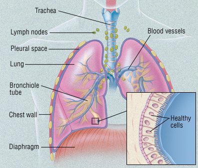
One of the most common cancers, lung cancer usually occurs when a cancer-causing agent, or carcinogen, triggers the growth of abnormal cells in the lung. These cells multiply out of control and eventually form a tumor. As the tumor grows, it destroys nearby areas of the lung. Eventually, tumor cells can spread (metastasize) to nearby lymph nodes and other parts of the body. These include the

  • liver
  • bones
  • adrenal glands
  • brain.

In most cases, the carcinogens that trigger lung cancer are chemicals found in cigarette smoke. However, more and more lung cancers are being diagnosed in people who have never smoked.

Lung Cancer


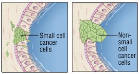
Lung cancers are divided into two groups, based on how their cells look under the microscope: non-small cell lung cancer and small cell lung cancer. Non-small cell lung cancer may be localized. This means that it is limited to the lung or that it hasn’t spread beyond the chest. As a result, it can usually be treated with surgery. Small cell lung cancer is rarely localized, even when it is detected early. It is rarely treated with surgery. Knowing whether the cancer has spread is critical, because it affects treatment decisions.

However, even when doctors think that the cancer is localized, it often comes back shortly after surgery. This means cancer cells had started to spread before surgery, but they couldn’t yet be detected.

Non-small cell lung cancer

Non-small cell lung cancer is more likely than small cell cancer to be localized at the time of diagnosis. It also is more likely than small cell cancer to be treatable with surgery. It often responds poorly to chemotherapy (anticancer drugs). However, sophisticated genetic tests can help predict which patients may show favorable responses to particular treatments, including chemotherapy.

Non-small cell lung cancer accounts for about 85% of all lung cancers. These cancers are divided into subgroups, based on how their cells look under a microscope:

  • Adenocarcinoma. This is the most common type of lung cancer. Although it is related to smoking, it is the most common type of lung cancer in nonsmokers. It is also the most common form of lung cancer in women and in people younger than 45. It usually develops near the edge of the lung. It can also involve the pleura, the membrane covering the lung.
  • Squamous cell carcinoma. This type of lung cancer tends to form a mass near the center of the lungs. As the mass gets larger, it can bulge into one of the larger air passages, or bronchi. In some cases, the tumor forms a cavity in the lungs.
  • Large cell carcinoma. Like adenocarcinoma, large cell carcinoma tends to develop at the edge of the lungs and spread to the pleura. Like squamous cell carcinoma, it can form a cavity in the lungs.
  • Adenosquamous carcinoma, undifferentiated carcinoma, and bronchioloalveolar carcinoma.These are relatively rare non-small cell lung cancers. Adenosquamous carcinoma has a worse prognosis compared to either adenocarcinoma or squamous cancer.

Small cell lung cancer 

At the time of diagnosis, small cell lung cancer is more likely than non-small cell cancer to have spread beyond the lung. This makes it almost impossible to cure with surgery. However, it can be managed with chemotherapy or radiation therapy. Small cell cancers account for about 15% of all lung cancers.

Risk factors

Your risk of all types of lung cancer increase if you

  • smoke. Smoking cigarettes is by far the leading risk factor for lung cancer. In fact, cigarette smokers are 13 times more likely to develop lung cancer than nonsmokers. Cigar and pipe smoking are almost as likely to cause lung cancer as cigarette smoking.
  • breathe tobacco smoke. Nonsmokers who inhale fumes from cigarette, cigar, and pipe smoking have an increased risk of lung cancer.
  • are exposed to radon gas. Radon is a colorless, odorless radioactive gas formed in the ground. It seeps into the lower floors of homes and other buildings and can contaminate drinking water. Radon exposure is the second leading cause of lung cancer. It’s not clear whether elevated radon levels contribute to lung cancer in nonsmokers. But radon exposure does contribute to lung cancer in smokers and in people who regularly breathe high amounts of the gas at work (miners, for example). You can test radon levels in your home with a radon testing kit.
  • are exposed to asbestos. Asbestos is a mineral used in insulation, fireproofing materials, floor and ceiling tiles, automobile brake linings, and other products. People exposed to asbestos on the job (miners, construction workers, shipyard workers, and some auto mechanics) have a higher-than-normal risk of lung cancer. People who live or work in buildings with asbestos-containing materials that are deteriorating also have an increased risk of lung cancer. The risk is even higher in people who also smoke. Asbestos exposure also increases the risk of developing mesothelioma. It’s a relatively rare and usually fatal cancer that starts in the lining of the lungs.
  • are exposed to other cancer-causing agents at work. These include uranium, arsenic, vinyl chloride, nickel chromates, coal products, mustard gas, chloromethyl ethers, gasoline, and diesel exhaust.

Symptoms

In some cases, lung cancer is detected when a person with no symptoms has a chest x-ray or computed tomography (CT) scan for another reason. But most people with lung cancer have one or more of these symptoms:

  • a cough that doesn’t go away
  • coughing up blood or mucus
  • wheezing
  • shortness of breath
  • trouble breathing
  • chest pain
  • fever
  • discomfort when swallowing
  • hoarseness
  • weight loss
  • poor appetite.
  • an irregular heart beat if the cancer is located close to the heart

If the cancer has spread beyond the lungs, it can cause other symptoms. For example, you may have bone pain if it has spread to your bones.

Certain small cell lung cancers may secrete chemicals that can alter the body’s chemical composition. For example, levels of sodium and calcium may be abnormal. This can lead to the diagnosis of small cell lung cancer.

Many of these symptoms can be caused by other conditions. See your doctor if you have symptoms so that the problem can be diagnosed and properly treated.

Diagnosis

Your doctor may suspect lung cancer based on

  • your symptoms
  • your smoking history
  • whether you live with a smoker
  • your exposure to asbestos and other cancer-causing agents.

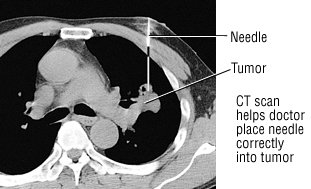
To look for evidence of cancer, your doctor will examine you, paying special attention to your lungs and chest. He or she will order imaging tests to check your lungs for masses. In most cases, a chest x-ray will be done first. If the x-ray shows anything suspicious, a CT scan will be done. As the scanner moves around you, it takes many pictures. A computer then combines the images. This creates a more detailed image of the lungs, allowing doctors to confirm the size and location of a mass or tumor.

You may also have a magnetic resonance imaging (MRI) scan or a positron emission tomography (PET) scanMRI scans provide detailed pictures of the body’s organs, but they use radio waves and magnets to create the images, not x-rays. PET scans look at the function of tissue rather than anatomy. Lung cancer tends to show intense metabolic activity on a PET scan. Some medical centers offer combined PET-CT scanning.

If cancer is suspected based on these images, more tests will be done to make the diagnosis, determine the type of cancer, and see if it has spread. These tests may include the following:

  • Sputum sample. Coughed up mucus is checked for cancer cells.
  • Biopsy. A sample of abnormal lung tissue is removed and examined under a microscope in a laboratory. If the tissue contains cancer cells, the type of cancer can be determined by the way the cells look under the microscope. The tissue is often obtained during a bronchoscopy. However, surgery may be necessary to expose the suspicious area.
  • Bronchoscopy. During this procedure, a tube-like instrument is passed down the throat and into the lungs. A camera on the end of the tube allows doctors to look for cancer. Doctors can remove a small piece of tissue for a biopsy.
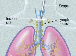
  • Mediastinoscopy. In this procedure, a tube-like instrument is used to biopsy lymph nodes or masses between the lungs. (This area is called the mediastinum.) A biopsy obtained this way can diagnose the type of lung cancer and determine whether the cancer has spread to lymph nodes.
  • Fine-needle aspiration. With a CT scan, a suspicious area can be identified. A tiny needle is then inserted into that part of the lung or pleura. The needle removes a bit of tissue for examination in a laboratory. The type of cancer can then be diagnosed.
  • Thoracentesis. If there is fluid build-up in the chest, it can be drained with a sterile needle. The fluid is then checked for cancer cells.
  • Video-assisted thoracoscopic surgery (VATS). In this procedure, a surgeon inserts a flexible tube with a video camera on the end into the chest through an incision. He or she can then look for cancer in the space between the lungs and the chest wall and on the edge of the lung. Abnormal lung tissue can also be removed for a biopsy.
  • Bone scans, MRI scans and CT scans. These imaging tests can detect lung cancer that has spread to the bones, brain, or other parts of the body.

After the cancer has been diagnosed, it is assigned a “stage.” The stages differ for non-small cell lung cancer and small cell lung cancer.

Non-small cell lung cancer

Stages of non-small cell lung cancer reflect the tumor’s size and how far the cancer has spread. Stages I through III are further divided into A and B categories.

  • Stage I tumors are small and have not invaded the surrounding tissue or organs.
  • Stage II and III tumors have invaded surrounding tissue and/or organs and have spread to lymph nodes.
  • Stage IV tumors have spread beyond the chest.

Small Cell Lung Cancer

Many experts divide small cell lung cancers into two groups:

  • Limited stage. These cancers involve only one lung and the nearby lymph nodes.
  • Extensive stage. These cancers have spread beyond the lung to other areas of the chest or to distant organs.

Knowing the type of cancer and its stage helps doctors determine the best treatment. Limited stage cancer, for example, may be treated with surgery and/or chemotherapy. Extensive stage cancer is much less likely to be cured.

However, many doctors now stage small cell lung cancers like non-small cell lung cancers. This more formal method may make the terms limited stage and extensive stage obsolete.

Expected Duration

Lung cancer will continue to grow and spread until it is treated.

Prevention

To reduce your risk of lung cancer,

  • Don’t smoke. If you already smoke, talk to your doctor about getting the help you need to quit.
  • Avoid secondhand smoke. Choose smoke-free restaurants and hotels. Ask guests to smoke outdoors, especially if there are children in your home.
  • Reduce exposure to radon. Have your home checked for radon gas. A radon level above 4 picocuries/liter is unsafe. If you have a private well, have your drinking water checked, too. Kits to test for radon are widely available.
  • Reduce exposure to asbestos. Because there is no safe level of asbestos exposure, any exposure is too much. If you have an older home, check to see if any insulation or other asbestos-containing material is exposed or deteriorating. The asbestos in these areas must be professionally removed or sealed up. If the removal isn’t done properly, you may be exposed to more asbestos than you would have been if it had been left alone. People who work with asbestos-containing materials should use approved measures to limit their exposure and to prevent bringing asbestos dust home on their clothing.
  • Have a 30 pack-year smoking history (pack years is calculated by multiplying the number of cigarettes smoked per day times the number of years you smoked), AND
  • Are currently smoke or have quit within the past 15 years, AND
  • Are healthy enough to undergo lung cancer surgery.

Treatment

After lung cancer has been diagnosed, the type of treatment depends on the type of cancer and how much the tumor has spread (its stage).

Non-small cell lung cancer

Surgery is the main treatment for non-small cell lung cancers that have not spread beyond the chest. The type of surgery will depend on the extent of the cancer. It will also depend on whether other lung conditions, such as emphysema, are present.

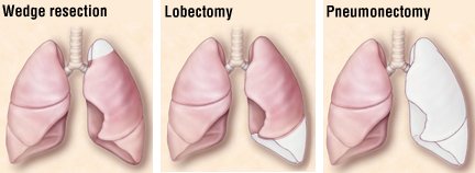
There are three types of surgery:

  • Wedge resection removes only a small part of the lung.
  • Lobectomy removes one lobe of the lung.
  • Pneumonectomy removes an entire lung.

Lymph nodes are also removed and examined to see if the cancer has spread.

Some surgeons use video-assisted thoracoscopy (VATS) to remove small, early-stage tumors, especially if the tumors are near the outer edge of the lung. (VATS can also be used to diagnose lung cancer.) Because the incisions for VATS are small, this technique is less invasive than a traditional “open” procedure.

Because surgery will remove part or all of a lung, breathing may be more difficult afterwards, especially in patients with other lung conditions (emphysema, for example). Doctors can test lung function prior to surgery and predict how it might be affected by surgery.

Depending on how far the cancer has spread, treatment may include chemotherapy (the use of anticancer drugs) and radiation therapy. These may be given before and/or after surgery.

When the tumor has spread significantly, chemotherapy may be recommended to slow its growth, even if it cannot cure the disease. Chemotherapy has been shown to ease symptoms and prolong life in cases of advanced lung cancer.

Radiation therapy can relieve symptoms, too. It is often used to treat lung cancer that has spread to the brain or bones and is causing pain. It can also be used alone or with chemotherapy to treat the lung cancer that is confined to the chest.

People who may not withstand surgery due to other serious medical problems may receive radiation therapy, with or without chemotherapy, as an alternative to surgery. Advances in radiation have made it possible for prolonged survival in some people, with results similar to surgery.

In specialized cancer centers, cancerous tissue may be tested for specific genetic abnormalities (mutations). Doctors may then be able to treat the cancer with a “targeted therapy.” These therapies can derail the cancer’s growth by preventing or changing chemical reactions linked to particular mutations. For example, some target therapies prevent cancer cells from receiving chemical “messages” telling them to grow.

Knowing about specific genetic mutations can help predict which therapy will be best. This strategy can be especially helpful in certain patients, such as women with adenocarcinoma of the lung who have never smoked.

Small cell lung cancer

The treatment of small cell lung cancer depends on its stage:

  • Limited stage. Treatments include various combinations of chemotherapy, radiation and, rarely, surgery, with or without radiation to the brain to prevent cancer spread. While small cell lung cancer often responds well to chemotherapy, it very often returns months or even years later.
  • Extensive stage. Treatments include chemotherapy, with or without brain radiation, or radiation treatments to areas of existing metastases in the brain, spine or other bones. Even if the imaging tests show that the cancer has not spread to the brain, many experts suggest treating the brain anyway. That’s because cancer cells may be there even if they haven’t yet shown up on the imaging tests. The question of whether or not to use brain radiation must be considered carefully; many patients experience memory loss afterwards. The decision to use brain radiation is a very crucial one, since many patients may experience a decrease in memory function after radiation therapy, with or without chemotherapy.

Gastroparesis: Causes, Symptoms &Treatments

  • Uncontrolled diabetes
  • Gastric surgery with injury to the vagus nerve
  • Medications such as narcotics and some antidepressants
  • Parkinson’s disease
  • Multiple sclerosis
  • Rare conditions such as: amyloidosis (deposits of protein fibers in tissues and organs) and scleroderma (a connective tissue disorder that affects the skin, blood vessels, skeletal muscles, and internal organs).

What Are the Symptoms of Gastroparesis?

There are many symptoms of gastroparesis, including:

  • Heartburn or GERD
  • Nausea
  • Vomiting undigested food
  • Feeling full quickly when eating
  • Abdominal bloating
  • Poor appetite and weight loss
  • Poor blood sugar control

What Are the Complications of Gastroparesis?

Some of the complications of gastroparesis include:

  • Food that stays in the stomach too long can ferment, which can lead to the growth of bacteria.
  • Food in the stomach can harden into a solid collection, called a bezoar. Bezoars can cause obstructions in the stomach that keep food from passing into the small intestine.
  • People who have both diabetes and gastroparesis may have more difficulty because blood sugar levels rise when food finally leaves the stomach and enters the small intestine, making blood sugar control more of a challenge.

How Is Gastroparesis Diagnosed?

To diagnose gastroparesis, your doctor will review your symptoms and medical history. He or she will also give you a physical exam and may order certain blood tests, including blood sugar levels. Other tests used to diagnose and evaluate gastroparesis may include:

  • Barium X-ray : You drink a liquid (barium), which coats the esophagus, stomach, and small intestine and shows up on X-ray. This test is also known as an upper GI (gastrointestinal) series or a barium swallow.
  • Radioisotope gastric-emptying scan (gastric scintigraphy): You eat food that contains a very small amount of radioisotope (a radioactive substance), then lie under a scanning machine; if the scan shows that more than 10% of food is still in your stomach 4 hours after eating, you are diagnosed with gastroparesis.
  • Gastric manometry: A thin tube that is passed through your mouthand into the stomach measures the stomach’s electrical and muscular activity to determine the rate of digestion.
  • Electrogastrography: This test measures electrical activity in the stomach using electrodes placed on the skin.
  • The smart pill: This is a small electronic device that is swallowed. It sends back information about how fast it is traveling as it moves through the digestive system.
  • Ultrasound : This is an imaging test that uses sound waves to create pictures of body organs. Your doctor may use ultrasound to eliminate other diseases.
  • Upper endoscopy : This procedure involves passing a thin tube (endoscope) down the esophagus to examine the lining of the stomach.

What Is the Treatment for Gastroparesis?

Gastroparesis is a chronic (long-lasting) condition. This means that treatment usually doesn’t cure the disease. But there are steps you can take to manage and control the condition.

Some patients may benefit from medications, including:

  • Reglan (metoclopramide): You take this drug before eating and it causes the stomach muscles to contract and move food along. Reglan also decreases the incidence of vomiting and nausea. Side effects include diarrhea, drowsiness, anxiety, and, rarely, a serious neurological disorder.
  • Erythromycin: This is an antibiotic that also causes stomach contractions and helps move food out. Side effects include diarrhea and development of resistant bacteria from prolonged exposure to the antibiotic.
  • Antiemetics: These are drugs that help control nausea.

People who have diabetes should try to control their blood sugar levels to minimize the problems of gastroparesis.

Dietary Modifications for Gastroparesis

One of the best ways to help control the symptoms of gastroparesis is to modify your daily eating habits. For instance, instead of three meals a day, eat six small meals. In this way, there is less food in the stomach; you won’t feel as full, and it will be easier for the food to leave your stomach. Another important factor is the consistency of food; liquids and low residue foods are encouraged (for example, applesauce should replace whole apples with intact skins).

You should also avoid foods that are high in fat (which can slow down digestion) and fiber (which is difficult to digest).

Other Treatment Options for Gastroparesis

In a severe case of gastroparesis, a feeding tube, or jejunostomy tube, may be used. The tube is inserted through the abdomen and into the small intestine during surgery. To feed yourself, put nutrients into the tube, which go directly into the small intestine; this way, they bypass the stomach and get into the bloodstream more quickly.

Using an instrument through a small incision, botulinum toxin (such as Botox) can be injected into the pylorus, the valve that leads from the stomach to the small intestine. This can relax the valve, keeping it open for a longer period of time to allow the stomach to empty.

Another treatment option is intravenous or parenteral nutrition. This is a feeding method in which nutrients go directly into the bloodstream through a catheter placed into a vein in your chest. Parenteral nutrition is intended to be a temporary measure for a severe case of gastroparesis.

Gastritis: Causes, Symptoms & Treatments

What Is Gastritis?

What Causes Gastritis?

Gastritis can be caused by irritation due to excessive alcohol use, chronic vomiting, stress, or the use of certain medicationssuch as aspirin or other anti-inflammatory drugs. It may also be caused by any of the following:

  • Helicobacter pylori (H. pylori): A bacteria that lives in the mucous lining of the stomach; without treatment, the infection can lead to ulcers, and in some people, stomach cancer.
  • Bile reflux: A backflow of bile into the stomach from the bile tract (that connects to the liver and gallbladder)
  • Infections caused by bacteria and viruses

If gastritis is left untreated, it can lead to a severe loss of blood and may increase the risk of developing stomach cancer.

What Are the Symptoms of Gastritis?

Symptoms of gastritis vary among individuals, and in many people there are no symptoms. However, the most common symptoms include:

  • Nausea or recurrent upset stomach
  • Abdominal bloating
  • Abdominal pain
  • Vomiting
  • Indigestion
  • Burning or gnawing feeling in the stomach between meals or at night
  • Hiccups
  • Loss of appetite
  • Vomiting blood or coffee ground-like material
  • Black, tarry stools

How Is Gastritis Diagnosed?

To diagnose gastritis, your doctor will review your personal and family medical history, perform a thorough physical evaluation, and may recommend any of the following tests:

  • Upper endoscopy. An endoscope, a thin tube containing a tiny camera, is inserted through your mouth and down into your stomach to look at the stomach lining. The doctor will check for inflammation and may perform a biopsy, a procedure in which a tiny sample of tissue is removed and then sent to a laboratory for analysis.
  • Blood tests. The doctor may perform various blood tests, such as checking your red blood cell count to determine whether you have anemia, which means that you do not have enough red blood cells. He or she can also screen for H. pylori infection and pernicious anemia with blood tests.
  • Fecal occult blood test (stool test). This test checks for the presence of blood in your stool, a possible sign of gastritis.

What Is the Treatment for Gastritis?

Treatment for gastritis usually involves:

  • Taking antacids and other drugs (such as proton pump inhibitors or H-2 blockers) to reduce stomach acid
  • Avoiding hot and spicy foods
  • For gastritis caused by H. pylori infection, your doctor will prescribe a regimen of several antibiotics plus an acid blocking drug (used for heartburn)
  • If the gastritis is caused by pernicious anemia, B12 vitamin shots will be given.
  • Eliminating irritating foods from your diet such as lactose from dairy or gluten from wheat

Once the underlying problem disappears, the gastritis usually does, too.

You should talk to your doctor before stopping any medicine or starting any gastritis treatment on your own.

Leg Fatty Tumors: Causes and Treatments

Leg Fatty TumorsTt

Fatty tumors in legs come in two different types–benign and malignant. There are three different kinds of benign fat tissue tumors–lipomas, the most common; lipoblastomas, occurring in infants and young children; and hibernomas, which are like lipomas but much less common. A malignant or cancerous soft tissue tumor is called a sarcoma. There are two types: osteosarcomas, which develop from bone, and soft tissue sarcomas which can develop from fat, muscle, nerves, fibrous tissues, blood vessels or deep skin tissues. They can be found in any part of the body, but most occur in the arms or legs. Sarcomas are not common and most are called carcinomas.


Causes, Diagnosis and Treatment of Benign Fatty Tumors

A lipoma is a slow-growing, fatty lump usually situated between your skin and muscle layer. It moves easily with finger pressure, is doughy to the touch and generally not tender. Lipomas can become painful if they grow and press on nerves. Most are detected during middle age. The exact cause of lipomas is not known, but they do tend to run in families, indicating perhaps a genetic predisposition. Diagnosis of this type tumor is usually done with a physical exam or a tissue sample (biopsy) for lab findings. Sometimes an ultrasound or MRI or CT scan is used if the tumor is large, has unusual features or seems to go deeper than the fatty tissue. In a small number of cases the lipoma is actually a form of cancer called a liposarcoma, but these are usually very painful. There is usually no treatment for a lipoma unless it bothers you, is painful or starts to grow. Surgical removal is the most common form of treatment. Steroid injections will shrink the tumor but usually doesn’t eliminate it. Liposuction uses a needle and syringe to remove the lump, but it is difficult to remove the entire lipoma with this method.

Pages:1...19202122232425...81