Global Treatment Services Pvt. Ltd.

Global Treatment Services

Multiple myeloma – A rare blood cancer

Overview

Multiple myeloma is a rare blood cancer that affects plasma cells in the bone marrow.  it is a blood cancer related to lymphoma and leukemia.

Multiple myeloma happens when healthy cells turn into abnormal cells that multiply and produce abnormal antibodies called M proteins. This change starts a cascade of medical issues and conditions that can affect your bones, your kidneys and your body’s ability to make healthy white and red blood cells and platelets. As multiple myeloma gets worse, those plasma cells begin to spill out of your bone marrow and spread through your body.

Some people have multiple myeloma without symptoms but blood tests show signs of conditions that may become multiple myeloma. In this case, healthcare providers may recommend watchful waiting or monitoring your overall health rather than starting treatment. Healthcare providers can’t cure multiple myeloma, but they can treat related conditions and symptoms and slow its progress.

Causes

Multiple myeloma symptoms develop over time and may resemble other medical conditions or diseases. Here are some of the conditions linked to multiple myeloma and their causes:

  • Anemia: You don’t have enough red blood cells because multiplying abnormal plasma cells don’t leave room for your red blood cells.
  • Bacterial infection, particularly pneumonia: Like red blood cells, abnormal plasma cells are multiplying and crowding healthy white blood cells that fight infection.
  • Thrombocytopenia: You don’t have enough platelets — the cells that help your blood to clot — because abnormal plasma cells crowd out those platelets and keep your bone marrow from making enough platelets.
  • Bone pain/bone fracture: Bone pain and/or fracture can happen if abnormal plasma cells destroy bone tissue, causing a soft spot in your bone. These are osteolytic lesions.
  • Kidney problems/failure: Your kidneys filter waste and toxins. Abnormal plasma cells make M proteins that block the filtering process and damage your kidneys.
  • Amyloidosis: This disease happens when abnormal proteins (amyloid proteins) build up in your organs.
  • Hypercalcemia: This condition happens when damaged or weakened bones release too much calcium into your bloodstream.
  • Hyperviscosity syndrome: M proteins — proteins made by abnormal plasma cells — thicken your blood. Your heart has to work harder to pump blood through your body.
  • Cryoglobulinemia: Multiple myeloma can make proteins in your blood clump together when it’s cold.

Symptoms

Multiple myeloma causes many symptoms, but bone pain often is the first symptom people notice. Other symptoms include:

  • Weakness in your arms and legs and/or a sensation of numbness in your arms and legs. Multiple myeloma can affect the bones in your spine, causing them to collapse and press on your spinal cord.
  • Having fatigue — feeling so tired you can’t manage daily activities — and feeling weak. These are signs of anemia.
  • Nausea and vomiting. This may be a sign of hypercalcemia.
  • Not having an appetite and/or feeling thirstier than usual. These may be signs of hypercalcemia.
  • Unexplained weight loss.
  • Unexplained fever. This may be a symptom of a bacterial infection.
  • Bruising or bleeding more easily. This may be a sign abnormal plasma cells prevent your body from producing enough platelets. Platelets help your blood to clot.
  • Feeling confused or “foggy.”

To confirm a diagnosis, you might have blood tests including:

  • Complete blood count (CBC): This test measures your red and white blood cell numbers, the amount of hemoglobin in your red blood cells and your red blood cell concentration level.
  • Blood chemistry test: This test shows your creatine levels (how well your kidneys work), albumin levels (this is a protein), calcium levels and lactic dehydrogenase levels (LDH). LDH is a tumor marker. A tumor marker is a substance in cancerous cells or healthy cells responding to cancer.
  • Quantitative immunoglobulin test: This blood test measures the levels of certain antibodies in your blood.
  • Electrophoresis: This test looks for M proteins in your blood.
  • Urine tests: Healthcare providers may ask you to collect your urine at home over a 24-hour period so they can test for Bence Jones protein, another sign of multiple myeloma.
  • X-rays: Healthcare providers use X-rays to look for bones damaged by multiple myeloma.
  • Computed tomography (CT) scan: This is another way to look for bone damage.
  • Magnetic resonance imaging (MRI): This test uses radio waves and strong magnets to create detailed images of your bones and spine. Healthcare providers may use this test to look for plasmacytomas. These are single groups of abnormal plasma cells.
  • Positron emission tomography (PET) scan: This is another test for plasmacytomas.
  • Bone marrow biopsies: Healthcare providers may do bone marrow biopsies to analyze the percentage of normal and abnormal plasma cells in your bone marrow. They may also test your bone marrow sample for changes in your DNA that may drive cancer growth.

Multiple myeloma can cause problems including:

  • Bone problems. Your bones can become weaker, leading to fractures.
  • Blood problems. You might get anemia, which means your body doesn’t have enough red blood cells. This can make you tired and pale and cause heart problems. You might also have too few platelets, which makes it harder for your blood to clot.
  • Infections. When you have myeloma, your body produces a lot of weak antibodies that crowd out healthy ones, making it harder for you to fight infection. A lack of white blood cells can also weaken your immune system.
  • Kidney damage. Myeloma can clog your kidneys so they don’t filter the way they should. This might lead to kidney failure.

The staging process for multiple myeloma starts with placing the condition in one of four classifications:

  • MGUS: Blood tests show small amounts of M protein. About 1 % to 2% of people with MGUS develop multiple myeloma.
  • Solitary or isolated plasmacytomas: These are single groups of abnormal plasma cells.
  • SMM: This is a pre-cancerous form of multiple myeloma. People who have SMM may have mild symptoms along with tests showing small amounts of M protein in their blood and an increased number of plasma cells in their bone marrow.
  • Multiple myeloma: This classification means tests show multiple groups of abnormal plasma cells, high M protein levels in blood or urine and a high percentage of abnormal plasma cells in bone marrow. People in this classification often have anemia, hypercalcemia, kidney failure and osteolytic lesions.

Healthcare providers use the Revised International Staging System (R-ISS) to stage multiple myeloma. The RISS system is based in part on blood tests that show the levels of albumin, beta-2 microglobulin and lactate dehydrogenase. Healthcare providers also consider tests for specific gene abnormalities.

Management and Treatment

Some treatments healthcare providers may use if you have symptoms caused by multiple myeloma include:

  • Pain medications:People often have significant bone pain.
  • Antibiotics:A weakened immune system can lead to more infections. Your doctor may prescribe antibiotics to clear up these infections.
  • Steroids: High doses of steroids can kill cancer cells and reduce inflammation.
  • Chemotherapy: Healthcare providers use chemotherapy to reduce the number of abnormal plasma cells.
  • Immunotherapy:This treatment stimulates your immune system so it develops more cancer-fighting cells.
  • Radiation therapy: This treatment kills cancer cells and reduces bone tumors.
  • Stem cell transplants: Stem cells are specialized cells in your bone marrow or blood that can help produce healthy new plasma cells. Healthcare providers typically recommend autologous stem cell transplants. This treatment replaces damaged or unhealthy stem cells with healthy stem cells from your body. Rarely, you may receive healthy plasma stem cells from a donor. This is an allogenic stem cell transplant.

Conclusion

Multiple myeloma happens when healthy plasma cells become abnormal cells that multiply and produce abnormal antibodies. People live 10 years or more with multiple myeloma. As with most types of cancer, early diagnosis and treatment help people live longer. While there is no cure for multiple myeloma, it can  treat related conditions and symptoms and slow its progression.

Further if you or your loved ones godforbid come across any such kind of disease we global treatment services can assist you with right doctors and hospital for the best treatment, what you need  do is send us all the latest reports of the patient on +91 +9880149003 or you can email us on query@gtsmeditour.com

We shall assist you get the right treatment abroad with all needed support.

Happy to assist..!

Than you

Pituitary Tumor/Pituitary adenoma

Overview:

The pituitary gland is a small pea-sized organ in the brain. located behind the nose at the base of the brain, your pituitary gland controls a lot of important stuff- like your growth, heart rate, and ability to have kids. It’s sometimes called the MASTER GLAND because it tells your other glands when to make more hormones. Those hormones then move throughout your body and tell your organs what they need to do to keep everything in working order. Treatments may require surgery and medication to block excess hormone production or shrink the tumour. In some cases, radiation also be used.

A pituitary gland tumor is a group of abnormal cells that grows out of control in your pituitary gland. Most of these tumors are not cancerous or benign, Still, the tumors can cause serious problems, either because of their size (large tumors) or because they make extra hormones your body doesn’t need (functioning tumors).

pitu3

Causes:

When you have a genetic condition, you have a defect in one or more genes. Some conditions may make you more likely to get pituitary tumors, such as:

  • Carney complex (rare genetic disorder that causes many noncancerous tumors)
  • Familial isolated pituitary adenoma, or FIPA (rare condition that makes your body grow larger than normal)
  • Isolated familial acromegaly (similar to FIPA)
  • McCune-Albright syndrome (rare condition of that shows up as abnormalities in your bones and skin)
  • Multiple endocrine neoplasia, type I and type IV (MEN1, MEN4) (Disorders that can cause tumors in your glands)

Symptoms:

Sometimes, a pituitary tumor may press on or damage parts of the pituitary gland, causing it to stop making one or more hormones. Too little of a certain hormone will affect the work of the gland or organ that the hormone controls. The following signs and symptoms may occur:

  • problems with vision and certain physical changes.
  • Headache
  • loss of body hair
  • In women, less frequent or no menstrual periods or no milk from the breasts.
  • In men, loss of facial hair, growth of breast tissue, and impotence.
  • In women and men, lower sex drive. In children, slowed growth and sexual development.
  • Dizziness
  • Seizures
  • Nausea and vomiting
  • Runny or “drippy” nose (cerebrospinal fluid that surrounds the brain and spinal cord leaks into the nose).

Diagnosis:

  • MRI of the pituitary. This test uses large magnets, radio waves, and a computer to make detailed images of organs and structures in your body without the use of X-rays.
  • Blood, urine tests & Biopsy. During this test, the provider removes a tissue sample with a needle or during surgery. It is then checked under a microscope. A biopsy can tell if cancer or other abnormal cells are present.

Treatment:

Treatment will depend on your symptoms, age, and general health. It will also depend on how severe the condition is.

Treatment may include:

  • Surgery to remove the tumor. Surgery is often a better choice for smaller tumors.
  • External radiation (external beam therapy). This treatment sends high levels of radiation right to the tumor cells. Special shields may be used to protect the tissue around the treatment area. These treatments are painless and often last a few minutes.
  • Radiosurgery (stereotactic radiosurgery) or gamma knife treatment. This is often 1 high dose of radiation sent right into the tumor tissue. It causes less damage to nearby tissues. It is not actually surgery. But like surgery, it is a 1-session treatment that removes the tumor.
  • Medicine. Different types of medicine may be used to control how much growth hormone, prolactin, or other hormone is made by the tumor.


Thank you..!


Lung Cancer

robina-weermeijer-Pw9aFhc92P8-unsplashThe month of November brings lung cancer into focus and brings awareness to the ways we can all strive to improve our lung health and empower those impacted by the disease.Lung cancer is the most common cause of death due to cancer in both men and women throughout the world. Nearly 9 out of 10 lung cancers are caused by smoking cigarettes.

What Is Lung Cancer?

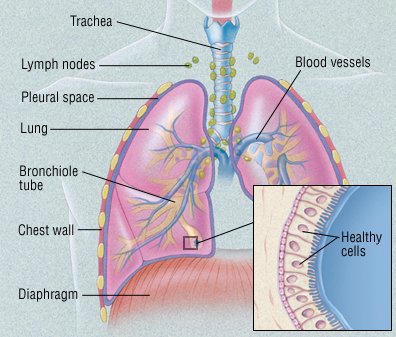
Cancer is a disease in which cells in the body grow out of control. When cancer starts in the lungs, it is called lung cancer.One of the most common cancers, lung cancer usually occurs when a cancer-causing agent, or carcinogen, triggers the growth of abnormal cells in the lung. These cells multiply out of control and eventually form a tumor. As the tumor grows, it destroys nearby areas of the lung. Eventually, tumor cells can spread (metastasize) to nearby lymph nodes and other parts of the body. These include the

  • liver
  • bones
  • adrenal glands
  • brain.

In most cases, the carcinogens that trigger lung cancer are chemicals found in cigarette smoke. However, more and more lung cancers are being diagnosed in people who have never smoked.

Lung Cancer

Types of Lung Cancer:

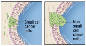
Lung cancers are divided into two groups, based on how their cells look under the microscope:

  • Non-small cell lung cancer (NSCLC)
  • Small cell lung cancer.(SCLC)

Non-small cell lung cancer may be localized. This means that it is limited to the lung or that it hasn’t spread beyond the chest. As a result, it can usually be treated with surgery. Small cell lung cancer is rarely localized, even when it is detected early. It is rarely treated with surgery. Knowing whether the cancer has spread is critical, because it affects treatment decisions.

However, even when doctors think that the cancer is localized, it often comes back shortly after surgery. This means cancer cells had started to spread before surgery, but they couldn’t yet be detected.

Non-small cell lung cancer

Non-small cell lung cancer is more likely than small cell cancer to be localized at the time of diagnosis. It also is more likely than small cell cancer to be treatable with surgery. It often responds poorly to chemotherapy (anticancer drugs). However, sophisticated genetic tests can help predict which patients may show favorable responses to particular treatments, including chemotherapy.

Non-small cell lung cancer accounts for about 85% of all lung cancers. These cancers are divided into subgroups, based on how their cells look under a microscope:

  • Adenocarcinoma. This is the most common type of lung cancer. Although it is related to smoking, it is the most common type of lung cancer in nonsmokers. It is also the most common form of lung cancer in women and in people younger than 45. It usually develops near the edge of the lung. It can also involve the pleura, the membrane covering the lung.
  • Squamous cell carcinoma. This type of lung cancer tends to form a mass near the center of the lungs. As the mass gets larger, it can bulge into one of the larger air passages, or bronchi. In some cases, the tumor forms a cavity in the lungs.
  • Large cell carcinoma. Like adenocarcinoma, large cell carcinoma tends to develop at the edge of the lungs and spread to the pleura. Like squamous cell carcinoma, it can form a cavity in the lungs.
  • Adenosquamous carcinoma, undifferentiated carcinoma, and bronchioloalveolar carcinoma.These are relatively rare non-small cell lung cancers. Adenosquamous carcinoma has a worse prognosis compared to either adenocarcinoma or squamous cancer.

Small cell lung cancer 

At the time of diagnosis, small cell lung cancer is more likely than non-small cell cancer to have spread beyond the lung. This makes it almost impossible to cure with surgery. However, it can be managed with chemotherapy or radiation therapy. Small cell cancers account for about 15% of all lung cancers.

Causes and Risk factors:

Risk of all types of lung cancer increase if you:

  • smoke. Smoking cigarettes is by far the leading risk factor for lung cancer. In fact, cigarette smokers are 13 times more likely to develop lung cancer than nonsmokers. Cigar and pipe smoking are almost as likely to cause lung cancer as cigarette smoking.
  •  Exposed to radon gas. Radon is a colorless, odorless radioactive gas formed in the ground. It seeps into the lower floors of homes and other buildings and can contaminate drinking water. Radon exposure is the second leading cause of lung cancer. It’s not clear whether elevated radon levels contribute to lung cancer in nonsmokers. But radon exposure does contribute to lung cancer in smokers and in people who regularly breathe high amounts of the gas at work (miners, for example). You can test radon levels in your home with a radon testing kit.
  • Exposed to asbestos. Asbestos is a mineral used in insulation, fireproofing materials, floor and ceiling tiles, automobile brake linings, and other products. People exposed to asbestos on the job (miners, construction workers, shipyard workers, and some auto mechanics) have a higher-than-normal risk of lung cancer. People who live or work in buildings with asbestos-containing materials that are deteriorating also have an increased risk of lung cancer. The risk is even higher in people who also smoke. Asbestos exposure also increases the risk of developing mesothelioma. It’s a relatively rare and usually fatal cancer that starts in the lining of the lungs.
  • Exposed to other cancer-causing agents at work. These include uranium, arsenic, vinyl chloride, nickel chromates, coal products, mustard gas, chloromethyl ethers, gasoline, and diesel exhaust.
  • Passive smoking: Passive smoking or the inhalation of tobacco smoke by non-smokers who share living or working quarters with smokers, also is an established risk factor for the development of lung cancer.
  • Air pollution : Air pollution from vehicles, industry, and power plants can raise the likelihood of developing lung cancer in exposed individuals. Up to 1%-2% of lung cancer deaths are attributable to breathing polluted air, and experts believe that prolonged exposure to highly polluted air can carry a risk for the development of lung cancer similar to that of passive smoking.

  • Exposure to diesel exhaust: Exhaust from diesel engines contains gases and soot (particulate matter). Many occupations, such as truck drivers, tollbooth workers, forklift and other heavy machinery operators, railroad and dock workers, miners, garage workers and mechanics, and some farm workers are frequently exposed to diesel exhaust. Studies of workers exposed to diesel exhaust have shown a small but significant increase in the risk of developing lung cancer.
  • Prior history of lung cancer:  Survivors of lung cancer have a greater risk of developing a second lung cancer than the general population has of developing a first lung cancer. Survivors of non-small-cell lung cancers (NSCLCs) have an additive risk of 1%-2% per year for developing a second lung cancer. In survivors of small-cell lung cancers (SCLCs), the risk for development of second lung cancers approaches 6% per year.

Symptoms:

In some cases, lung cancer is detected when a person with no symptoms has a chest x-ray or computed tomography (CT) scan for another reason. But most people with lung cancer have one or more of these symptoms:

  • a cough that doesn’t go away
  • coughing up blood or mucus
  • wheezing
  • shortness of breath
  • trouble breathing
  • chest pain
  • fever
  • discomfort when swallowing
  • hoarseness
  • weight loss
  • poor appetite.
  • an irregular heart beat if the cancer is located close to the heart

If the cancer has spread beyond the lungs, it can cause other symptoms. For example, you may have bone pain if it has spread to your bones.

Certain small cell lung cancers may secrete chemicals that can alter the body’s chemical composition. For example, levels of sodium and calcium may be abnormal. This can lead to the diagnosis of small cell lung cancer.

Many of these symptoms can be caused by other conditions. See your doctor if you have symptoms so that the problem can be diagnosed and properly treated.

Diagnosis

Your doctor may suspect lung cancer based on

  • your symptoms
  • your smoking history
  • whether you live with a smoker
  • your exposure to asbestos and other cancer-causing agents.

To look for evidence of cancer, your doctor will examine you, paying special attention to your lungs and chest. He or she will order imaging tests to check your lungs for masses. In most cases, a chest x-ray will be done first. If the x-ray shows anything suspicious, a CT scan will be done. As the scanner moves around you, it takes many pictures. A computer then combines the images. This creates a more detailed image of the lungs, allowing doctors to confirm the size and location of a mass or tumor.

You may also have a magnetic resonance imaging (MRI) scan or a positron emission tomography (PET) scanMRI scans provide detailed pictures of the body’s organs, but they use radio waves and magnets to create the images, not x-rays. PET scans look at the function of tissue rather than anatomy. Lung cancer tends to show intense metabolic activity on a PET scan. Some medical centers offer combined PET-CT scanning.

If cancer is suspected based on these images, more tests will be done to make the diagnosis, determine the type of cancer, and see if it has spread. These tests may include the following:

  • Sputum sample. Coughed up mucus is checked for cancer cells.
  • Biopsy. A sample of abnormal lung tissue is removed and examined under a microscope in a laboratory. If the tissue contains cancer cells, the type of cancer can be determined by the way the cells look under the microscope. The tissue is often obtained during a bronchoscopy. However, surgery may be necessary to expose the suspicious area.
  • Bronchoscopy. During this procedure, a tube-like instrument is passed down the throat and into the lungs. A camera on the end of the tube allows doctors to look for cancer. Doctors can remove a small piece of tissue for a biopsy.
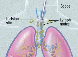
  • Mediastinoscopy. In this procedure, a tube-like instrument is used to biopsy lymph nodes or masses between the lungs. (This area is called the mediastinum.) A biopsy obtained this way can diagnose the type of lung cancer and determine whether the cancer has spread to lymph nodes.
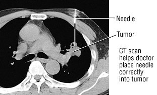
  • Fine-needle aspiration. With a CT scan, a suspicious area can be identified. A tiny needle is then inserted into that part of the lung or pleura. The needle removes a bit of tissue for examination in a laboratory. The type of cancer can then be diagnosed.
  • Thoracentesis. If there is fluid build-up in the chest, it can be drained with a sterile needle. The fluid is then checked for cancer cells.
  • Video-assisted thoracoscopic surgery (VATS). In this procedure, a surgeon inserts a flexible tube with a video camera on the end into the chest through an incision. He or she can then look for cancer in the space between the lungs and the chest wall and on the edge of the lung. Abnormal lung tissue can also be removed for a biopsy.
  • Bone scans, MRI scans and CT scans. These imaging tests can detect lung cancer that has spread to the bones, brain, or other parts of the body.

Stages:

After the cancer has been diagnosed, it is assigned a “stage.” The stages differ for non-small cell lung cancer and small cell lung cancer.

Non-small cell lung cancer

Stages of non-small cell lung cancer reflect the tumor’s size and how far the cancer has spread. Stages I through III are further divided into A and B categories.

  • Stage I tumors are small and have not invaded the surrounding tissue or organs.
  • Stage II and III tumors have invaded surrounding tissue and/or organs and have spread to lymph nodes.
  • Stage IV tumors have spread beyond the chest.

Small Cell Lung Cancer

Many experts divide small cell lung cancers into two groups:

  • Limited stage. These cancers involve only one lung and the nearby lymph nodes.
  • Extensive stage. These cancers have spread beyond the lung to other areas of the chest or to distant organs.

Knowing the type of cancer and its stage helps doctors determine the best treatment. Limited stage cancer, for example, may be treated with surgery and/or chemotherapy. Extensive stage cancer is much less likely to be cured.

However, many doctors now stage small cell lung cancers like non-small cell lung cancers. This more formal method may make the terms limited stage and extensive stage obsolete.

Treatment

After lung cancer has been diagnosed, the type of treatment depends on the type of cancer and how much the tumor has spread i.e, its stage.

Non-small cell lung cancer

Surgery is the main treatment for non-small cell lung cancers that have not spread beyond the chest. The type of surgery will depend on the extent of the cancer. It will also depend on whether other lung conditions, such as emphysema, are present.

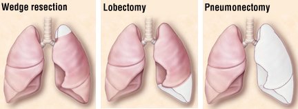
There are three types of surgery:

  • Wedge resection removes only a small part of the lung.
  • Lobectomy removes one lobe of the lung.
  • Pneumonectomy removes an entire lung.

Lymph nodes are also removed and examined to see if the cancer has spread.

Some surgeons use video-assisted thoracoscopy (VATS) to remove small, early-stage tumors, especially if the tumors are near the outer edge of the lung. (VATS can also be used to diagnose lung cancer.) Because the incisions for VATS are small, this technique is less invasive than a traditional “open” procedure.

Because surgery will remove part or all of a lung, breathing may be more difficult afterwards, especially in patients with other lung conditions (emphysema, for example). Doctors can test lung function prior to surgery and predict how it might be affected by surgery.

Depending on how far the cancer has spread, treatment may include chemotherapy (the use of anticancer drugs) and radiation therapy. These may be given before and/or after surgery.

When the tumor has spread significantly, chemotherapy may be recommended to slow its growth, even if it cannot cure the disease. Chemotherapy has been shown to ease symptoms and prolong life in cases of advanced lung cancer.

Radiation therapy can relieve symptoms, too. It is often used to treat lung cancer that has spread to the brain or bones and is causing pain. It can also be used alone or with chemotherapy to treat the lung cancer that is confined to the chest.

People who may not withstand surgery due to other serious medical problems may receive radiation therapy, with or without chemotherapy, as an alternative to surgery. Advances in radiation have made it possible for prolonged survival in some people, with results similar to surgery.

In specialized cancer centers, cancerous tissue may be tested for specific genetic abnormalities (mutations). Doctors may then be able to treat the cancer with a “targeted therapy.” These therapies can derail the cancer’s growth by preventing or changing chemical reactions linked to particular mutations. For example, some target therapies prevent cancer cells from receiving chemical “messages” telling them to grow.

Knowing about specific genetic mutations can help predict which therapy will be best. This strategy can be especially helpful in certain patients, such as women with adenocarcinoma of the lung who have never smoked.

Small cell lung cancer

The treatment of small cell lung cancer depends on its stage:

  • Limited stage. Treatments include various combinations of chemotherapy, radiation and, rarely, surgery, with or without radiation to the brain to prevent cancer spread. While small cell lung cancer often responds well to chemotherapy, it very often returns months or even years later.
  • Extensive stage. Treatments include chemotherapy, with or without brain radiation, or radiation treatments to areas of existing metastases in the brain, spine or other bones. Even if the imaging tests show that the cancer has not spread to the brain, many experts suggest treating the brain anyway. That’s because cancer cells may be there even if they haven’t yet shown up on the imaging tests. The question of whether or not to use brain radiation must be considered carefully; many patients experience memory loss afterwards. The decision to use brain radiation is a very crucial one, since many patients may experience a decrease in memory function after radiation therapy, with or without chemotherapy.

Prevention of lung cancer

KEY POINTS

  • Avoiding risk factors and increasing protective factors may help prevent lung cancer.
  • The following are risk factors for lung cancer:
    • Cigarette, cigar, and pipe smoking
    • Secondhand smoke
    • Family history
    • Environmental risk factors
    • Beta carotene supplements in heavy smokers
  • The following are protective factors for lung cancer:
    • Not smoking
    • Quitting smoking
    • Lower exposure to workplace risk factors
    • Lower exposure to radon.

Home remedies for lung cancer symptoms

Home remedies and homeopathic remedies won’t cure cancer. But certain home remedies may help relieve some of the symptoms associated with lung cancer and side effects of treatment.

Ask your doctor if you should take dietary supplements and if so, which ones. Some herbs, plant extracts, and other home remedies can interfere with treatment and endanger your health. Be sure to discuss all complementary therapies with your doctor to make sure they’re safe for you.

Options may include:

  • Massage: With a qualified therapist, massage can help relieve pain and anxiety. Some massage therapists are trained to work with people with cancer.
  • Acupuncture: When performed by a trained practitioner, acupuncture may help ease pain, nausea, and vomiting. But it’s not safe if you have low blood counts or take blood thinners.
  • Meditation: Relaxation and reflection can reduce stress and improve overall quality of life in cancer patients.
  • Hypnosis: Helps you relax and may help with nausea, pain, and anxiety.
  • Yoga: Combining breathing techniques, meditation, and stretching, yoga can help you feel better overall and improve sleep.

Diet

There’s no diet specifically for lung cancer. It is important to get all the nutrients your body needs.

If you’re deficient in certain vitamins or minerals, your doctor can advise you which foods can provide them. Otherwise, you’ll need a dietary supplement. Don’t take supplements without talking to your doctor because some can interfere with treatment.

Here are a few dietary tips:

  • Eat whenever you have an appetite.
  • If you don’t have a major appetite, try eating smaller meals throughout the day.
  • If you need to gain weight, supplement with low sugar, high-calorie foods and drinks.
  • Use mint and ginger teas to soothe your digestive system.
  • If your stomach is easily upset or you have mouth sores, avoid spices and stick to bland food.
  • If constipation is a problem, add more high-fiber foods.

As you progress through treatment, your tolerance to certain foods may change. So can your side effects and nutritional needs. It’s worth discussing nutrition with your doctor often. You can also ask for a referral to a nutritionist or dietician.

There’s no diet known to cure cancer, but a well-balanced diet can help you fight side effects and feel better.

Lung Cancer and Survival rate:

Once cancer enters the lymph nodes and bloodstream, it can spread anywhere in the body. The outlook is better when treatment begins before cancer spreads outside the lungs.

Other factors include age, overall health, and how well you respond to treatment. Because early symptoms can be easily overlooked, lung cancer is usually diagnosed in later stages.

Survival rates and other statistics provide a broad picture of what to expect. There are significant individual differences, though. Your doctor is in the best position to discuss your outlook.

Current survival statistics don’t tell the whole story. In recent years, new treatments have been approved for stage 4 non-small cell lung cancer (NSCLC). Some people are surviving much longer than previously seen with traditional treatments.

The following are the estimated five-year survival rates for NSCLC by SEER stage:

  • Localized: 60 percent
  • Regional: 33 percent
  • Distant: 6 percent
  • All SEER stages: 23 percent

Small-cell lung cancer (SCLC) is very aggressive. For limited stage SCLC, the five-year survival rate is 14 percentTrusted Source. Median survival is 16 to 24 months. Median survival for extensive stage SCLC is six to 12 months.

Long-term disease-free survival is rare. Without treatment, median survival from diagnosis of SCLC is only two to four months.

The relative five-year survival rate for mesothelioma, a type of cancer caused by asbestos exposure, is 5 to 10 percent.

Outlook

Lung cancer is the most common cancer in the world. This disease carries a higher risk of death than breast, colon, and prostate cancer combined if not detected and treated early. It is also largely preventable through not smoking. Lung cancer is more treatable if diagnosed in its early stages.

The prognosis and outlook for lung cancer depend upon the cancer type and the spread. People should talk to their doctor who can provide a more accurate prognosis, taking into account the individual’s overall health and the extent of their cancer.

Takeaway

Lung cancer is  the most common, serious and potentially deadly disease that affects a person’s ability to breath.

However, early diagnostic screening of individuals who are at high risk for lung cancer can help them receive treatment in earlier and more treatable stages. Treatment options include surgery to remove sections or all of the lung, chemotherapy, and radiation therapy, as well as targeted drug therapy.

If a person has concerns that they could be at risk for lung cancer, they should consult a doctor immediately.

Breast Cancer Awareness

Global Hand Wash Day (13)October is termed as Breast cancer Awareness month across the globe.After skin & lung cancer, breast cancer is the most common cancer diagnosed in women and rarely in men. Breast cancer is cancer that forms in the cells of the breasts.Its treatment depends on the stage of cancer. It may consist of chemotherapy, radiation, hormone therapy and surgery.

On average, 1 in 8 women will develop breast cancer in their lifetimes. About two-thirds of women with breast cancer are 55 or older. Most of the rest are between 35 and 54.

Fortunately, breast cancer is very treatable if you spot it early. Localized cancer (meaning it hasn’t spread outside your breast) can usually be treated before it spreads.

Once the cancer begins to spread, treatment becomes more complicated. It can often control the disease for years.

What is Breast Cancer?

The term “breast cancer” refers to a malignant tumor that has developed from cells in the breast. Usually breast cancer either begins in the cells of the lobules, which are the milk-producing glands, or the ducts, the passages that drain milk from the lobules to the nipple. Less commonly, breast cancer can begin in the stromal tissues, which include the fatty and fibrous connective tissues of the breast.

A tumor can be benign (not dangerous to health) or malignant (has the potential to be dangerous). Benign tumors are not considered cancerous: their cells are close to normal in appearance, they grow slowly, and they do not invade nearby tissues or spread to other parts of the body. Malignant tumors are cancerous. Left unchecked, malignant cells eventually can spread beyond the original tumor to other parts of the body.

Breast cancer is always caused by a genetic abnormality (a “mistake” in the genetic material). However, only 5-10% of cancers are due to an abnormality inherited from your mother or father. Instead, 85-90% of breast cancers are due to genetic abnormalities that happen as a result of the aging process and the “wear and tear” of life in general.

Causes Of Breast Cancer: How Did This Happen?

When you’re told that you have breast cancer, it’s natural to wonder what may have caused the disease. But no one knows the exact causes of breast cancer. Doctors seldom know why one woman develops breast cancer and another doesn’t, and most women who have breast cancer will never be able to pinpoint an exact cause. What we do know is that breast cancer is always caused by damage to a cell’s DNA.

Symptoms:

Different people have different symptoms of breast cancer. Some people do not have any signs or symptoms at all.

Few warning signs of breast cancer are—

  • New lump in the breast or underarm (armpit).
  • Thickening or swelling of part of the breast.
  • Irritation or dimpling of breast skin.
  • Redness or flaky skin in the nipple area or the breast.
  • Pulling in of the nipple or pain in the nipple area.
  • Nipple discharge other than breast milk, including blood.
  • Any change in the size or the shape of the breast.
  • Pain in any area of the breast.

Keep in mind that these symptoms can happen with other conditions that are not cancer.

Stages:

A doctor stage cancer according to the size of the tumor and whether it has spread to lymph nodes or other parts of the body.

There are different ways of staging breast cancer. One way is from stage 0–4, with subdivided categories at each numbered stage. Descriptions of the four main stages are listed below, though the specific substage of a cancer may also depend on other specific characteristics of the tumor, such as HER2 receptor status.

  • Stage 0: Known as ductal carcinoma in situ (DCIS), the cells are limited to within the ducts and have not invaded surrounding tissues.
  • Stage 1: At this stage, the tumor measures up to 2 centimeters (cm) across. It has not affected any lymph nodes, or there are small groups of cancer cells in the lymph nodes.
  • Stage 2: The tumor is 2 cm across, and it has started to spread to nearby nodes, or is 2–5 cm across and has not spread to the lymph nodes.
  • Stage 3: The tumor is up to 5 cm across, and it has spread to several lymph nodes or the tumor is larger than 5 cm and has spread to a few lymph nodes.
  • Stage 4: The cancer has spread to distant organs, most often the bones, liver, brain, or lungs.

    Risk Factors:

    UnControlable:

  • Age. Women over 50 are more likely to get breast cancer than younger women.
  • Race: African American women are more likely than white women to get breast cancer before  menopause.
  • Dense breasts. If your breasts have more connective tissue than fatty tissue, it can be hard to see tumors on a mammogram.
  • Personal history of cancer. Your odds go up slightly if you have certain benign breast conditions. They go up more sharply if you’ve had breast cancer before.
  • Family history. If a first-degree female relative (mother, sister, or daughter) had breast cancer, you’re two times more likely to get the disease. Having two or more first-degree relatives with a history of breast cancer increases your risk at least three times. This is especially true if they got cancer before menopause or if it affected both breasts. The risk can also rise if your father or brother was diagnosed with breast cancer.
  • Genes. Changes to two genes, BRCA1 and BRCA2, are responsible for some cases of breast cancer in families. About 1 woman in 200 has one of these genes. While they make you more likely to get cancer, they don’t mean you definitely will. If you have a BRCA1 or BRCA2 mutation, you have a 7 in 10 chance of being diagnosed with breast cancer by age 80. These genes also raise your odds of ovarian cancer, and they’re linked to pancreatic cancer and male breast cancer. Other gene mutations linked to breast cancer risk include mutations of the PTEN gene, the ATM gen, the TP53 gene, the CHEK2 gene, the CDH1 gene, the STK11 gene, and the PALB2 gene. These carry a lower risk for breast cancer development than the BRCA genes.
  • Menstrual history. Your breast cancer odds go up if:
    • Your periods start before age 12.
    • Your periods don’t stop until after you’re 55.
  • Radiation. If you had treatment for cancers like Hodgkin’s lymphoma before age 40, you have an increased risk of breast cancer.
  • Diethylstilbestrol (DES). Doctors used this drug between 1940 and 1971 to prevent miscarriage. If you or your mother took it, your breast cancer odds go up.

    Controlable:

  • Physical activity The less you move, the higher your chances.
  • Weight and diet. Being overweight after menopause raises your odds.
  • Alcohol. Regular drinking — especially more than one drink a day — increases the risk of breast cancer.
  • Reproductive history.

You have your first child after age 30.

You don’t breastfeed.

You don’t have a full-term pregnancy.

  • Taking hormones. Your chances can go up if you:

Use hormone replacement therapy that includes both estrogen and progesterone during menopause for more than 5 years. This increase in breast cancer risk returns to normal 5 years after you stop treatment.

Use certain birth control methods including birth control pills, shots, implants, IUDS, skin patches, or vaginal rings that contain hormones.

  • Still, most women who are at high risk for breast cancer don’t get it. On the other hand, 75% of women who develop breast cancer have no known risk factors.

Types:

There are several different types of breast cancer, including:

  • Ductal carcinoma: This begins in the milk duct and is the most common type.
  • Lobular carcinoma: This starts in the lobules.

Invasive breast cancer occurs when the cancer cells break out from inside the lobules or ducts and invade nearby tissue. This increases the chance of cancer spreading to other parts of the body.

Noninvasive breast cancer develops when the cancer remains inside its place of origin and has not yet spread. However, these cells can sometimes progress to invasive breast cancer.

Diagnosis:

If you feel a lump or if something shows up on a mammogram, your doctor will begin the breast cancer diagnosis process.

They’ll ask about your personal and family healthy history. Then, they’ll do a breast exam and order tests that include:

Imaging tests. Your doctor will use these to learn more about your breast.

  • Ultrasound. This test uses sound waves to make a picture of your breast.
  • Mammogram. This detailed X-ray gives doctors a better view of lumps and other problems.
  • Magnetic resonance imaging (MRI). This body scan uses a magnet linked to a computer to create detailed images of the insides of your breasts.
  • Biopsy. For this test, the doctor removes tissue or fluid from your breast. They look at it under a microscope to check for if cancer cells and, if they’re there, learn which type they are. Common procedures include:
    • Fine-needle aspiration. This is for easy-to-reach lumps or those that might be filled with fluid.
    • Core-needle biopsy. This type uses a bigger needle to remove a piece of tissue.
    • Surgical (open) biopsy. A surgeon removes the entire lump along with nearby breast tissue.
    • Lymph node biopsy. The doctor removes a part of the lymph nodes under your arm to see if the cancer has spread.
    • Image-guided biopsy. The doctor uses imaging to guide the needle.The doctor can test your biopsy sample for:
      • Tumor features. Is it invasive or in situ, ductal, or lobular? Has it spread to your lymph nodes? The doctor also measures the margins or edges of the tumor and their distance from the edge of the biopsy tissue, which is which is called margin width.
      • Estrogen receptors (ER) and progesterone receptors (PR). This tells the doctor if the hormones estrogen or progesterone make your cancer grow. This affects how likely it is that your cancer will come back and what type of treatment is most likely to prevent that.
      • HER2. This test looks for the gene human epidermal growth factor receptor 2. It can help cancer grow more quickly. If your cancer is HER2-positive, targeted therapy could be a treatment option for you.
      • Grade. This tells the doctor how different the cancer cells look from healthy cells and whether they seem to be slower-growing or faster-growing.
      • Oncotype Dx. This test evaluates 16 cancer-related genes and five reference genes to estimate the risk of the cancer coming back within 10 years of diagnosis.
      • Breast Cancer Index. This test can help your doctor decide how you need endocrine therapy.
      • MammaPrint. This test uses information from 70 genes to predict the risk of the cancer coming back.
      • PAM50 (Prosigna). This test uses information from 50 genes to predict if the cancer will spread.

      You might get blood tests including:

      • Complete blood count (CBC). This measures the number of different types of cells, like red and white blood cells, in your blood. It lets your doctor know if your bone marrow is working like it should.
      • Blood chemistry. This shows how well your liver and kidneys are working.
      • Hepatitis tests. These are sometimes done to check for hepatitis B and hepatitis C. If you have an active hepatitis B infection, you may need medication to fight the virus before you get chemotherapy. Without it, chemo can cause the virus to grow and damage your liver.

Treatment:

Treatment will depend on several factors, including:

  • the type and stage of the cancer
  • the person’s sensitivity to hormones
  • the age, overall health, and preferences of the individual

The main treatment options include:

radiation therapy

  • surgery
  • biological therapy, or targeted drug therapy
  • hormone therapy
  • chemotherapy

Factors affecting the type of treatment a person has will include the stage of the cancer, other medical conditions, and their individual preference.

Surgery

If surgery is necessary, the type will depend on both the diagnosis and individual preference. Types of surgery include:

Lumpectomy: This involves removing the tumor and a small amount of healthy tissue around it.

A lumpectomy can help prevent the spread of the cancer. This may be an option if the tumor is small and easy to separate from its surrounding tissue.

Mastectomy: A simple mastectomy involves removing the lobules, ducts, fatty tissue, nipple, areola, and some skin. In some types, a surgeon will also remove the lymph nodes and muscle in the chest wall.

Sentinel node biopsy: If breast cancer reaches the sentinel lymph nodes, which are the first nodes to which a cancer can spread, it can spread into other parts of the body through the lymphatic system. If the doctor does not find cancer in the sentinel nodes, then it is usually not necessary to remove the remaining nodes.

Axillary lymph node dissection: If a doctor finds cancer cells in the sentinel nodes, they may recommend removing several lymph nodes in the armpit. This can prevent the cancer from spreading.

Reconstruction: Following mastectomy, a surgeon can reconstruct the breast to look more natural. This can help a person cope with the psychological effects of breast removal.

The surgeon can reconstruct the breast at the same time as performing a mastectomy or at a later date. They may use a breast implant or tissue from another part of the body.

Radiation therapy

A person may undergo radiation therapy around a month after surgery. Radiation involves targeting the tumor with controlled doses of radiation that kill any remaining cancer cells.

Chemotherapy

A doctor may prescribe cytotoxic chemotherapy drugs to kill cancer cells if there is a high risk of recurrence or spread. When a person has chemotherapy after surgery, doctors call it adjuvant chemotherapy.

Sometimes, a doctor may choose to administer chemotherapy before surgery to shrink the tumor and make its removal easier. Doctors call this neoadjuvant chemothera

Hormone blocking therapy

Doctors use hormone blocking therapy to prevent hormone sensitive breast cancers from returning after treatment. Hormone therapy may be used to treat estrogen receptor (ER)-positive and progesterone receptor (PR)-positive cancers.

They usually administer hormone blocking therapy after surgery but might sometimes use it beforehand to shrink the tumor.

Hormone blocking therapy may be the only option for people who are not suitable candidates for surgery, chemotherapy, or radiotherapy.

Doctors may recommend a person has hormone therapy for 5–10 years after surgery. However, the treatment will not affect cancers that are not sensitive to hormones.

Examples of hormone blocking therapy medications may include:

  • tamoxifen
  • aromatase inhibitors
  • ovarian ablation or suppression
  • Goserelin, which is a luteinizing hormone-releasing agonist drug that suppresses the ovaries

Hormone treatment may affect fertility.

Biological treatment

Targeted drugs can destroy specific types of breast cancer. Examples include:

  • trastuzumab (Herceptin)
  • lapatinib (Tykerb)
  • bevacizumab (Avastin)

Treatments for breast and other cancers can have severe adverse effects. When deciding on a treatment, people should discuss the potential risks with a doctor and look at ways to minimize the side effects.

Facts to know about Breasts

  1. Breasts contain fat cells, not muscles
  2. Breast size cannot be altered by any excercise. it can only  be altered by painful surgery.
  3. Breast size depends upon a lot of factors such as harmonalchanges, bodyfat, genetics etc.
  4. Most of the time when body gains or losses fat, breast size changes.

Conclusion

There are steps every person can take to help the body stay as healthy as possible, such as eating a balanced diet, maintaining a healthy weight, not smoking, limiting alcohol, and exercising regularly . While these may have some impact on your risk of getting breast cancer, they cannot eliminate the risk.

Developing breast cancer is not your or anyone’s fault. Feeling guilty, or telling yourself that breast cancer happened because of something you or anyone else did, is not productive.

Regular checks and screening can help detect symptoms early. Women should discuss their options with a doctor.There is no way to prevent breast cancer. However, certain lifestyle decisions can significantly reduce the risk of breast cancer as well as other types.