Global Treatment Services Pvt. Ltd.

Global Treatment Services

Marfan Syndrome: Causes, Symptoms & Treatments

What is Marfan syndrome?

Marfan syndrome is a heritable condition that affects the connective tissue. The primary purpose of connective tissue is to hold the body together and provide a framework for growth and development. In Marfan syndrome, the connective tissue is defective and does not act as it should. Because connective tissue is found throughout the body, the syndrome can affect many body systems, including the skeleton, eyes, heart and blood vessels, nervous system, skin, and lungs.

Who gets this syndrome?

Marfan syndrome affects men, women, and children, and has been found among people of all races and ethnic backgrounds.

What are the signs and symptoms?

It affects different people in different ways. Some people have only mild symptoms, while others are more severely affected. In most cases, the symptoms progress as the person ages. The body systems most often affected by the syndrome are:

  • Skeleton. People with the syndrome are typically very tall, slender, and loose-jointed. Because Marfan syndrome affects the long bones of the skeleton, a person’s arms, legs, fingers, and toes may be disproportionately long in relation to the rest of the body. A person with the syndrome often has a long, narrow face, and the roof of the mouth may be arched, causing the teeth to be crowded. Other skeletal problems include a sternum (breastbone) that is either protruding or indented, curvature of the spine (scoliosis), and flat feet.
  • Eyes. More than half of all people with the condition experience dislocation of one or both lenses of the eye. The lens may be slightly higher or lower than normal, and may be shifted off to one side. The dislocation may be minimal, or it may be pronounced and obvious. One serious complication that may occur with this disorder is retinal detachment. Many people with the condition are also nearsighted (myopic), and some can develop early glaucoma (high pressure within the eye) or cataracts (the eye’s lens loses its clearness).

Picture of Syringomas on the Eyelids of a Marfan Syndrome Patient

  • Heart and blood vessels (cardiovascular system). Marfan syndrome patients have problems associated with the heart and blood vessels. Because of faulty connective tissue, the wall of the aorta (the large artery that carries blood from the heart to the rest of the body) may be weakened and stretch, a process called aortic dilatation. Aortic dilatation increases the risk that the aorta will tear (aortic dissection) or rupture, causing serious heart problems or sometimes sudden death. Sometimes, defects in heart valves can also cause problems. In some cases, certain valves may leak, creating a “heart murmur,” which a doctor can hear with a stethoscope. Small leaks may not result in any symptoms, but larger ones may cause shortness of breath, fatigue, and palpitations (a very fast or irregular heart rate).
  • Nervous system. The brain and spinal cord are surrounded by fluid contained by a membrane called the dura, which is composed of connective tissue. Marfan syndrome patients grow older, the dura often weakens and stretches, then begins to weigh on the vertebrae in the lower spine and wear away the bone surrounding the spinal cord. This is called dural ectasia. These changes may cause only mild discomfort; or they may lead to radiated pain in the abdomen; or to pain, numbness, or weakness in the legs.
  • Skin. Many people with Marfan syndrome develop stretch marks on their skin, even without any weight change. These stretch marks can occur at any age and pose no health risk. However, people with Marfan syndrome are also at increased risk for developing an abdominal or inguinal hernia, in which a bulge develops that contains part of the intestines.
  • Lungs. Although connective tissue problems make the tiny air sacs within the lungs less elastic, people with Marfan syndrome generally do not experience noticeable problems with their lungs. If, however, these tiny air sacs become stretched or swollen, the risk of lung collapse may increase. Rarely, people with Marfan syndrome may have sleep-related breathing disorders such as snoring, or sleep apnea (which is characterized by brief periods when breathing stops).

Is Marfan syndrome genetic or inherited (causes)?

Caused by a defect, or mutation, in the gene that determines the structure of fibrillin-1, a protein that is an important part of connective tissue. A person with Marfan syndrome is born with the disorder, even though it may not be diagnosed until later in life.

The defective gene that causes Marfan syndrome can be inherited: The child of a person who has Marfan syndrome has a 50 percent chance of inheriting the disease. Sometimes a new gene defect occurs during the formation of sperm or egg cells, making it possible for two parents without the disease to have a child with the disease. But this is rare. Two unaffected parents have only a 1 in 10,000 chance of having a child with Marfan syndrome. Possibly 25 percent of cases are due to a spontaneous mutation at the time of conception.

Although everyone with Marfan syndrome has a defect in the same gene, different mutations are found in different families, and not everyone experiences the same characteristics to the same degree. In other words, the defective gene expresses itself in different ways in different people. This phenomena is known as variable expression. Scientists do not yet understand why variable expression occurs in people with Marfan syndrome.

How Is Marfan Syndrome Treated?

Marfan syndrome requires a treatment plan that is individualized to the patient’s needs. Some people need regular follow-up appointments with their doctor, and during the growth years, routine cardiovascular, eye, and orthopedic exams. Others may need medications or surgery. The approach depends on the structures affected and the severity:

Medications:

Medications are typically not used to treat Marfan syndrome. However, your doctor may prescribe a beta-blocker, which decreases the forcefulness of the heartbeat and the pressure within the arteries, thus preventing or slowing the enlargement of the aorta. Beta-blocker therapy is usually started when the person with Marfan syndrome is young.

Some people are unable to take beta-blockers because they have asthma or because of the medication’s side effects, which may include drowsiness or weakness, headaches, slowed heartbeat, swelling of the hands and feet or trouble breathing and sleeping. In these cases, another medication called a calcium channel blocker may be recommended.

Surgery:

The goal of surgery for Marfan syndrome is to prevent aortic dissection or rupture and to treat problems affecting the heart’s valves, which control the flow of blood in and out of the heart and between the heart’s chambers.

The decision to perform surgery is based on the size of the aorta, expected normal size of the aorta, rate of aortic growth, age, height, gender, and family history of aortic dissection. Surgery involves replacing the dilated portion of the aorta with a graft, a piece of man-made material that is inserted to replace the damaged or weak area of the blood vessel.

A leaky aortic or mitral valve (the valve that controls the flow of blood between the two left chambers of the heart) can damage the left ventricle (the lower chamber of the heart that is the main pumping chamber) or cause heart failure. In these cases, surgery to replace or repair the affected valve is necessary. If surgery is performed early, before the valves are damaged, the aortic or mitral valve may be repaired and preserved. If the valves are damaged, they may need to be replaced.

If surgery is needed, you should consult with a surgeon who is experienced in surgery for Marfan syndrome. People who have surgery for Marfan syndrome still require life-long follow-up care to prevent future complications associated with the disease.

How Does Marfan Sydrome Affect Lifestyle Choices?

  • Activity. Most people with Marfan syndrome can participate in certain types of physical and/or recreational activities. Those with dilation of the aorta will be asked to avoid high intensity team sports, contact sports, and isometric exercises (such as weight lifting). Ask your cardiologist about activity guidelines for you.
  • Pregnancy . Genetic counseling should be performed prior to pregnancy because Marfan syndrome is an inherited condition. Pregnant women with Marfan syndrome are also considered high-risk cases. If the aorta is normal size, the risk for dissection is lower, but not absent. Those with even slight enlargement are at higher risk and the stress of pregnancy may cause more rapid dilation. Careful follow-up, with frequent blood pressure checks and monthly echocardiograms is required during pregnancy. If there is rapid enlargement or aortic regurgitation, bed rest may be required. Your doctor will discuss with you the best method of delivery with you.
  • Endocarditis prevention. People with Marfan syndrome who have heart or valve involvement or who have had heart surgery may be at increased risk for bacterial endocarditis. This is an infection of the heart valves or tissue which occurs when bacteria enters the blood stream. To prevent this, antibiotics may be needed prior to dental or surgical procedures. Ask your doctor whether you need antibiotics, and if so, how much and what kind should be taken. A wallet card may be obtained from the American Heart Association with specific antibiotic guidelines.
  • Emotional considerations. Learning you have Marfan syndrome may cause you to feel angry, frightened or sad. You may need to make changes in your lifestyle and adjust to having careful medical follow-up the rest of your life. You may have financial concerns. You also need to consider the risk to your future children. The National Marfan Foundation can provide support.

Cystic fibrosis in Pancreas : Causes, Symptoms & Treatments

What is the pancreas?

The pancreas is an organ approximately six inches long that is located in the abdomen behind the stomach and in front of the spine and aorta. The pancreas is divided into three regions: the head, the body, and the tail. The head of the pancreas is located on the right side of the abdomen adjacent to the duodenum. The tail is on the left side of the abdomen, and the body lies between the head and the tail.

There are two functional parts to the pancreas, referred to as the exocrine and endocrine parts. The majority of the cells of the pancreas produce digestive juices which contain the enzymes necessary for digesting food in the intestine. The enzymes are secreted into smaller collecting ducts within the pancreas (side branches). The side branches empty into a larger duct, the main pancreatic duct, which empties into the intestine through the papilla of Vater in the duodenum. During passage through the ducts, bicarbonate is added to the digestive enzymes to make the pancreatic secretion alkaline. The cells and ducts producing the digestive juices comprise the exocrine part of the pancreas.

Just before the main pancreatic duct enters the duodenum, it usually merges with the common bile duct that collects bile (a fluid that helps to digest fat) produced by the liver. The common bile duct usually joins the pancreatic duct in the head of the pancreas. The union of these two ducts forms the ampulla of Vater which drains both the bile and pancreatic fluid into the duodenum through the papilla of Vater.

Buried within the tissue of the pancreas, primarily in the head, are small collections of cells, termed the Islets of Langerhans. The cells of the Islets produce several hormones, for example, insulin, glucagon, and somatostatin; that are released into the blood (the islets do not connect with the pancreatic ducts) and travel in the blood to other parts of the body. These hormones have effects throughout the body, for example, insulin, which helps to regulate blood sugar levels. The hormone-secreting portion of the pancreas – the Islets – is the endocrine part of the pancreas.

Illustration of the Pancreas

What are pancreatic cysts?

Pancreatic cysts are collections (pools) of fluid that can form within the head, body, and tail of the pancreas. Some pancreatic cysts are true cysts (non-inflammatory cysts), that is, they are lined by a special layer of cells that are responsible for secreting fluid into the cysts. Other cysts are pseudocysts (inflammatory cysts) and do not contain specialized lining cells. Often these pseudocysts contain pancreatic digestive juices because they are connected to the pancreatic ducts. Pancreatic cysts can range in size from several millimeters to several centimeters. Many pancreatic cysts are small and benign and produce no symptoms, but some cysts become large and cause symptoms, and others are cancerous or precancerous. (Precancerous cysts are benign cysts that have the potential to become cancerous.)

Different types of cysts contain different types of fluids. For example, pseudocysts that form after an attack of acute pancreatitis contain digestive enzymes such as amylase in high concentrations. Mucinous cysts contain mucus (a proteinaceous liquid) produced by the mucinous cells that form the inside lining of the cyst.

What are the symptoms of pancreatic cysts?

  • The symptoms of pancreatic cysts depend on their size and location. Small (less than two cm) cysts usually cause no symptoms. Large pancreatic cysts can causeabdominal pain and back pain presumably by exerting pressure on the surrounding tissues and nerves.
  • Small or large cysts in the head of the pancreas also may cause jaundice(yellowing of the skin and eyes with darkening of the urine) due to obstruction of the common bile duct. (Obstruction causes bile to back up and forces bilirubin–the chemical that produces jaundice–back into the bloodstream and forces it to be excreted in the urine.)
  • If the cysts become infected, it may result in fever, chills, and sepsis.
  • On rare occasions, large pseudocysts can compress the stomach or the duodenum leading to obstruction to the movement of food in the intestines, resulting in abdominal pain and vomiting.
  • If a cyst becomes malignant and begins to invade the surrounding tissues, it may lead to the same type of pain as pancreatic cancer, pain that usually is constant and felt in the back and upper abdomen.

What are the causes of pancreatic cysts?

There are two major types of pancreatic cysts; pseudocysts (inflammatory cysts) and true cysts (non-inflammatory cysts). Inflammatory cysts are benign, whereas non-inflammatory cysts can be benign, precancerous, or cancerous.

Pseudocysts

Most of the inflammatory cysts of the pancreas are pancreatic pseudocysts. Pseudocysts of the pancreas result from pancreatitis (inflammation of the pancreas). The common causes of pancreatitis include alcoholism, gallstones, trauma, and surgery. The fluid inside the pseudocysts represents liquefied dead pancreatic tissue, cells of inflammation, and a high concentration of digestive enzymes that are present in pancreatic exocrine secretions. (Most pseudocysts have connections with the pancreatic ducts.) Most pseudocysts caused by acute pancreatitis resolve spontaneously (without treatment) within several weeks. Pseudocysts that need treatment are those that persist beyond six weeks and are causing symptoms such as pain, obstruction of the stomach or duodenum, or have become infected.

True cysts

  • Serous cyst adenomas: These cysts are mostly benign and commonly occur in middle-aged women. They usually are located in the body or tail of the pancreas. Typically they are small and cause no symptoms although rarely they may cause abdominal pain.
  • Mucinous cyst adenomas: Thirty percent of these cysts contain cancer, and those that do not contain cancer are considered precancerous. They also commonly occur in middle-aged women and are usually located in the body or tail of the pancreas.
  • Intraductal papillary mucinous neoplasm (IPMN): These cysts have a high likelihood of being or becoming cancerous. At the time of diagnosis, there is a 40% to 50% chance of the cyst already being cancerous. These cysts are more common in middle-aged men and are more commonly located in the head of the pancreas. The cysts typically produce large amounts of mucous which sometimes can be seen draining out of the papilla of Vater at the time of endoscopic retrograde cholangio-pancreatography (ERCP), a test that visualizes the ampulla of Vater and the pancreatic duct. These cysts can cause abdominal pain, jaundice, and pancreatitis. Increased risk for cancer occurs with older age of the patient, presence of symptoms, involvement of the main pancreatic duct, dilation of the main pancreatic duct over 10 mm, the presence of nodules in the wall of the duodenum, and size over 3 cm for side-branch IPMN.
  • Solid pseudopapillary tumor of the pancreas: These are rare tumors that have both solid and cystic components and are found mainly in young Asian and black women. They may reach a large size and can become malignant. Prognosis is excellent after complete surgical resection of these tumors.

What is the treatment for pancreatic cysts?

The most important aspect of management of pancreatic cysts is the determination of whether a cyst is benign (and usually needs no treatment) or if it is precancerous or cancerous and must be removed.

The second most important aspect of management is to determine whether a patient with a precancerous or cancerous pancreatic cyst is a suitable surgical candidate. In medical centers experienced in performing pancreatic surgery, surgical removal of precancerous or cancerous cysts results in a high rate of cure.

Very small cysts can be followed to detect an increase in size that may indicate cancer or an increased risk of developing cancer. Not all cysts need to have endoscopic ultrasound or be aspirated; some may have characteristics so suggestive of malignancy that surgery is recommended without endoscopic ultrasound. Others may have characteristics so suggestive of a non-cancerous cyst that no endoscopic ultrasound needs to be done although imaging studies (ultrasound, CT, MRI) may be repeated periodically. There are not yet standard recommendations for managing pancreatic cysts. Different medical centers have adopted different approaches to diagnosis and treatment. Management decisions must be individualized for each patient after discussions with a doctor familiar with the patient’s health status. The following are examples of how a doctor might manage pancreatic cysts.

  1. Pancreatic pseudocysts need treatment if they persist beyond six weeks after acute pancreatitis, especially if they reach a large size and cause symptoms such as obstruction of the stomach or common bile duct, abdominal pain, or become infected. Small pancreatic cysts (for example, cysts smaller than one cm) will have little chance of being cancerous. Nevertheless, even these small cysts can grow in size and turn cancerous in the future. Thus, these patients are monitored with yearly scans (for example, yearly ultrasound or MRI). The cysts do not have to be evaluated with endoscopic ultrasound and fine needle aspiration. If the cysts grow in size and/or cause symptoms, the patient will be evaluated further using endoscopic ultrasound and fine needle aspiration.
  2. Pancreatic cysts larger than 2 cm in young, healthy individuals usually are treated with surgical removal, especially if the cysts produce symptoms.
  3. Pancreatic cysts larger than 2 cm in elderly patients can be studied with endoscopic ultrasound and fine needle aspiration. If fluid cytology, CEA measurements or analysis of DNA suggest cancerous or precancerous changes, the patients can be evaluated for pancreatic surgery.

Friedreich’s ataxia : causes, symptoms & treatments

Friedreich’s ataxia is an inherited (genetic) disorder that causes certain nerve cells to deteriorate over time. In many cases, this disorder also affects the heart, certain bones and cells in the pancreas that produce insulin. The illness typically begins with difficulty walking. People with Friedreich’s ataxia develop clumsy, shaky movements of the legs (called gait ataxia) during childhood or early adolescence. In rare cases, symptoms appear in infants and in middle-aged adults. As the disease gets worse, people may develop bony deformities of the spine and feet, loss of sensation in the limbs, speech problems, abnormal eye movements, heart disease and diabetes.

Scientists believe that many symptoms of Friedreich’s ataxia are related to abnormally low levels of frataxin, a protein that helps to protect cells from “free radicals,” which are toxic (poisonous) byproducts of the cells’ energy production. In a person with Friedreich’s ataxia, a segment of the genetic code on chromosome number 9 can have as many as 1,000 repetitions, instead of the normal range of 7 to 22. These repetitions produce an error that leads to a decreased production of frataxin. As free radicals accumulate within cells, and more and more cells are destroyed or altered, the long-term effects of Friedreich’s ataxia lead to a thinner spinal cord, enlarged heart muscle, disturbances in speech and eye movement, and loss of the pancreas’s ability to regulate blood sugar. Ultimately, almost everyone with Friedreich’s ataxia is confined to a wheelchair, and a large percentage of people develop serious heart problems, including heart failure.

Friedreich’s ataxia is a recessive disorder, which means that 2 copies of the abnormal ninth chromosome must be inherited (1 from each parent). People who inherit only one abnormal copy (approximately 1 of every 90 Americans of European ancestry) don’t have the disease, but are “carriers” who can pass the abnormal chromosome to their children.

Symptoms

Because Friedreich’s ataxia affects many organs, it can produce a variety of symptoms:

Neuromuscular symptoms involving the limbs include clumsy, shaking movements (ataxia) of the arms and legs, difficulty walking, paralysis of the leg muscles, difficulty moving the arms, and loss of sensation (especially vibration and sense of position) in the limbs.

Neurological problems include difficulty speaking (usually seen as a slow, hesitating speech pattern), rapid, involuntary, jerky movements of the eyeballs (nystagmus), reduced vision and hearing loss.

Bony deformities of the spine and feet (usually triggered by neuromuscular problems) include curvature of the spine (scoliosis), high-arched foot, clubfoot, deformities of the toes and foot inversion (foot turns inward).

Cardiac symptoms may include shortness of breath (especially with exertion), chest pain, abnormally rapid or irregular heartbeat, and symptoms of heart failure (leg swelling, difficulty breathing while lying flat, waking from sleep to urinate).

Symptoms of diabetes (in 10% of cases) include extreme thirst, frequent urination, weight loss, fatigue and blurry vision.

In most cases, people with a very high number of repetitions tend to develop the illness earlier than others. They also have more severe symptoms. People with a relatively low number of repetitions may not develop symptoms until age 30 or 40 and may not experience severe heart problems.

Diagnosis

A doctor will review your symptoms, medical history and any family history of neuromuscular disorders. You will have a thorough physical exam, with special attention paid to your heart, and a neurological examination, with special attention paid to your legs, arms and eyes. Then, depending on the findings, your doctor may order one or more of the following diagnostic tests:

Nerve conduction studies – Determines whether nerve cell damage has slowed the transmission of nerve impulses.

Electromyogram – Looks for muscle damage.

Electrocardiogram – Checks for abnormalities in the heartbeat.

Echocardiogram – Assesses heart function, measures the thickness of the heart muscle and determines the size of the heart chambers.

Magnetic resonance imaging – Scans the brain and spinal cord to look for signs of deterioration, especially loss of thickness in the spinal cord.

Blood tests and urinalysis – Checks for high blood sugar, and tries to rule out other illnesses that may mimic Friedreich’s ataxia.

Holter monitor – A continuous 24-hour electrocardiogram recording of the heart’s rhythm to look for potentially dangerous irregular heartbeats.

Genetic testing can confirm the chromosomal abnormality that causes Friedreich’s ataxia.

Expected Duration

Friedreich’s ataxia is an inherited (genetic) problem that is present at birth and persists throughout life.

Prevention

There is no way to prevent Friedreich’s ataxia. Through genetic testing and genetic counseling, people can get information about their risk of passing Friedreich’s ataxia on to their children.

Treatment

There is no way to correct or remove the extra repetitions that cause Friedreich’s ataxia. Treatment focuses on relieving symptoms, keeping the condition from getting worse and prolonging life. Treatment may include:

  • Physical therapy and occupational therapy
  • Bracing or surgery to correct bony deformities – If scoliosis is severe, surgery is usually done at a relatively early age (if possible), because heart disease that typically develops later in the illness makes the operation more dangerous later on.
  • Medication for heart disease – Medications such as beta blockers and ACE inhibitors are often used to treat the symptoms of the heart disease related to this condition. Also there is some evidence that these medications might slow down the progressive worsening of heart failure.
  • Antioxidant substances, such as vitamin E, coenzyme Q10, and idebenone may potentially delay disease progression.
  • Treatment to lower blood sugar – This includes a modified diet, together with oral anti-diabetic drugs or insulin.

DVT: Causes, Symptoms & Treatments

What is deep vein thrombosis?

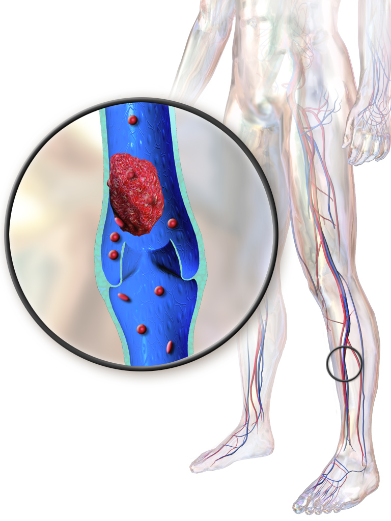
Deep vein thrombosis (DVT) is a serious condition that occurs when a blood clot forms in a vein located deep inside your body. A blood clot is a clump of blood that is in a gelatinous, solid state. Deep vein blood clots typically form in your thigh or lower leg, but they can also develop in other areas of your body. Other names associated with this condition include thromboembolism, post-thrombotic syndrome, and post-phlebitic syndrome. You to see Hematology doctor.


RISK FACTORS:Who is at risk for deep vein thrombosis?

DVT occurs most commonly in people who are over 50 years in age. Certain conditions that alter how your blood moves through your veins can raise your risk of developing clots. These include:

  • having an injury that damages your veins such as a bone fracture
  • being overweight, which puts more pressure on the veins in your legs and pelvis
  • having a family history of DVT
  • having a catheter placed in a vein
  • taking birth control pills or undergoing hormone therapy
  • smoking (especially heavily)
  • staying seated for a long time while you’re in a car or on a plane, especially if you already have at least one other risk factor

Some diseases and disorders can increase your risk of having blood clots. These include hereditary blood clotting disorders, especially when you have at least one other risk factor. Cancer and inflammatory bowel disease can also increase the risk of developing a blood clot. Heart failure, a condition that makes it more difficult for your heart to pump blood, also occurs with an increased risk of clots.

Surgery

DVT is a major risk associated with surgery. This is especially true if you’re having a surgery in the lower extremities, such as joint replacement surgery. Your doctor will discuss the risk of DVT if you need joint replacement surgery.

Pregnancy

Being pregnant increases your risk of DVT. Increased hormone levels, and a slower blood flow as your uterus expands and restricts blood flowing back from your lower extremities, contribute to this risk. This elevated risk continues until about six weeks after giving birth. Being on bed rest or having a cesarean delivery also increases your risk of having DVT.

What are the symptoms of deep vein thrombosis?

Common symptoms include:

  • swelling in your foot, ankle, or leg, usually on one side
  • cramping pain in your affected leg that usually begins in your calf
  • severe, unexplained pain in your foot and ankle
  • an area of skin that feels warmer than the skin on the surrounding areas
  • skin over the affected area turning pale or a reddish or bluish color

People may not find out that they have deep vein thrombosis until they’ve gone through emergency treatment for a pulmonary embolism. A pulmonary embolism is a life-threatening complication of DVT in which an artery in the lung becomes blocked.

What are the treatment options for deep vein thrombosis?

DVT treatments focus on keeping the clot from growing. In addition, treatment will attempt to prevent a pulmonary embolism and lower your risk of having more clots.

Medication

Your doctor might prescribe medications that thin your blood, such as heparin, warfarin, enoxaparin, or fondaparinux. This makes it harder for your blood to clot. It also keeps existing clots as small as possible and decreases the chance that you’ll develop more clots.

If blood thinners don’t work or if you have a severe case of DVT, your doctor might use thrombolytic drugs. Thrombolytic drugs work by breaking up clots. You’ll receive these intravenously.

Compression stockings

Wearing compression stockings can prevent swelling and may lower your chance of developing clots. They don’t demonstrate a reduction in recurrent DVT.

Compression stockings reach just below your knee or right above it. Your doctor may recommend you wear these every day.

Find a great selection of compression stockings here.

Filters

You might need to have a filter put inside the large abdominal vein called the vena cava if you aren’t able to take blood thinners. This form of treatment helps prevent pulmonary embolisms by stopping clots from entering your lungs.

However, there is a risk to filters being placed. If left long term, they can actually cause DVT. They should be used short term until the risk of thromboembolism is reduced and anticoagulation can be used.

 What are the complications associated with deep vein thrombosis?
A major complication of DVT is a pulmonary embolism. You can develop a pulmonary embolism if a blood clot moves to your lungs and blocks a blood vessel. This can cause serious damage to your lungs and other parts of your body. You should get immediate medical help if you have signs of a pulmonary embolism. These signs include:
  • dizziness
  • sweating
  • chest pain that gets worse with coughing or inhaling deeply
  • rapid breathing
  • coughing up blood
  • rapid heart rate

Tetralogy of fallot: causes, symptoms & treatments

Tetralogy of Fallot  is a rare condition caused by a combination of four heart defects that are present at birth (congenital).

These defects, which affect the structure of the heart, cause oxygen-poor blood to flow out of the heart and to the rest of the body. Infants and children with tetralogy of Fallot usually have blue-tinged skin because their blood doesn’t carry enough oxygen.

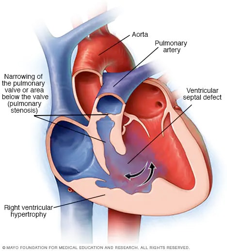
 Illustration showing components of tetralogy of Fallot

Tetralogy of Fallot is often diagnosed during infancy or soon after. However, tetralogy of Fallot might not be detected until later in life in some adults, depending on the severity of the defects and symptoms.

With early diagnosis followed by appropriate surgical treatment, most children and adults who have tetralogy of Fallot live relatively normal lives, though they’ll need regular medical care throughout life and might have restrictions on exercise.

Symptoms

Tetralogy of Fallot symptoms vary, depending on the extent of obstruction of blood flow out of the right ventricle and into the lungs. Signs and symptoms may include:

  • A bluish coloration of the skin caused by blood low in oxygen (cyanosis)
  • Shortness of breath and rapid breathing, especially during feeding or exercise
  • Loss of consciousness (fainting)
  • Clubbing of fingers and toes — an abnormal, rounded shape of the nail bed
  • Poor weight gain
  • Tiring easily during play or exercise
  • Irritability
  • Prolonged crying
  • A heart murmur

Tet spells

Sometimes, babies who have tetralogy of Fallot will suddenly develop deep blue skin, nails and lips after crying or feeding, or when agitated.

These episodes are called tet spells and are caused by a rapid drop in the amount of oxygen in the blood. Tet spells are most common in young infants, around 2 to 4 months old. Toddlers or older children might instinctively squat when they’re short of breath. Squatting increases blood flow to the lungs.

When to see a doctor

Seek medical help if you notice that your baby has the following symptoms:

  • Difficulty breathing
  • Bluish discoloration of the skin
  • Passing out or seizures
  • Weakness
  • Unusual irritability

If your baby becomes blue (cyanotic), place your baby on his or her side and pull your baby’s knees up to his or her chest. This helps increase blood flow to the lungs.

Causes

Tetralogy of Fallot occurs during fetal growth, when the baby’s heart is developing. While factors such as poor maternal nutrition, viral illness or genetic disorders might increase the risk of this condition, in most cases the cause of tetralogy of Fallot is unknown.

The four abnormalities that make up the tetralogy of Fallot include:

  • Pulmonary valve stenosis. Pulmonary valve stenosis is a narrowing of the pulmonary valve — the valve that separates the lower right chamber of the heart (right ventricle) from the main blood vessel leading to the lungs (pulmonary artery).Narrowing (constriction) of the pulmonary valve reduces blood flow to the lungs. The narrowing might also affect the muscle beneath the pulmonary valve. In some severe cases, the pulmonary valve doesn’t form properly (pulmonary atresia) and causes reduced blood flow to the lungs.
  • Ventricular septal defect. A ventricular septal defect is a hole (defect) in the wall (septum) that separates the two lower chambers of the heart — the left and right ventricles. The hole allows deoxygenated blood in the right ventricle — blood that has circulated through the body and is returning to the lungs to replenish its oxygen supply — to flow into the left ventricle and mix with oxygenated blood fresh from the lungs.Blood from the left ventricle also flows back to the right ventricle in an inefficient manner. This ability for blood to flow through the ventricular septal defect reduces the supply of oxygenated blood to the body and eventually can weaken the heart.
  • Overriding aorta. Normally the aorta — the main artery leading out to the body — branches off the left ventricle. In tetralogy of Fallot, the aorta is shifted slightly to the right and lies directly above the ventricular septal defect.In this position the aorta receives blood from both the right and left ventricles, mixing the oxygen-poor blood from the right ventricle with the oxygen-rich blood from the left ventricle.
  • Right ventricular hypertrophy. When the heart’s pumping action is overworked, it causes the muscular wall of the right ventricle to thicken. Over time this might cause the heart to stiffen, become weak and eventually fail.

Some children or adults who have tetralogy of Fallot may have other heart defects, such as a hole between the heart’s upper chambers (atrial septal defect), a right aortic arch or abnormalities of the coronary arteries.

Risk factors

While the exact cause of tetralogy of Fallot is unknown, various factors might increase the risk of a baby being born with this condition. These risk factors include:

  • A viral illness during pregnancy, such as rubella (German measles)
  • Alcoholism during pregnancy
  • Poor nutrition during pregnancy
  • A mother older than age 40
  • A parent who has tetralogy of Fallot
  • The presence of Down syndrome or DiGeorge syndrome

Complications

All babies who have tetralogy of Fallot need corrective surgery. Without treatment, your baby might not grow and develop properly.

Your baby may also be at an increased risk of serious complications, such as infective endocarditis — an infection of the inner lining of the heart or heart valve caused by a bacterial infection.

Untreated cases of tetralogy of Fallot usually develop severe complications over time, which might result in death or disability by early adulthood.

Treatment

Surgery is the only effective treatment for tetralogy of Fallot. Surgical options include intracardiac repair or a temporary procedure that uses a shunt. However, most babies and older children have intracardiac repair.

Your or your child’s doctors will determine the most appropriate surgery and the timing of the surgery based on your or your child’s condition.

In some cases your child may need medicine to keep the opening between two large blood vessels in the heart open. This can help to maintain blood flow from the heart to the lungs before intracardiac repair.

Intracardiac repair

This open-heart surgery is usually done during the first year after birth and involves several repairs. Adults with tetralogy of Fallot rarely may undergo this procedure if they didn’t have surgical repair as children.

The surgeon places a patch over the ventricular septal defect to close the hole between the lower chambers of the heart (ventricles).

He or she also repairs or replaces the narrowed pulmonary valve and widens the pulmonary arteries to increase blood flow to the lungs.

Because the right ventricle won’t need to work as hard to pump blood after this procedure, the right ventricle wall will go back to its normal thickness. After intracardiac repair, the oxygen level in the blood increases and symptoms will lessen.

Temporary surgery

Occasionally babies need to undergo a temporary (palliative) surgery before having intracardiac repair in order to improve blood flow to the lungs. This procedure may be done if your baby was born prematurely or has pulmonary arteries that are undeveloped (hypoplastic).

In this procedure, the surgeon creates a bypass (shunt) between a large artery that branches off from the aorta and the pulmonary artery.

When your baby is ready for intracardiac repair, the surgeon removes the shunt during the procedure for intracardiac repair.

After surgery

While most babies and adults do well after intracardiac repair, long-term complications are common. Complications may include:

  • Chronic pulmonary regurgitation, in which blood leaks through the pulmonary valve back into the pumping chamber (right ventricle)
  • Other heart valve problems, such as blood leaking back through the tricuspid valve
  • Holes in the wall between the ventricles (ventricular septal defects) that may continue to leak after repair or may need re-repair
  • Enlarged right ventricle or left ventricle that isn’t working properly
  • Irregular heartbeats (arrhythmias)
  • Coronary artery disease
  • Aortic root dilation, in which the ascending aorta enlarges
  • Sudden cardiac death

Complications can continue throughout childhood, adolescence and adulthood for people with tetralogy of Fallot. Most adults with repaired tetralogy of Fallot may require another procedure or intervention during their lifetimes. It’s very important to have regular follow-up with a cardiologist trained in caring for people with congenital heart disease (pediatric cardiologist or adult congenital cardiologist) who can evaluate you and determine the appropriate timing of another intervention or procedure.

Sometimes blood flow to the lungs may still be restricted after intracardiac repair. Infants, children or adults with these complications might require additional surgeries. More commonly, there is leakage through the repaired pulmonary valve. Most adults with repaired tetralogy of Fallot may have pulmonary valve leakage (regurgitation) and may need to have the pulmonary valve replaced during their lifetimes. Your cardiologist will determine the most appropriate timing for this procedure.

Arrhythmias are common after repair and may be treated with medications, a procedure to treat the arrhythmias (ablation) or a special pacemaker device that treats life-threatening heart rhythms (implantable cardioverter-defibrillator).

In addition, as with any surgery, there’s a risk of infection, unexpected bleeding or blood clots.

Ongoing care

After surgery you or your child will need lifelong care with a cardiologist trained in treating congenital heart disease (adult congenital cardiologist or pediatric cardiologist), including routine follow-up appointments to make sure that the initial operation or procedure was successful and to monitor for any new complications.

Your or your child’s doctor may conduct a physical examination and order tests in regular follow-up appointments to evaluate and monitor your or your child’s condition.

The doctor might also recommend that you or your child limit strenuous physical activity, particularly if there’s any pulmonary valve leakage or obstruction, or arrhythmias.

Sometimes, antibiotics are recommended during dental procedures to prevent infections that might cause endocarditis — an inflammation of the lining of the heart. Antibiotics are especially important for those who have had prior endocarditis, have artificial valves or have had repair with prosthetic material. Ask the doctor what’s right for you or your child.

PVD: Causes, Symptoms & Treatments

Peripheral Vascular disease (PVD)?

Peripheral vascular disease (PVD) refers to diseases of the blood vessels (arteries and veins) located outside the heart and brain. While there are many causes of peripheral vascular disease, doctors commonly use the term peripheral vascular disease to refer to peripheral artery disease (peripheral arterial disease, PAD), a condition that develops when the arteries that supply blood to the internal organs, arms, and legs become completely or partially blocked as a result of atherosclerosis.

Are atherosclerosis and peripheral vascular disease related?

Atherosclerosis is a gradual process whereby hard cholesterol substances (plaques) are deposited in the walls of the arteries. This buildup of cholesterol plaques causes hardening of the artery walls and narrowing of the inner channel (lumen) of the artery. When this happens in the peripheral circulation, peripheral vascular disease is the result. The atherosclerosis process begins early in life (as early as teens in some people). When atherosclerosis is mild and the arteries are not substantially narrowed, atherosclerosis causes no symptoms. Therefore, many adults typically are unaware that their arteries are gradually accumulating cholesterol plaques. However, when atherosclerosis becomes advanced with aging, it can cause critical narrowing of the arteries resulting in tissue ischemia (lack of blood and oxygen).

Arteries that are narrowed by advanced atherosclerosis can cause diseases in different organs. For example, advanced atherosclerosis of the coronary arteries (arteries that supply heart muscles) can lead to angina and heart attacks. Advanced atherosclerosis of the carotid and cerebral arteries (arteries that supply blood to the brain) can lead to strokes and transient ischemic attacks (TIAs). Advanced atherosclerosis in the lower extremities can lead to painwhile walking or exercising (claudication), deficient wound healing, and/or leg ulcers.

Picture of carotid artery disease and plaque buildup.

Picture of Carotid Artery Disease and Plaque Buildup

Picture of a heart attack (myocardial infarction).

Picture of a Heart Attack (Myocardial Infarction) – Buildup of Cholesterol Plaque and Blood Clot

Atherosclerosis is often generalized, meaning it affects arteries throughout the body. Therefore, patients with heart attacks are also more likely to develop strokes and peripheral vascular disease, and vice versa.

What are the signs and symptoms of peripheral artery disease (PVD)?

Approximately half of people with peripheral artery disease do not experience any symptoms. For patients with symptoms, the most common symptoms are intermittent claudication and rest pain.

  • Intermittent claudication refers to arm or leg pain or cramping in the arms or legs that occurs with exercise and goes away with rest. The severity and location of the pain of intermittent claudication vary depending upon the location and extent of blockage of the involved artery. The most common location of intermittent claudication is the calf muscle of the leg, leading to calf or leg pain while walking. The pain in the calf muscle occurs only during exercise such as walking, and the pain steadily increases with continued walking until the patient has to stop due to intolerable pain. Then the pain quickly subsides during rest. Intermittent claudication can affect one or both legs.
  • Rest pain in the legs occurs when the artery occlusion is so critical that there is not enough blood and oxygen supply to the legs even at rest and represents a more serious form of the condition. The pain typically affects the feet, is usually severe, and occurs at night when the patient is lying down, face up.

Other symptoms and signs of peripheral artery disease include:

  • Numbness of the legs or feet
  • Weakness and atrophy (diminished size and strength) of the calf muscle
  • A feeling of coldness in the legs or feet
  • Changes in color of the feet; feet turn pale when they are elevated, and turn dusky red in dependent position
  • Hair loss over the top of the feet and thickening of the toenails
  • Poor wound healing in the legs or feet
  • Painful ulcers and/or gangrene in areas of the feet where blood supply is lost; typically in the toes

What are the management and treatment guidelines for peripheral vascular disease (PVD)?

Treatment goals for peripheral artery disease include:

  1. Relieve the pain of intermittent claudication.
  2. Improve exercise tolerance by increasing the walking distance before the onset of claudication.
  3. Prevent critical artery occlusion that can lead to foot ulcers, gangrene, and amputation.
  4. Prevent heart attacks and strokes.

Treatment of peripheral artery disease includes lifestyle measures, supervised exercises, medications, angioplasty, and surgery.

Lifestyle changes

  • Smoking cessation eliminates a major risk factor for disease progression, and it lowers the incidences of pain at rest and amputations. Smoking cessation  also is important to prevent heart attacks and strokes.
  • A healthy diet can help lower blood cholesterol and other lipid levels and may help control blood pressure.
  • Keep other risk factors, such as diabetes, lipid levels, and blood pressure under control by following medical advice regarding medications and lifestyle changes.

Supervised exercise

Proper exercise can condition the muscles to use oxygen effectively and can speed the development of collateral circulation. Clinical trials have shown that regular supervised exercise can reduce symptoms of intermittent claudication and allow individuals to walk longer before the onset of claudication. Ideally, your doctor should prescribe an exercise program tailored to your specific needs.

Rehabilitation programs supervised by healthcare professionals such as nurses or physical therapists may help. Exercise at least three times a week, each session lasting longer than 30 to 45 minutes for the best results. Exercise usually involves walking on a monitored treadmill until claudication develops; walking time is then gradually increased with each session. Patients are also monitored for the development of chest pain or heart rhythm irregularities during exercise.


Trigeminal neuralgia :Causes, symptoms & treatments

The brain is connected to the body by the spinal cord with spinal nerves sending and receiving impulses and messages to and from the brain. However, there are twelve cranial nerves that directly connect to the body. These nerves are involved with the muscle and sensory function of the head and neck. (The exception is cranial nerve X or the vagus nerve, which is also responsible for the parasympathetic system of the chest and abdomen).

12 Cranial Nerves
Cranial Nerve Name Function
I Olvactory Smell
II Optic Vision
III, IV, VI Oculomoter, Trochlear, Abducens Eye movement
V Trigeminal Facial sensation, chewing
VII Facial Facial movement
VIII Auditory Hearing
IX Glossopharyngeal Taste, swallowing
X Vagus Swallowing, voice modulation, parasympathetic tone of the body
XI Accessory Neck muscles
XII Hypoglossal Swallowing, speech articulation

The trigeminal nerve (cranial nerve V) is so named because it has three (tri) branches responsible for face sensation; one branch also regulates chewing.

  • The ophthalmic branch (V1) is responsible for sensation from the scalp, forehead, upper eyelid and tip of the nose.
  • The maxillary branch (V2) sensation covers the lower eyelid, the side of the nose, the upper lip and cheek, and the upper teeth and gums.
  • The mandibular branch (V3) is responsible for sensation of the lower teeth and gums, lower lip, chin, jaw, and part of the ear. It is also responsible for supplying the muscles involved with chewing (mastication), those muscles involved with chewing.

What is trigeminal neuralgia (TN)?

Trigeminal neuralgia is inflammation of the trigeminal nerve, causing intense facial pain. It is also known as tic douloureax because the intense pain can cause patients to contort their face into a grimace and cause the head to move away from the pain. The obvious movement is known as a tic.

The pain of trigeminal neuralgia is intense and may be an isolated episode or may be occur every few hours, minutes, or seconds. There can be months or years between attacks, but in some patients whose pain is not well controlled; it can lead to a chronic pain syndrome, affecting activities of daily life and cause depression.

Though it can affect people of any age, trigeminal neuralgia tends to afflict people older than 60 years of age. It affects the right side of the face five times more often than the left.

What causes trigeminal neuralgia?

Most often, the cause of trigeminal neuralgia is idiopathic, meaning the cause is not known. There are some instances when the nerve can be compressed by nearby blood vessels, aneurysms, or tumors.

There are inflammatory causes of trigeminal neuralgia because of systemic diseases includingmultiple sclerosis, sarcoidosis, and Lyme disease. There also is an association with collagen vascular diseases including scleroderma and systemic lupus erythematosus.

What are the symptoms of trigeminal neuralgia?

 Symptoms of trigeminal neuralgia include an acute onset of sharp, stabbing pain to one side of the face. It tends to begin at the angle of the jaw and radiate along the junction lines; between the ophthalmic branchV1 and maxillary branch V2, or the maxillary branch V2 and the mandibular branch V3.

The pain is severe and described as an electric shock. It may be made worse by light touch, chewing, or cold exposure in the mouth. In the midst of an attack, affected individuals shield their face trying to protect it from being touched. This is an important diagnostic sign because with many other pain syndromes like a toothache, the person will rub or hold the face to ease the pain.

While there may be only one attack of pain, the person may experience recurrent sharp pain every few hours or every few seconds. Between the attacks, the pain resolves completely and the the person has no symptoms. However, because of fear that the intense pain might return, people can be quite distraught. Trigeminal neuralgia tends not to occur when the person is asleep, and this differentiates it from migraines, which often waken the person.

After the first episode of attacks, the pain may subside for months or years but there is always the risk that trigeminal neuralgia will recur without warning.

What is the treatment for trigeminal neuralgia?

  • Idiopathic trigeminal neuralgia most often is treated with good success using a single anticonvulsant medication such as carbamazepine (Tegretol).
  • Gabapentin (Neurontin, Gabarone), baclofen and phenytoin (Dilantin, Dilantin-125) may be used as second line drugs, often in addition to carbamazepine. In many patients, as time progresses, carbamazepine becomes less effective and these drugs can be used in combination to control the pain.
  • Should pain persist and medication fail to be effective, surgery or radiation therapy may be other treatment options.
  • Lamotrigine (Lamictal) may be prescribed for multiple sclerosis patients who develop trigeminal neuralgia.

Malaria: causes symptoms treatment and prevention

What is malaria?

Malaria is an infection of the blood that is carried from person to person by mosquitoes. The disease has been recognized for thousands of years and once was found almost everywhere except in the most northern areas of the world.  However, it remains a serious problem in much of the tropical and subtropical world.

Millions of people continue to be infected every year and close to one million of them die.

Malaria symptoms

With malaria you develop a high fever, which comes and goes every other day or few days. How often a fever returns varies with each species of malaria.

  • Many infections do not show this classic pattern of returning fevers at all. In many people the infection will seem more like flu with high fever and body aches.
  • People also will complain of headache, nausea, shaking chills (rigors),sweating and weakness.
  • As the infection progresses the fevers get less severe and you seem to recover. However, the infection can remain in many people for several years, particularly for those with a long history of exposure to malaria.
  • These people can develop some immunity and may be infected for many years while only rarely having symptoms.

The different types of malaria each bring on their own complications.

  • P falciparum: You can develop severe haemolytic anaemia (the red blood cells actually break down), kidney failure, coma and death. Treatment is a medical emergency. Drug resistance has become widespread. Current information on disease patterns, prevention for travellers and drug resistance can always be found through a travel health clinic or your GP surgery.
  • P ovale: This species also can cause anaemia, but this infection is rarely life threatening.
  • P vivax: You can develop anaemia and rupture of the spleen, which can become life threatening. People with P vivax or P ovale may relapse several months after the initial illness because of the persistence of dormant forms (called hypnozoites) remaining in the liver. These should be eradicated with medical treatment.
  • P malariae: This infection is rarely life threatening, but a long-standing disease can lead to kidney failure. If untreated, this infection can last throughout your life.

Malaria causes

Malaria is caused by protozoan of the genus Plasmodium.

  • Infection begins with a bite from an infected mosquito.
  • The parasite travels from the mosquito to your liver, where the parasite begins to reproduce.
  • The parasite leaves the liver and travels to the bloodstream, where it infects red blood cells. The parasite reproduces in the red blood cells, which destroys the cells and releases more parasites into the bloodstream.
  • If another mosquito bites an infected person, that mosquito can then carry the infection to someone else.

There are four species of Plasmodium that infect humans:

  • P vivax – Most common in India and Central and South America but found worldwide. It has an incubation period of 8-13 days. Infections can sometimes lead to life-threatening rupture of the spleen. In people treated only with chloroquine, this type of malaria can hide in the liver and return later.
  • P ovale – Rarely found outside Africa. This form of malaria has an incubation period of 8-17 days and can hide in the liver of partially treated people and return later.
  • P malariae – Found worldwide but less common than the other forms. This form of malaria has an incubation of 2-4 weeks. If untreated, the infection can last many years.
  • P falciparum – Common worldwide, this is the most life-threatening form of malaria. This parasite has an incubation period of 5-12 days. Resistance to many of the medicines used to treat or prevent malaria is increasing.
  • Although most people acquire malaria through mosquito bites, in some foreign countries the disease can have other sources.
    • Every year a handful of people are infected through blood transfusions or organ transplants.
    • IV drug users can develop malaria from sharing needles.
    • Each year a few babies are born to mothers who did not know they were infected. The babies then develop malaria.

When to seek medical care

People who recently have traveled to a country in which malaria exists and who develop a high fever or other symptoms that may be malaria should seek immediate medical attention at the GP’s surgery or at a hospital.

Examinations and tests

A healthcare professional will perform blood tests to determine if you have malaria and, if so, which type.

Malaria treatment

  • Your doctor will prescribe a medicine or a combination of medicines straight away, depending on the type of strain and where you were infected.
  • In much of the world, malaria is treated at home with oral medication and fluids. Severe infections require IV drug therapy.
  • The most important aspect of home care is to make sure you drink lots of fluids and not to become dehydrated.

Next steps follow-up

  • People infected with P vivax or P ovale will need to take chloroquine for several weeks after being treated in order to kill the parasites hiding in the liver.
  • Report any recurrent fever or symptoms to your doctor because treatment failures are fairly common, and additional treatment will be indicated.
  • Do not donate blood for several years after having been exposed to malaria.

Prevention

  • For people travelling to areas where malaria exists, prevention is perhaps the most important aspect of managing the disease. Seek advice well before you travel, because some medications need to be started before you travel. DEET, or diethyltoluamide, is one of the most commonly used repellents that the NHS says is effective in sprays, roll-ons, sticks and creams. However, London School of Hygiene and Tropical Medicine researchers have found mosquitoes are able to ignore the smell of one of the most popular insect repellent ingredients a few hours after first being exposed to it, highlighting the importance of taking additional precautions such as the use of mosquito nets.
  • Several medications are used to prevent infections during foreign travel. The pattern of resistance to these medications is constantly changing.
  • In some parts of the world, P falciparum is resistant to all these medications.

Type 2 Diabetes: Cause, Symptoms & Treatments

What Causes Diabetes?

Your pancreas makes a hormone called insulin. It’s what lets your cells turn glucose from the food you eat into energy. People with type 2 diabetes make insulin, but their cells don’t use it as well as they should. Doctors call this insulin resistance.

At first, the pancreas makes more insulin to try to get glucose into the cells. But eventually it can’t keep up, and the sugar builds up in your blood instead.

Usually a combination of things cause type 2 diabetes, including:

Genes. Scientists have found different bits of DNA that affect how your body makes insulin.

Extra weight. Being overweight or obese can cause insulin resistance, especially if you carry your extra pounds around the middle. Now type 2 diabetes affects kids and teens as well as adults, mainly because of childhood obesity.

Metabolic syndrome. People with insulin resistance often have a group of conditions including high blood glucose, extra fat around the waist, high blood pressure, and high cholesterol and triglycerides.

Too much glucose from your liver. When your blood sugar is low, your liver makes and sends out glucose. After you eat, your blood sugar goes up, and usually the liver will slow down and store its glucose for later. But some people’s livers don’t. They keep cranking out sugar.

Bad communication between cells. Sometimes cells send the wrong signals or don’t pick up messages correctly. When these problems affect how your cells make and use insulin or glucose, a chain reaction can lead to diabetes.

Broken beta cells. If the cells that make the insulin send out the wrong amount of insulin at the wrong time, your blood sugar gets thrown off. High blood glucose can damage these cells, too.

Risk Factors and Prevention

While certain things make getting diabetes more likely, they won’t give you the disease. But the more that apply to you, the higher your chances of getting it are.

Some things you can’t control.

  • Age: 45 or older
  • Family: A parent, sister, or brother with diabetes

Some things are related to your health and medical history. Your doctor may be able to help.

  • Prediabetes
  • Heart and blood vessel disease
  • High blood pressure, even if it’s treated and under control
  • Low HDL (“good”) cholesterol
  • High triglycerides
  • Being overweight or obese
  • Having a baby that weighed more than 9 pounds
  • Having gestational diabetes while you were pregnant
  • Polycystic ovary syndrome (PCOS)
  • Acanthosis nigricans, a skin condition with dark rashes around your neck or armpits
  • Depression

Other risk factors have to do with your daily habits and lifestyle. These are the ones you can really do something about.

  • Getting little or no exercise
  • Smoking
  • Stress
  • Sleeping too little or too much

Because you can’t change what happened in the past, focus on what you can do now and going forward. Take medications and follow your doctor’s suggestions to be healthy. Simple changes at home can make a big difference, too.

Lose weight. Dropping just 7% to 10% of your weight can cut your risk of type 2 diabetes in half.Get active. Moving muscles use insulin. Thirty minutes of brisk walking a day will cut your risk by almost a third.Eat right. Avoid highly processed carbs, sugary drinks, and trans and saturated fats. Limit red and processed meats.Quit smoking. Work with your doctor to avoid gaining weight, so you don’t create one problem by solving another.

Symptoms

The symptoms of type 2 diabetes can be so mild you don’t notice them. In fact, about 8 million people who have it don’t know it.

  • Being very thirsty
  • Peeing a lot
  • Blurry vision
  • Being irritable
  • Tingling or numbness in your hands or feet
  • Feeling worn out
  • Wounds that don’t heal
  • Yeast infections that keep coming back

Long-Term Effects

Over time, high blood sugar can damage and cause problems with your:

  • Heart and blood vessels
  • Kidneys
  • Eyes
  • Nerves, which can lead to trouble with digestion, the feeling in your feet, and your sexual response
  • Wound healing
  • Pregnancy

The best way to avoid these complications is to manage your diabetes well.

  • Take your diabetes medications or insulin on time.
  • Check your blood glucose.
  • Eat right, and don’t skip meals.
  • See your doctor regularly to check for early signs of trouble.

Fabry disease: causes, symptoms & treatments

What Is Fabry Disease?

Fabry disease runs in families. It can have lots of different symptoms, including pain in the hands and feet and a specific kind of rash.

When you have Fabry disease, a certain type of fatty substance builds up in your body. It narrows your blood vessels, which can hurt your skin, kidneys, heart, brain, and nervous system.

Your doctor may call Fabry disease a “storage disorder.” It usually starts in childhood and is much more common in men than women.

There are treatments that can make a difference in how you feel, day-to-day. Getting support from your family and friends is key, too.

Causes

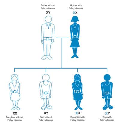
You get Fabry disease from your parents. It’s passed down through genes.

The problem is that your body can’t make an enzyme called alpha-galactosidase A, which you need to break down fatty substances like oils, waxes, and fatty acids. When you have Fabry disease, you either were born without that enzyme or it doesn’t work right.

Symptoms

You may notice things like:

  • Pain and burning in your hands and feet that get worse with exercise, fever, and hot weather or when you are tired
  • Small, dark red spots usually found between your belly button and knees
  • Cloudy vision
  • Hearing loss
  • Ringing in the ears
  • Sweating less than normal
  • Stomach pain, bowel movements right after eating

Fabry disease can lead to more serious problems, especially in men. These can include:

  • Higher chance of heart attack or stroke
  • Serious kidney problems, including kidney failure
  • High blood pressure
  • Heart failure
  • Enlarged heart
  • Osteoporosis

Treatment

Your doctor will probably recommend enzyme replacement therapy (ERT). It replaces the enzyme that is missing or not working correctly so that your body can break down fatty substances the way it should.

ERT is the only FDA-approved treatment for Fabry disease. It will help ease the pain and other symptoms that Fabry disease causes.

You will probably visit an outpatient center every few weeks to get the enzyme injected into a vein.

Your doctor may recommend that you also take:

  • Drugs to relieve pain (prescription or nonprescription)
  • Medicine for stomach problems
  • Blood thinners or other drugs for irregular heartbeat or other heart problems
  • Blood pressure medicine, which also helps protect your kidneys

You may need dialysis or a kidney transplant if Fabry disease has caused serious kidney damage.

You can also expect to get regular tests to keep track of how you’re doing. These may include:

  • Blood, urine, and thyroidtests
  • EKG (electrocardiogram). A nurse or other medical professional will attach soft, sticky patches to different parts of your body. These patches measure electrical signals from your heart and can tell how fast your heart is beating and if it has a healthy rhythm.
  • Echocardiogram. This is an ultrasound of your heart. It can show if all the parts of your heart are healthy and if it’s pumping well.
  • Brain MRI. An MRI, or magnetic resonance imaging, makes pictures of organs and structures inside your body.
  • CT of your head. CT, or computed tomography, is a powerful X-ray that makes detailed pictures of the inside of your body.
  • Hearing and eye exams
  • Lung function test to see how much air you breathe in and out, and how much oxygen is getting to your blood

Taking Care of Yourself

Be good to yourself. Do things that you enjoy, spend time with people who are good company, and save your energy for the things that really matter to you. Part of that may involve saying “no” more often, and letting people know what would help you. That’s OK to do!

Pages:123