Global Treatment Services Pvt. Ltd.

Global Treatment Services

Vasculitis: Symptoms, causes & Tratments

Vasculitis is a general term for a group of uncommon diseases that feature inflammation of the blood vessels. The blood vessels of the body are referred to as the vascular system. The blood vessels are comprised of arteries that pass oxygen-rich blood to the tissues of the body and veins that return oxygen-depleted blood from the tissues to the lungs for oxygen. Vasculitis is characterized by inflammation in and damage to the walls of various blood vessels.

Each of the vasculitis diseases is defined by certain patterns of distribution of blood vessel involvement, particular organ involvement, and laboratory test abnormalities. As a group, these diseases are referred to as vasculitides.

The word vasculitis is derived from the Latin “vasculum”, vessel + “- itis”, inflammation. Another term for vasculitis is angiitis. When arteries are the inflamed blood vessels, the condition is also referred to as arteritis. When the veins are inflamed, it is referred to as venulitis.

What causes vasculitis, and what are examples of diseases with vasculitis?

The actual cause of these vasculitis diseases is usually not known. However, immune system abnormality and inflammation of blood vessels are common features. Each form of vasculitis has its own characteristic pattern of symptoms, much of which depends on what particular organs are affected.

Examples of vasculitis include:

  • Kawasaki disease,
  • Behçet’s disease,
  • polyarteritis nodosa,
  • Wegener’s granulomatosis,
  • Cryoglobulinemia,
  • Takayasu’s arteritis,
  • Churg-Strauss syndrome,
  • Giant cell arteritis (temporal arteritis), and
  • Henoch-Schönlein purpura.

Vasculitis can also accompany:

  • Infections (such as hepatitis B),
  • Exposure to chemicals (such as amphetamines and cocaine),
  • Medications,
  • Cancers (such as lymphomas and multiple myeloma), and
  • Rheumatic diseases (such as rheumatoid arthritis and systemic lupus erythematosus

Treatments:

The treatment of the various forms of vasculitis is based on the severity of the illness and the organs involved. Treatments are generally directed toward stopping the inflammation and suppressing the immune system. Typically, cortisone-related medications, such as prednisone , are used. Additionally, other immune suppression drugs, such as cyclophosphamide (Cytoxan) and others, are considered. Additionally, affected organs (such as the heart or lungs) may require specific medical treatment when the disease is active.

The management of vasculitis is an evolving field in medicine. The ideal programs for monitoring and treatment will continue to improve as disease patterns and causes are defined by medical research.

Craniosynostosis : Symptoms, Causes & Treatments

Craniosynostosis (kray-nee-o-sin-os-TOE-sis) is a birth defect in which one or more of the joints between the bones of your baby’s skull close prematurely, before your baby’s brain is fully formed. When your baby has craniosynostosis, his or her brain can’t grow in its natural shape and the head is misshapen.

Craniosynostosis can affect one or more of the joints in your baby’s skull. In some cases, craniosynostosis is associated with an underlying brain abnormality that prevents the brain from growing properly.

Treating craniosynostosis usually involves surgery to separate the fused bones. If there’s no underlying brain abnormality, the surgery allows your baby’s brain adequate space to grow and develop.

Craniosynostosis symptoms:

Signs of craniosynostosis include:

  • A misshapen skull, with the shape depending on which of the cranial sutures are affected
  • An abnormal feeling or disappearing “soft spot” (fontanel) on your baby’s skull
  • Slow or no growth of the head as your baby grows
  • Development of a raised, hard ridge along affected sutures
  • Increased pressure within the skull (intracranial pressure)

The signs of craniosynostosis may not be noticeable at birth, but they become apparent during the first few months of your baby’s life.

Craniosynostosis types and characteristics

There are many different types of craniosynostosis. The term given to each type depends on what sutures are affected. Some of the most common types of craniosynostosis are:

  • Sagittal synostosis (scaphocephaly). Premature fusion of the suture at the top of the head (sagittal suture) forces the head to grow long and narrow, rather than wide. Scaphocephaly is the most common type of craniosynostosis.
  • Coronal synostosis (anterior plagiocephaly). Premature fusion of a coronal suture — one of the structures that run from each ear to the sagittal suture on top of the head — may force your baby’s forehead to flatten on the affected side. It may also raise the eye socket and cause a deviated nose and slanted skull.
  • Bicoronal synostosis (brachycephaly). When both of the coronal sutures fuse prematurely, your baby may have a flat, elevated forehead and brow.

Causes:

Craniosynostosis is often classified as nonsyndromic or syndromic. Nonsyndromic craniosynostosis is the most common type of craniosynostosis, and its cause is unknown.

However, syndromic craniosynostosis is a complication caused by certain genetic syndromes, such as Apert syndrome, Pfeiffer syndrome and Crouzon syndrome, which can affect your baby’s skull development. Besides craniosynostosis, other conditions also may accompany such syndromes — including hand and foot deformities, dental abnormalities, and heart problems.


Treatments and drugs:

Mild cases of craniosynostosis — those that involve only one suture and no underlying syndrome — may require no treatment. Skull abnormalities may become less obvious as your baby grows and develops hair. Or your doctor might recommend a specially molded helmet to help reshape your baby’s head and allow room for your baby’s brain growth.

Surgery:

For most babies, however, surgery is the primary treatment for craniosynostosis. The type and timing of surgery depend on the type of craniosynostosis and whether there’s an underlying syndrome that needs treatment.

The purpose of surgery is to relieve pressure on the brain, create room for the brain to grow normally and improve your child’s appearance. A team that includes a specialist in surgery of the head and face (craniofacial surgeon) and a specialist in brain surgery (neurosurgeon) often performs the procedure.

  • Traditional surgery. The surgeon makes an incision in your baby’s scalp and cranial bones, then reshapes the affected portion of the skull. Sometimes plates and screws, often made of material that is absorbed over time, are used to hold the bones in place. Surgery, which is performed during general anesthesia, usually takes hours.After surgery, your baby remains in the hospital for at least three days. Some children may require a second surgery later because the craniosynostosis recurs. Also, children with facial deformities often require future surgeries to reshape their faces.
  • Endoscopic surgery. This less invasive form of surgery isn’t an option for everyone. But in certain cases, the surgeon may use a lighted tube (endoscope) inserted through one or two small scalp incisions over the affected suture. The surgeon then opens the suture to enable your baby’s brain to grow normally. Endoscopic surgery usually takes about an hour, causes less swelling and blood loss, and shortens the hospital stay, often to one day after surgery.

If your baby has an underlying syndrome, your doctor may recommend regular follow-up visits after surgery to monitor head growth and check for increased intracranial pressure. Head growth will be routinely monitored at well-child visits.

Aneurysm: Symptoms, treatment & Prevention

What is an aneurysm?

An aneurysm is an arterial condition in which the wall of an artery weakens, creating a bulge, or distension of the artery.

Diagrams of arteries and blood flow.
A comparison between an artery with an aneurysm and two different types of artery. Note the cross-section showing a thinner artery wall in the third diagram for aneurysm, compared with normal and narrowed arteries.

An aneurysm can occur in important arteries such as those supplying blood to the brain, and the aorta; the large artery that originates at the left ventricle of the heart and passes down through the chest and abdominal cavities.

The normal diameter of the aorta is around 0.8 inches. This width can bulge to beyond 2 inches with an aneurysm, a width that would typically necessitate surgical treatment.

An aneurysm can also occur in peripheral arteries – usually behind the knee (popliteal aneurysms) – although rupture of these is relatively uncommon.

The two most important common locations for aneurysms are:

  • In the artery directly leaving the heart – an aortic aneurysm (including thoracic and, further down, abdominal aortic aneurysms)
  • In an artery in the brain – a cerebral aneurysm.

Thoracic aortic aneurysm is often abbreviated to TAA, and abdominal aortic aneurysm to AAA. Brain aneurysms are often termed intracranial aneurysms, as well as “berry aneurysms” on account of their size and shape.

Two other examples of aneurysm are mesenteric artery aneurysm (affecting the artery supplying the intestines of the gut) and splenic artery aneurysm (occurring in the spleen, an abdominal organ).

Symptoms of aneurysm:

Most aneurysms do not themselves cause any symptoms.

Even if an aneurysm does not rupture, however, a large aneurysm may obstruct circulation to other tissues. An aneurysm can also contribute to the formation of blood clots that then obstruct smaller blood vessels, potentially causing ischemic stroke or other serious problems; this is known as thromboembolism.

Back pain can be a symptom of an aneurysm although most aneurysms are asymptomatic.

Abdominal aneurysms are sometimes associated with symptoms if they grow rapidly. Some people report abdominal or lower back pain, or a pulsating sensation in the abdomen.

Similarly, thoracic aneurysms can cause symptoms by affecting nearby tissues, including nerves and other blood vessels.

If an aneurysm compresses the laryngeal or vagus nerve, it can cause chest or back pain and symptoms such as coughing, wheezing and difficulty swallowing. Compression of the coronary artery can also cause chest pain.

Otherwise, aneurysms tend to produce symptoms only when there are complications such as rupture.

Symptoms can also be related to the cause of the aneurysm rather than the aneurysm itself. In the case of infection or vasculitis (blood vessel inflammation), for example, there may be fever, malaise or weight loss.

Complications of aneurysm

If an aneurysm has remained undetected, the first sign of it could be when there is a complication – in particular, a rupture – with symptoms resulting from this rather than the aneurysm itself.

The majority of people living with an aneurysm do not suffer any of the complications in the following list. Managing the risk factors is important, however, because all of these possibilities are serious.

Complications of aneurysm include:

Image of a stroke.
Brain aneurysm can lead to subarachnoid hemorrhage; a symptom of this stroke bleed is a sudden extreme headache.
  • Thromboembolism – depending on where the clot has traveled to, thromboembolism can cause pain in the extremities or the abdomen. If a clot travels to the brain, it can cause a stroke
  • Dissection of the aorta – see below for more detail
  • Severe chest and/or back pain – if a silent or diagnosed aortic aneurysm in the chest ruptures, severe chest or back pain may arise. Such symptoms may help hospital medical staff diagnose an aneurysm.
  • Angina – certain types of aneurysm can lead to angina, another type of chest pain; the pain is related to narrowed arteries supplying the heart itself (causing myocardial ischemia and possibly heart attack).
  • Sudden extreme headache – if a brain aneurysm leads to subarachnoid hemorrhage (a kind of stroke), the main symptom is sudden extreme headache; often so severe that it is unlike any previous experience of head pain.
  • Other symptoms – with any aneurysm rupture there may be pain, low blood pressure, a rapid heart rate, and light-headedness.

Again, most people with an aneurysm will not suffer any of these complications, and the risk can be reduced by taking preventive steps.

Treatments for aneurysm

Not all cases of unruptured aneurysm need active treatment, but when an aneurysm ruptures, emergency surgery is needed.

More details follow for the treatment options against the two main types of aneurysm.

Aortic aneurysm treatment options

An aortic aneurysm, whether abdominal or thoracic, may not need any active treatment and instead may just be monitored regularly. Medications and preventive measures may form part of conservative management or they may accompany active surgical treatment – through open or endovascular surgery.

Aortic aneurysm surgery can be performed as open surgery or as endovascular surgery.

Aneurysms that rupture require emergency surgery. Without immediate repair, a ruptured aneurysm is always fatal in the thoracic aorta, and almost always fatal in the abdominal aorta.

The decision to operate on an unruptured aneurysm in the aorta depends on a number of factors related to the individual patient, as well as to features of the aneurysm itself:

  • Individual factors – age, general health, coexisting conditions and personal choice
  • The aneurysm’s characteristics – size relative to location in the thorax or abdomen and the rate of growth
  • Other factors – chronic abdominal pain or risk of thromboembolism may make surgery a good option.

An aortic aneurysm that has a larger diameter (about 2 inches or 5 cm) is more likely to prompt a recommendation of surgery, as is an aneurysm which is growing more quickly (a little less than 1/4 inch over the last 6-12 months).

For both elective unruptured and emergency ruptured cases, surgery can take one of two forms:

  • Open surgery to fit a synthetic or stent graft
  • Endovascular stent-graft surgery.

Endovascular surgery involves accessing the blood vessels through a small incision near the hip. In stent-graft surgery, an endovascular graft is inserted through this incision using a catheter and is positioned in the aorta to seal off the aneurysm.

In the emergency of a ruptured abdominal aortic aneurysm, the decision over which procedure to perform will be urgent. For elective operations to repair unruptured aneurysms, a review published in 2014 sought to determine the relative risks and benefits of open vs. endovascular surgery.

The review found that while there was a lack of robust studies comparing the two options directly, similar rates of complication were reported for both types of surgery.

The evidence suggested that endovascular aneurysm repair (EVAR) was “less invasive,” but the procedure did “not always significantly alter the postoperative course or length of hospital stay for patients.”

The authors concluded that open surgery still had a role for those patients who were not suitable for endovascular surgery.

Endovascular surgery has become the generally preferred method of aortic aneurysm surgery because of:

  • Shorter operative times
  • Often shorter hospital stays
  • Better levels of surgical experience
  • A perceived lower risk of disease or death surrounding surgery compared with laparoscopic surgery (although the review found this could be more perception than reality).

Endovascular surgery for the repair of aortic aneurysms does carry the following risks however, in addition to the usual risks of surgery:

  • Bleeding around the graft (which requires additional surgery)
  • Bleeding before or after the procedure
  • Blockage of the stent
  • Nerve damage, resulting in weakness, pain or numbness in the leg
  • Kidney failure
  • Reduced blood supply to the legs, kidneys or other organs
  • Erectile dysfunction
  • Unsuccessful surgery that then requires open surgery
  • Slippage of the stent.

Cerebral aneurysm treatment options

Surgical treatment of an aneurysm in the brain is reserved for cases that present a high risk of rupture.

Aneurysms that are less likely to burst are not treated surgically because of the potential risk of brain-damage resulting from possible surgical complications.

In lieu of surgery, patients are given guidance on how to monitor and modify, where possible, the risk factors for rupture of a brain aneurysm. This will likely involve monitoring blood pressure – see prevention below.

Where a ruptured brain aneurysm has led to a subarachnoid hemorrhage (a medical emergency), a patient may be admitted to hospital and undergo brain surgery.

Such a procedure would aim to close off the artery that ruptured due to the aneurysm, in the hope of preventing another bleed.

Prevention of aneurysm

The single most important way a person can reduce their risk of developing an aneurysm is to not smoke or to quit if already smoking. Smoking is not only the strongest risk factor for the development of an aortic aneurysm, it is also one of the greatest risk factors for an aneurysm growing and rupturing.

Smoking is the strongest risk factor for both the development and rupture of aneurysms.

Smoking is a greater risk factor for aneurysm than it is for atherosclerosis, the cardiovascular disease where fatty deposits accumulate on the arterial wall and which can weaken artery walls.

The fact that smoking is also a big risk factor for atherosclerosis is directly relevant, however, because most aneurysms are caused by weakening of the artery wall brought on by atherosclerosis:

Studies have shown that continued smoking induces a significantly faster expansion of aortic aneurysms – by about 0.4 millimeters a year).

Brain aneurysms are not always possible to avoid to the same extent as aortic aneurysms because ruptured brain aneurysms causing subarachnoid hemorrhage stroke can be congenital – present at birth.

Brain aneurysm has both genetic and environmental risk factors, and a history of brain aneurysms in the family is an established risk factor, as is increasing age. However, known risk factors also include, alongside hypertension (high blood pressure), smoking.

Cigarette smoke is also a major source of free radicals which cause oxidative stress. Oxidative stress and resulting tissue damage can occur due to higher production or reduced removal of free radicals. Oxidative stress can also increase inflammation, which contributes to the formation and rupture of cerebral aneurysms.


TOS : Symptoms, causes and treatments

Thoracic outlet syndrome (TOS) is a condition whereby symptoms are produced (such as numbness in fingers, pain in shoulder, arm, and neck) due to compression of nerves and/or blood vessels in the upper chest. The passageway for these nerves and blood vessels to exit the chest and supply the upper extremities is referred to as the thoracic outlet. Muscle, bone, and other tissues border the thoracic outlet. Any condition that results in enlargement or movement of these tissues of or near the thoracic outlet can cause the thoracic outlet syndrome. These conditions include muscle enlargement (such as fromweight lifting), injuries, an extra rib extending from the neck (cervical rib), weight gain, and rare tumors at the top of the lung. Often no specific cause is detectable.

Thoracic outlet syndrome is a condition whereby symptoms are produced from compression of nerves or blood vessels, or both, because of an inadequate passageway through an area (thoracic outlet) between the base of the neck and the armpit.

Picture of thoracic outlet syndrome (TOS) showing symptom areas

Thoracic outlet syndrome symptoms include

  • neck pain,
  • shoulder pain,
  • arm pain,
  • numbness and tingling of the fingers, and
  • impaired circulation to the extremities (causing discoloration).

What causes thoracic outlet syndroms?

An inadequate passageway for nerves and blood vessels as they pass through an area (thoracic outlet) between the base of the neck and the armpit causes thoracic outlet syndrome. This can be constant or intermittent. Thoracic outlet syndrome can be caused by weight lifting, obesity, tumors in the chest, and extra ribs extending from the seventh cervical vertebra at the base of the neck.


What are thoracic outlet syndrome risk factors?

Risk factors include occupations that involve heavy usage of the upper extremities against resistance, including jack-hammer operators and dental hygienists, weight lifting, pregnancy, and obesity. Any condition that causes encroachment of the space for the brachial plexus at the thoracic outlet can lead to thoracic outlet syndrome, including poor posture.

What are thoracic outlet syndrome symptoms and signs?

Symptoms include neck, shoulder, and arm pain, numbness in the fingers, or impaired circulation and flushed sensations to the extremities (causing discoloration). The involved upper extremity can feel weak. Often symptoms are reproduced or worsened when the arm is positioned above the shoulder or extended. Patients can have a wide spectrum of symptoms from mild and intermittent to severe and constant. Pains can extend to the fingers and hands, causing weakness.

What types of doctors treat thoracic outlet syndrome?

Doctors who treat thoracic outlet syndrome include general physicians, such as general-medicine doctors, family medicine doctors, and internists, as well as rheumatologists, physical-medicine doctors, and chest surgeons.

What is the prognosis for thoracic outlet syndrome?

Most people with thoracic outlet syndrome can have complete resolution of symptoms with conservative measures, including exercises specific for thoracic outlet syndrome, physical therapy, and avoiding stressing the tissues of the thoracic outlet. It can be helpful to avoid sleeping with the arms extended above the head. Rarely, surgical intervention can be necessary to take pressure off of involved nerves and blood vessels. Complications include embolization to the hand and nerve damage to the extremity involved.

Is it possible to prevent thoracic outlet syndrome?

It’s possible to prevent thoracic outlet syndrome by maintaining relaxed tissues of the upper chest. This can involve prevention exercises, stretches, and therapies designed to loosen the tissues around the shoulders and neck. 


Endometrial Polyps: Symptoms, Causes & Treatments

Definition

Endometrial polyps or uterine polyps are growths attached to the interior wall of the uterus that expand into the uterine cavity. Excess growth of cells in the endometrium leads to the formation of endometrial polyps. They are by and large noncancerous, though they could turn out to be cancerous in future.

Uterine polyps vary in size from a few millimeters to a number of centimeters. They affix to the uterine wall by a big base or a thin stalk.

A woman can have one or several endometrial polyps. They usually stay enclosed within the uterus, but seldom, they slide down through the gap of the uterus into the vagina. Endometrial polyps generally occur in women who are in the course of or have attained menopause, though younger women can get them too.

Symptoms

Signs of endometrial polyps consist of:

  • Irregular period bleeding
  • Bleeding between the menstrual periods
  • Extreme heavy menstrual periods
  • Vaginal bleeding post menopause
  • Infertility

Risk Factors

Risk factors for developing uterine polyps include:

  • Being perimenopausal or postmenopausal
  • High blood pressure
  • Obesity
  • Intake of tamoxifen, a drug cure for breast cancer

Diagnosis

If the doctor suspects uterine polyps, one of the following will be performed:

  • Transvaginal ultrasound
  • Hysteroscopy
  • Endometrial biopsy

Most endometrial polyps are benign. Nevertheless, some precancerous changes of the uterus or uterine cancers appear as endometrial polyps. The doctor will suggest removal of the polyp and will send a tissue sample for lab examination.

Treatment

For endometrial polyps, the doctor might recommend:

Watchful Waiting: Undersized polyps without signs might resolve on their own. Treatment of small polyps is unnecessary unless there is a risk of uterine cancer.

Medication

Some hormonal medications, including progestins and gonadotropin-releasing hormone agonists, may reduce the symptoms. But taking such medications is usually a short-term solution at best as the symptoms reappear once medication is stopped.

Surgery

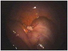
Polyps can be removed during hysteroscopy, and sent for histopathological analysis. Depending on the result, further action can be taken.

Sarcoglycanopathy: Symptoms & Treatments

Sarcoglycanopathy: A form of limb-girdle muscular dystrophy involving abnormalities of the sarcoglycan protein which results in progressive muscle weakness. The severity is greatly variably from wheelchair confinement at the age of 9 years to asymptomatic adults. Most tend to live to their third decade. More detailed information about the symptoms, causes, and treatmentsof Sarcoglycanopathy is available below.

 Symptoms of Sarcoglycanopathy:The list of signs and symptoms mentioned in various sources for Sarcoglycanopathyincludes the 18 symptoms listed below:

  • Delayed motor milestones
  • Calf muscle hypertrophy
  • Abnormal curvature of the spine
  • Weakness of pelvic muscles
  • Weakness of shoulder muscles
  • Wasting of pelvic muscles
  • High creatine kinase level
  • Toe walking
  • Muscle pain
  • Muscle cramps
  • Foot drop
  • Loss of mobility
  • Lordosis
  • Wasting of front of thigh
  • Wasting of back of thighs
  • Wasting of shoulder girdle muscles
  • Hip flexion contractures
  • Unusual smile


Treatments:

  • Family Practice
  • Pediatrics (Child Health Specialist)
  • Adolescent Medicine (Teen Health)
  • Geriatric Medicine (Senior Health)
  • Internal Medicine
  • Critical Care Medicine
  • Public Health

Doctors and specialists in areas related to Sarcoglycanopathy:

    • Muscle and Orthopedic Specialists:
      • Neuromusculoskeletal Medicine
      • Orthopedic Surgery


Pyeloplasy: Symptoms & Procedure

Pyeloplasty is an operation to correct a blockage that has occurred between the kidney and the ureter. The ureter is the tube which drains the kidney into the bladder. Removing the blockage allows urine to drain freely from the kidney to the bladder.
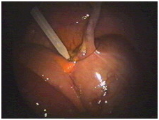
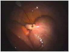
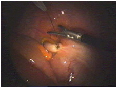
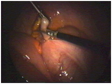
What is a Laparoscopic Pyeloplasty ? A Laparoscopic Pyeloplasty is a minimally invasive surgical procedure to correct a blockage or narrowing (puj obstruction) of the junction of the kidney to the tube draining urine.Why is Laparoscopic Pyeloplasty performed ?

Obstruction of the ureteropelvic junction (UPJ) can be caused by congenital abnormalities like horseshoe kidney, fibrous scarring due to stone or previous operation, a blood vessel which may cause it to kink or a stone that gets impacted in the upper part of ureter. This can cause damage to kidney tissue and eventually lead to pain, stones, infection, high blood pressure, deterioration of kidney function and kidney failure. Laparoscopic Pyeloplasty is the procedure of choice to relieve all these symptoms.

Who is an ideal candidate for Laparoscopic Pyeloplasty ?

If you have obstruction of the ureteropelvic junction (UPJ) which can be caused by congenital abnormalities like horseshoe kidney, fibrous scarring due to stone or previous operation, or if you have a blood vessel which may cause your ureteropelvic junction (UPJ) to kink or if you have a stone that gets impacted in the upper part of ureter, then you are an ideal candidate for Laparoscopic Pyeloplasty.

Preparation for surgery:
Medications to Avoid Prior to Surgery

Aspirin, Motrin, Ibuprofen, Advil, Alka Seltzer, Vitamin E, Ticlid, Coumadin, Lovenox, Celebrex, Voltaren, Vioxx, Plavix and some other arthritis medications can cause bleeding and should be avoided 1 week prior to the date of surgery (Please contact your surgeon’s office if you are unsure about which medications to stop prior to surgery. Do not stop any medication without contacting the prescribing doctor to get their approval).

Bowel Preparation and Clear Liquid Diet

Do not eat or drink anything after midnight the night before the surgery and drink one bottle of Magnesium Citrate (can be purchased at your local pharmacy) the evening before your surgery.Drink only clear fluids for a 24-hour period prior to the date of your surgery. Clear liquids are liquids that you are able to see through. Please follow the diet below.

Clear Liquid Diet
Remember not to eat or drink anything after midnight the evening before your surgery. Clear liquids are liquids that you are able to see through.

Please follow the diet below :

  • Water
  • Clear Broths (no cream soups, meat, noodles etc.)
  • Chicken broth
  • Beef broth
  • Juices (no orange juice or tomato juice)
  • Apple juice or apple cider
  • Grape juice
  • Cranberry juice
  • Tang
  • Hawaiian punch
  • Lemonade
  • Kool Aid
  • Gator Aid
  • Tea (you may add sweetener, but no cream or milk)
  • Coffee (you may add sweetener, but no cream or milk)
  • Clear Jello (without fruit)
  • Popsicles (without fruit or cream)
  • Italian ices or snowball (no marshmallow)
^ Back to Top

Q. How is the operation done ?

Laparoscopic Pyeloplasty India Surgery offers info on Laparoscopic Pyeloplasty India, Laparoscopic Pyeloplasty Surgery India


Figure : an illustration of the kidney and ureter before, during and following the procedure.


The first part of the operation is to give you an anaesthetic (put you to sleep) so that you will not be aware of anything whilst the operation is being performed.

The operation can take 3-4 hours, but can vary depending upon the individual. Three small cuts (approximately 1cm) are made in your stomach to perform the operation. A small plastic tube (stent) is placed inside the pipe leading from the kidney (ureter) at the beginning of the operation to bridge the surgical repair and help urine to drain freely from the kidney involved.

The tube will be left inside for 4-6 weeks and you will return as a day case to have this removed. This is done under local anaesthesia through the passage where you pass urine.

A tube (catheter) is placed into your bladder to allow urine to drain whilst you recover from your operation. The urine may have blood in it but this is normal and will clear in a day or two. The tube (catheter) will be removed once you are walking around, in a day or two.

You may also have a wound drain in your stomach to drain away any blood. This will be removed when there is little or no fluid draining from it.

Laparoscopic Pyeloplasty Surgery India, Cost Laparoscopic Pyeloplasty Surgery India


Figure: Traditional open kidney surgery is performed through an 8-12 inch incision extending from the ribs towards the abdomen. A portion of one of the ribs is usually removed as part of the surgery.


Laparoscopic Pyeloplasty India Surgery offers info on Laparoscopic Pyeloplasty India, Cost Laparoscopic Pyeloplasty Surgery India


Figure : An illustration of 3 incisions during the procedure


^ Back to Top
Advantages over open surgery
  • Less pain
  • Shorter hospital stay
  • Quicker recovery
  • Better cosmetic result

Q. What is the recovery period like following Laparoscopic Pyeloplasty ?

You will spend the next few hours following Laparoscopic Pyeloplasty, in the recovery room to monitor your vital signs and observe for any immediate post-operative complications. You will be hospitalized for 24 – 48 hours following Laparoscopic Pyeloplasty. Pain medication and antibiotics will be given post-operatively.

The stent will be removed 4 – 6 weeks after Laparoscopic Pyeloplasty. Avoid performing strenuous activities like lifting something heavy, jogging, treadmill or playing sports for at least 2 weeks after Laparoscopic Pyeloplasty. You will be instructed to move around, avoid getting constipated, do some very simple breathing exercises to help prevent respiratory infections. You will be able to perform all your routine daily activities in about 2 – 4 weeks time after Laparoscopic Pyeloplasty.

^ Back to Top
Potential Risks and Complications

Although this procedure has proven to be very safe, as in any surgical procedure there are risks and potential complications. The safety and complication rates are similar when compared to the open surgery.

Potential risks include : -

    • Bleeding : – Blood loss during this procedure is typically minor (less than 100 cc) and a blood transfusion is rarely required. If you are still interested in autologous blood transfusion (donating your own blood) prior to your surgery, you must make your surgeon aware. When the packet of information is mailed or given to you regarding your surgery, you will receive an authorization form for you to take to the Red Cross in your area.
    • Infection : – All patients are treated with broad-spectrum intravenous antibiotics prior to starting the surgery to decrease the chance of infection from occurring after surgery. If you develop any signs or symptoms of infection after the surgery (fever, drainage from your incision, urinary frequency, discomfort, pain or anything that you may be concerned about) please contact us at once.
    • Hernia : – Hernias at incision sites rarely occur since all keyhole incisions are closed carefully at the completion of your surgery.
    • Tissue / organ injury : – Although uncommon, possible injury to surrounding tissue and organs including bowel, vascular structures, spleen, liver, pancreas and gallbladder could require further surgery. Injury could occur to nerves or muscles related to positioning.
    • Conversion to open surgery : – this surgical procedure may require conversion to the standard open operation if extreme difficulty is encountered during the laparoscopic procedure. This could result in a larger standard open incision and possibly a longer recuperation period.
  • Failure to correct UPJ obstruction : – Roughly 3 % of patients undergoing this operation will have persistent blockage due to recurrent scarring. If this occurs additional surgery may be necessary.

Q. What is the outcome of Laparoscopic Pyeloplasty ?

Laparoscopic Pyeloplasty almost has a success rate of 95%, there is reduced hospital stay (one day in younger, healthy patients), faster healing and quicker return to normal activity and work.

Trigeminal neuralgia: Symptoms, Causes and Treatments

What Is Trigeminal Neuralgia?

Trigeminal neuralgia (TN), also called tic douloureux, is a condition that is characterized by intermittent, shooting pain in the face.

Trigeminal neuralgia affects the trigeminal nerve, one of the largest nerves in the head. The trigeminal nerve sends impulses of touch, pain, pressure, and temperature to the brain from the face, jaw, gums, forehead, and around the eyes.

What Causes Trigeminal Neuralgia?

The most frequent cause of trigeminal neuralgia is a blood vessel pressing on the nerve near the brain stem. Over time, changes in the blood vessels of the brain can result in blood vessels rubbing against the trigeminal nerve root. The constant rubbing with each heartbeat wears away the insulating membrane of the nerve, resulting in nerve irritation.

What Are the Symptoms of Trigeminal Neuralgia?

Trigeminal neuralgia causes a sudden, severe, electric shock-like, or stabbing pain that lasts several seconds. The pain can be felt on the face and around the lips, eyes, nose, scalp, and forehead. Symptoms can be brought on when a person is brushing the teeth, putting onmakeup, touching the face, swallowing, or even feeling a slight breeze.

Trigeminal neuralgia is often considered one of the most painful conditions seen in medicine. Usually, the pain is felt on one side of the jaw or cheek, but some people experience pain at different times on both sides. The attacks of pain may be repeated one after the other. They may come and go throughout the day and last for days, weeks, or months at a time. At times, the attacks can disappear for months or years. The disorder is more common in women than in men and rarely affects anyone younger than age 50.

How Is Trigeminal Neuralgia Diagnosed?

Magnetic resonance imaging (MRI) can be used to determine whether a tumor or multiple sclerosis is irritating the trigeminal nerve. Otherwise, no test can determine with certainty the presence of trigeminal neuralgia. Tests can, however, help rule out other causes of facial disorders. Trigeminal neuralgia usually is diagnosed based on the patient’s description of the symptoms.

How Is Trigeminal Neuralgia Treated?

Trigeminal neuralgia can be treated with antiseizure drugs such as Tegretol (carbamazepine) or Neurontin (gabapentin). The medications Klonopin (clonazepam) and Depakote (divalproex sodium) may also be effective and may be used in combination with other drugs to achieve pain relief. Some antidepressant drugs also have significant pain relieving effects.

If medications are ineffective or if they produce undesirable side effects, neurosurgical procedures are available to relieve pressure on the nerve or to reduce nerve sensitivity.

Surgery : Micro vascular decompression so patient will be free of all symptoms.

Some patients report having reduced or relieved pain by means of alternative medical therapies such as acupuncture, chiropractic adjustment, self-hypnosis, or meditation.

Myopathy : symptoms, causes and treatments

Signs and Symptoms of Myopathy:

Although symptoms depend on the type of myopathy, some generalizations can be made. Skeletal muscle weakness is the hallmark of most myopathies, with some noticeable exceptions, such as myotonia and paramyotonia congenita. In these two inheritable muscular disorders, the muscles become enlarged, rather than weakened and atrophied, and do not relax after contracting.In most myopathies, weakness occurs primarily in the muscles of the shoulders, upper arms, thighs, and pelvis (proximal muscles). In some cases, the distal muscles of the hands and feet may be involved during the advanced stage of disease.

Other typical symptoms of muscle disease include the following:

  1. Aching.
  2. Cramping
  3. Pain
  4. Stiffness
  5. Tenderness\
  6. Tightnes

Initially, individuals may feel fatigued doing very light physical activity. Walking and climbing stairs may be difficult because of weakness in the pelvic and leg muscles that stabilize the trunk.
Patients often find it difficult to rise from a chair. As the myopathy progresses, there may be muscle wasting.

Treatment for Myopathy:

Treatment for myopathies depends on the cause. The goals of myopathy treatment are to slow progression of the disease and relieve symptoms.

Treatments range from drug therapy for muscular dystrophies and inflammatory myopathies to avoiding situations that work the muscles too hard for metabolic myopathies. Some physicians recommend that patients with myopathy keep their weight down (a lighter body demands less work from the muscles) and avoid overexerting the muscles.
When breathing problems develop, an incentive spirometer can help improve breathing function in some patients. Unfortunately, there is no way to strengthen the breathing muscles.
Treatment for Muscular Dystrophies:
The goals of MD treatment are to slow progression of disease and relieve symptoms. Duchenne MD and Becker MD are the subjects of current medical research and clinical trials may be available for patients with either disease.
Corticosteroids (e.g., deflazacort, prednisone) seem to be the most effective medications. Both improve strength and walking ability for about 6 months in boys with Duchenne dystrophy. Following initial improvement, further progression of the disease may be delayed for 3 to 5 years in some cases.
Prolonged use of corticosteroids can cause severe side effects including the following:

  • Bone loss (osteoporosis)
  • Depression
  • High blood pressure (hypertension)
  • Thinning of the skin
  • Weight gain

Calcium supplements and antidepressants may be prescribed to counteract these side effects.
Preventive treatment for permanent contraction of a muscle (contractures) includes physical therapy and bracing. There are currently no drugs available to prevent or treat contractures.
Heel cord surgery (also called tendon release) and spine-straightening surgery (i.e., rod insertion) may be necessary in cases of severe contractures. Heel cord surgery is performed when the patient is still able to walk. Braces are usually required following surgery.

Treatment for Endocrine Myopathies:

Often, treating the underlying condition helps relieve muscle weakness and pain associated with endocrine myopathies.
Treatment for Inflammatory Myopathies:

Inflammatory myopathies, such as polymyositis and dermatomyositis, are usually treated with drugs that suppress the action of the immune system. Prednisone is most commonly used to treat inflammatory myopathies. It is used initially in high doses (up to 100mg/day) and then slowly tapered to the lowest possible dose that relieves symptoms. Long-term use of prednisone can cause severe side effects, including bone loss, depression, and high blood pressure.

Treatment for Metabolic Myopathies:

The primary goal in treating metabolic myopathies is to avoid situations that tax the muscles and promote muscle pain and weakness, like strenuous exercise.

Dengue fever: Symptoms, causes and treatments

Dengue Symptoms and Signs

Primary symptoms of dengue appear three to 15 days after the mosquito bite and include the following:

  • high fever and severe headache,
  • with severe pain behind the eyes that is apparent when trying to move the eyes.

Other associated symptoms are:

  • joint pain,
  • muscle and bone pain,
  • rash,
  • and mild bleeding.

Many affected people complain of high knee pain and low back pain.

Dengue infection is a leading cause of death and sickness in tropical and subtropical parts of the world. Dengue is caused by one of four viruses that are transmitted by the bite of an infected mosquito. Dengue hemorrhagic fever is a more serious form of dengue infection.

Primary symptoms of dengue appear three to 15 days after the mosquito bite and include high fever and severe headache, with severe pain behind the eyes that is apparent when trying to move the eyes. Other associated symptoms are joint pain, muscle and bone pain, rash, and mild bleeding. Many affected people complain of low back pain. The lymph nodes of the neck and groin may be swollen. Young children and people infected for the first time typically have milder symptoms than older children and adults.

Dengue hemorrhagic fever starts with the typical signs and symptoms of dengue as described above. The fever lasts from two to seven days. After the fever begins to abate, symptoms occur that are related to increased permeability of the capillary blood vessels. These symptoms can include severe abdominal pain, prolonged vomiting, and breathing problems. Bleeding tendencies, including easy bruising, nosebleeds, bleeding gums, skin hemorrhages, and even internal bleeding may occur. The disease may progress to failure of the circulatory system, leading to shock and death.

How is dengue fever contracted?

The virus is contracted from the bite of a striped Aedes aegypti mosquito that has previously bitten an infected person. The mosquito flourishes during rainy seasons but can breed in water-filled flower pots, plastic bags, and cans year-round. One mosquito bite can cause the disease.

The virus is not contagious and cannot be spread directly from person to person. It is mosquito-borne, so there must be a person-to-mosquito-to-another-person pathway. The full life cycle of the virus involves the mosquito as the vector (transmitter) and the human as the source of infection.

What is the incubation period for dengue fever?

After being bitten by a mosquito carrying the virus, the incubation period for dengue fever ranges from three to 15 (usually five to eight) days before the signs and symptoms of dengue appear in stages.

Treatments:

It is found that at present there is no specific vaccine which can kill the virus of dengue fever not even a single antivirus tablets have been manufactured.
Dengue fever is treated by a medicine which is known as Paracetamol, an anti-pyretic.

The pain during the dengue fever in the bones can be relaxed through the pain killing tablets or by analgesics.

Patient must be hospitalized when there is DHF and DSS. The rate mortality is found to be high, about 50%, in the case of not getting hospitalized timely. With the help of hospitals, the treatment makes the mortality rate down to near about 3%. There are some other supportive treatments which help in the treatment of patient suffer from dengue. It includes Fluid of intravenous replacement which helps in preventing the shock.

Now-a-days, vaccines start getting developed for the treatment against four serotypes of dengue. They proved to be the best way to cure the disease of dengue.

In many endemic countries, it is found that the measures in preventing from mosquitoes have been failed. Patients start getting more prone to dehydration after drinking plenty of liquid things. In some of the cases, the prevention can be done through oxygen. And the use of Steroids is not considered useful.

Dengue Fever Testing:

How is it used?

The testing of dengue fever is done to determine whether the recent potential exposure and symptoms of the person body has been infected by dengue or not. It is difficult to diagnose the infection without doing laboratory tests as the symptoms initially may resemble like diseases like malaria. There are 2 types of testing available:

  • Antibody tests— primarily these tests are used to help in diagnosing a recent or current infection. 2 different antibodies classes are detected by them that are produced by body in response to the infection of dengue fever, IgM and IgG. A combination of both these tests is required for diagnosis due to reason that the immune system of the body produces different antibodies levels during the time of illness. IgM antibodies are 1st produced and tests for it are most effective at the time when they are performed 7-10 days later the exposure. For some weeks, the blood levels rise and then decrease gradually. After some months, the IgM antibodies gets fall down below the detectable levels. They are produced slower than the rate of infection. The level rises typically with the acute infection, it stabilizes and then it persists long-term. All those who get exposed to it prior to current infection, maintains IgG antibodies level in the level that affects the diagnostic results’ interpretation.
  • Molecular testing (PCR, polymerase chain reaction)— this test detects dengue virus’s genetic material in blood till 5 daysof the symptom onset.

When is it ordered?

Testing might be ordered at the time when people have symptoms and signs associated with dengue at tropical locations where it is present. Main symptoms and signs include:

  • Low count of white blood cells
  • Easy bruising
  • Nose and gum bleeds
  • Bone, muscle or joint pain
  • Pain behind eyes or severe headache
  • Sudden high fever (40°C or 104°F)

Usually testing is ordered within 1 to 2 weeks of the starting of the symptoms for detecting the infection. If the antibody testing is done, an additional sample of the blood is collected after 2 weeks of the symptoms in order to determine whether the level of antibody is rising.

What does the test result mean?

  • Antibody testing— the test may have negative or positive result or might be reported as the antibody titer having an interpretation of the type of antibody (IgM or IgG) present.

Positive IgG and IgM tests detected for the dengue antibodies in the blood means that the person has become infected within recent weeks with the dengue virus. If IgG comes positive but IgM is negative or low, then it is probably that the person has got an infection earlier in past. If the titer of dengue IgG antibody increases from 1:4 to 1:64 between the initial samples then it is likely to have recent infection.

Negative IgG and/or IgM antibodies means that the person tested is not having any infection and the symptoms are because of any other reason or the antibody level may be very low for measuring. The individual might still have dengue infection- it might be just that it is very soon after the virus exposure to give a detectable antibody level.

The table as follows summarizes the results seen with the antibody testing:

IgM Result IgG Result Possible Interpretation
Positive Negative Current infection
Positive Positive Current infection
Low or negative or not tested Four-fold increase in samples taken 2-4 weeks apart Recent infection
Low or negative Positive Past infection
Negative Negative Too soon after initial exposure for antibodies to develop or symptoms due to another cause


  • Molecular testing— the most reliable of all diagnosis is the PCR test that is considered as it detects the virus presence in the body but it is not available widely. A positive PCR result is considered conclusive. A PCR test having negative result indicates no infection present or low level of virus to detect if the test gets performed after the period of 5-day window since the time when the virus being present in the blood sample is collected. If recent exposures are suspected, then repeating the tests may be warranted later.


Which are the food reduce the dengue fever:

  • Kiwi fruit, fig, Pomogranates, papaya etc( these will increase the blood count )
  • Juice made from young leaves of papaya.(daily have one time 5ml or 3ml- continue for 3-5 days).


Pages:1...66676869707172...92