


Breast cancer definition
Breast cancer is a malignant tumor (a collection of cancer cells) arising from the cells of the breast. Although breast cancer predominantly occurs in women, it can also affect men. This article deals with breast cancer in women.
What are the statistics on male breast cancer?
Breast cancer is rare in men but typically has a significantly worse outcome. This is partially related to the often late diagnosis of male breast cancer, when the cancer has already spread.
Symptoms are similar to the symptoms in women, with the most common symptom being a lump or change in skin of the breast tissue or nipple discharge. Although it can occur at any age, male breast cancer usually occurs in men over 60 years of age.
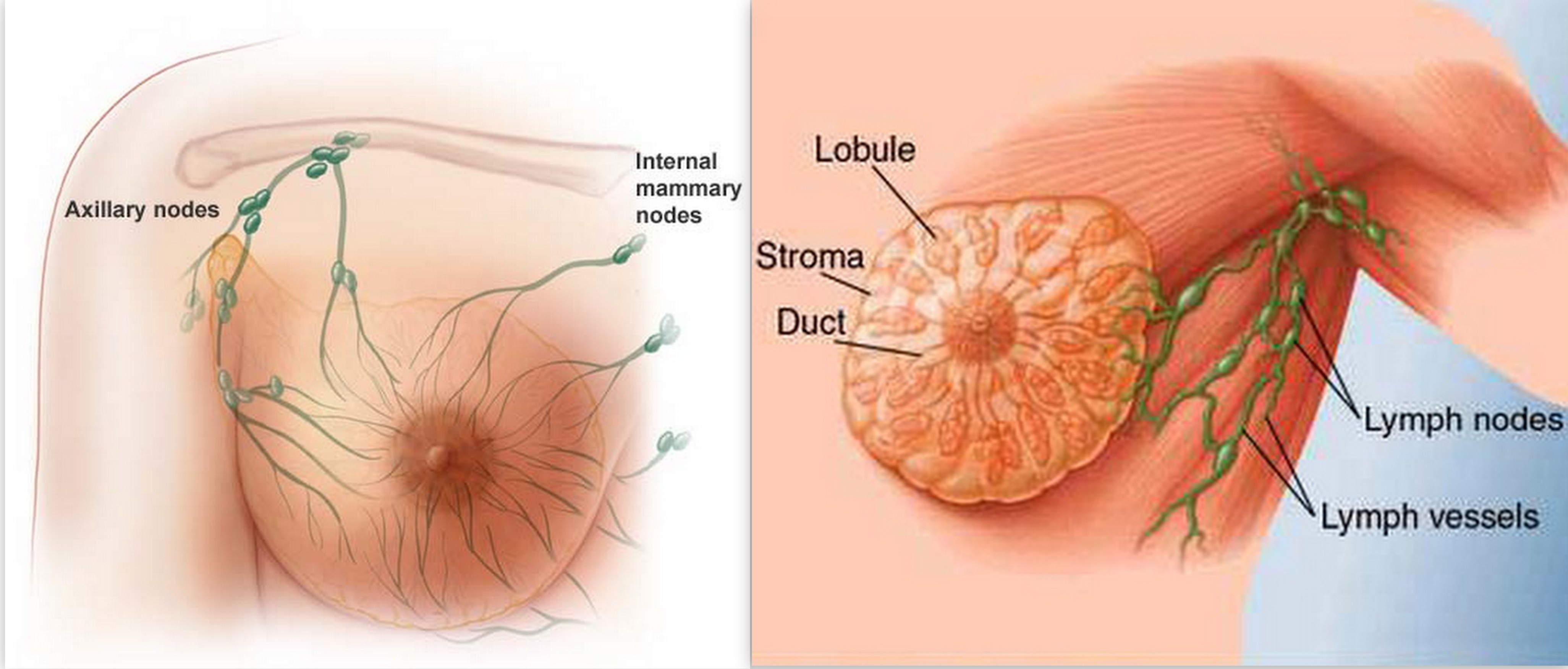
What are the different types of breast cancer? Where does breast cancer come from?
There are many types of breast cancer. Some are more common than others, and there are also combinations of cancers. Some of the most common types of cancer are as follows:
- Ductal carcinoma in situ: The most common type of noninvasive breast cancer is ductal carcinoma in situ (DCIS). This type of cancer has not spread and therefore usually has a very high cure rate.
- Invasive ductal carcinoma: This cancer starts in a duct of the breast and grows into the surrounding tissue. It is the most common form of breast cancer. About 80% of invasive breast cancers are invasive ductal carcinoma.
- Invasive lobular carcinoma: This breast cancer starts in the glands of the breast that produce milk. Approximately 10% of invasive breast cancers are invasive lobular carcinoma.
- The remainder of breast cancers are much less common and include the following:
- Mucinous carcinoma are formed from mucus-producing cancer cells. Mixed tumors contain a variety of cell types.
- Medullary carcinoma is an infiltrating breast cancer that presents with well-defined boundaries between the cancerous and noncancerous tissue.
- Inflammatory breast cancer: This cancer makes the skin of the breast appear red and feel warm (giving it the appearance of an infection). These changes are due to the blockage of lymph vessels by cancer cells.
- Triple-negative breast cancers: This is a subtype of invasive cancer with cells that lack estrogen and progesterone receptors and have no excess of a specific protein (HER2) on their surface. It tends to appear more often in younger women and African-American women.
- Paget’s disease of the nipple: This cancer starts in the ducts of the breast and spreads to the nipple and the area surrounding the nipple. It usually presents with crusting and redness around the nipple.
- Adenoid cystic carcinoma: These cancers have both glandular and cystic features. They tend not to spread aggressively and have a good prognosis.
- Lobular carcinoma in situ: This is not a cancer but an area of abnormal cell growth that can lead to invasive breast cancer later in life.

What causes breast cancer?
There are many risk factors that increase the chance of developing breast cancer. Although we know some of these risk factors, we don’t know the cause of breast cancer or how these factors cause the development of a cancer cell.
We know that normal breast cells become cancerous because of mutations in the DNA, and although some of these are inherited, most DNA changes related to breast cells are acquired during one’s life.
Proto-oncogenes help cells grow. If these cells mutate, they can increase growth of cells without any control. Such mutations are referred to as oncogenes. Such uncontrolled cell growth can lead to cancer.
What about antiperspirants or deodorants as causes of breast cancer?
Research has shown that parabens (a preservative used in deodorants) can build up in breast tissues. However, this study did not show that parabens cause breast cancer or that the parabens (which are found in many other products) were linked to the use of deodorants.
A 2002 study did not show any increased risk for breast cancer in women using an underarm deodorant or antiperspirant. A 2003 study showed an earlier age for breast cancer diagnosis in women who shaved their underarms more frequently and used underarm deodorants.
Treatments:
Patients with breast cancer have many treatment options. Most treatments are adjusted specifically to the type of cancer and the staging group. Treatment options are being adjusted frequently and your health care provider will have the information on the current standard of care available. Treatment options should be discussed with a health care team. The following are the basic treatment modalities used in the treatment of breast cancer.
Surgery
Most women with breast cancer will require surgery. Broadly, the surgical therapies for breast cancer can be divided into breast-conserving surgery and mastectomy.
Breast-conserving surgery
This surgery will only remove part of the breast (sometimes referred to as partial mastectomy). The extent of the surgery is determined by the size and location of the tumor.
In a lumpectomy, only the breast lump and some surrounding tissue is removed. The surrounding tissue (surgical margins) are inspected for cancer cells. If no cancer cells are found, this is called “negative” or “clear margins.” Frequently, radiation therapy is given after lumpectomies.
Mastectomy
During a mastectomy (sometimes also referred to as a simple mastectomy), all the breast tissue is removed. If immediate reconstruction is considered, a skin-sparing mastectomy is sometimes performed. In this surgery, all the breast tissue is removed as well, but the overlying skin is preserved.
Radical mastectomy
During this surgery, the surgeon removes the axillary lymph nodes as well as the chest wall muscle in addition to the breast. This procedure is done much less frequently than in the past, as in most cases, a modified radical mastectomy is as effective.
Modified radical mastectomy
This surgery removes the axillary lymph nodes in addition to the breast tissue. Depending on the stage of the cancer, a health care team might give someone a choice between a lumpectomy and a mastectomy. Lumpectomy allows sparing of the breast but usually requires radiation therapy afterward. If lumpectomy is indicated, long-term follow-up shows no advantage of a mastectomy over the lumpectomy.
Preventive surgery
For a small group of patients who have a very high risk of breast cancer, surgery to remove the breasts may be an option. Although this reduces the risk significantly, a small chance of developing cancer remains.
Double mastectomy is a surgical option to prevent breast cancer. This prophylactic (preventive) surgery can decrease the risk of breast cancer by about 90% for women at moderate to high risk for breast cancer.
Such an approach should be carefully discussed with a health care team.
The discussion about whether to undergo any preventive surgery should include
- genetic testing for BRCA1 or BRCA2 gene mutations,
- full review of risk factors,
- family history of cancer and specifically breast cancer, and
- other preventive options such as medications.
Radiation therapy
Radiation therapy destroys cancer cells with high energy rays. There are two ways to administer radiation therapy.
External beam radiation
This is the usual way radiation therapy is given for breast cancer. A beam of radiation is focused onto the affected area by an external machine. The extent of the treatment is determined by a health care team and is based on the surgical procedure performed and whether lymph nodes were affected or not.
The local area will usually be marked after the radiation team has determined the exact location for the treatments. Usually, the treatment is given five days a week for five to six weeks.
Brachytherapy
This form of delivering radiation uses radioactive seeds or pellets. Instead of a beam from the outside delivering the radiation, these seeds are implanted into the breast next to the cancer.
Chemotherapy
Chemotherapy is treatment of cancers with medications that travel through the bloodstream to the cancer cells. These medications are given either by intravenous injection or by mouth.
Chemotherapy can have different indications and may be performed in different settings as follows:
- Adjuvant chemotherapy: If surgery has removed all the visible cancer, there is still the possibility that cancer cells have broken off or are left behind. If chemotherapy is given to assure that these small amounts of cells are killed as well, it is called adjunct chemotherapy. Chemotherapy is not given in all cases, since some women have a very low risk of recurrence even without chemotherapy, depending upon the tumor type and characteristics.
- Neoadjuvant chemotherapy: If chemotherapy is given before surgery, it is referred to as neoadjuvant chemotherapy. Although there seems to be no advantage to long-term survival whether the therapy is given before or after surgery, there are advantages to see if the cancer responds to the therapy and by shrinking the cancer before surgical removal.
- Chemotherapy for advanced cancer: If the cancer has metastasized to distant sites in the body, chemotherapy can be used for treatment. With cases of metastatic breast cancer, the health-care team will need to determine the most appropriate length of treatment.
There are many different chemotherapeutic agents that are either given alone or in combination. Usually, these drugs are given in cycles with certain treatment intervals followed by a rest period. The cycle length and rest intervals differ from drug to drug.
Hormone therapy
This therapy is often used to help reduce the risk of cancer reoccurrence after surgery, but it can also be used as adjunct treatment.
Estrogen (a hormone produced by the ovaries) promotes the growth of a few breast cancers, specifically those containing receptors for estrogen (ER positive) or progesterone (PR positive). The following drugs are used in hormone therapy:
- Tamoxifen (Nolvadex): This drug prevents estrogen from binding to estrogen receptors on breast cells.
- Toremifene (Fareston) works similar to Tamoxifen and is only indicated in metastatic breast cancer.
- Fulvestrant (Faslodex): This drug eliminates the estrogen receptor and can be used even if tamoxifen is no longer useful.
- Aromatase inhibitors: They stop estrogen production in postmenopausal women. Examples are letrozole (Femara), anastrozole (Arimidex), and exemestane (Aromasin).
Targeted therapy
As we are learning more about gene changes and their involvement in causing cancer, drugs are being developed that specifically target the cancer cells. They tend to have fewer side effects than chemotherapy (as they target only the cancer cells) but usually are still used in adjunct with chemotherapy.
Alternative treatments
Whenever a disease has the potential for much harm and death, physicians search for alternative treatments. As a patient or the loved one of a patient, there may be an inclination to try everything and leave no option unexplored. The danger in this approach is usually found in the fact that the patient might not avail themselves of existing, proven therapies. One should discuss any interest in alternative treatments with a health care team and together explore the different options.
.jpg)


%2Band%2Bcarbuncles.jpg)
