Global Treatment Services Pvt. Ltd.

Global Treatment Services

Pulmonary Hypertension: Symptoms, Causes & Treatments

Pulmonary hypertension is a type of high blood pressure that affects the arteries in the lungs and the right side of your heart.

Pulmonary hypertension begins when tiny arteries in your lungs, called pulmonary arteries, and capillaries become narrowed, blocked or destroyed. This makes it harder for blood to flow through your lungs, and raises pressure within your lungs’ arteries. As the pressure builds, your heart’s lower right chamber (right ventricle) must work harder to pump blood through your lungs, eventually causing your heart muscle to weaken and eventually fail.

Pulmonary hypertension is a serious illness that becomes progressively worse and is sometimes fatal. Although pulmonary hypertension isn’t curable, treatments are available that can help lessen symptoms and improve your quality of life.

Respiratory system

Symptoms:

The signs and symptoms of pulmonary hypertension in its early stages may not be noticeable for months or even years. As the disease progresses, symptoms become worse.

Pulmonary hypertension symptoms include:

  • Shortness of breath (dyspnea), initially while exercising and eventually while at rest
  • Fatigue
  • Dizziness or fainting spells (syncope)
  • Chest pressure or pain
  • Swelling (edema) in your ankles, legs and eventually in your abdomen (ascites)
  • Bluish color to your lips and skin (cyanosis)
  • Racing pulse or heart palpitations

Causes:

Your heart has two upper and two lower chambers. Each time blood passes through your heart, the lower right chamber (right ventricle) pumps blood to your lungs through a large blood vessel (pulmonary artery). In your lungs, the blood releases carbon dioxide and picks up oxygen. The oxygen-rich blood then flows through blood vessels in your lungs (pulmonary arteries, capillaries and veins) to the left side of your heart.

Ordinarily, the blood flows easily through the vessels in your lungs, so blood pressure is usually much lower in your lungs. With pulmonary hypertension, the rise in blood pressure is caused by changes in the cells that line your pulmonary arteries. These changes cause extra tissue to form, eventually narrowing or completely blocking the blood vessels, making the arteries stiff and narrow. This makes it harder for blood to flow, raising the blood pressure in the pulmonary arteries.

Idiopathic pulmonary hypertension

When an underlying cause for high blood pressure in the lungs can’t be found, the condition is called idiopathic pulmonary hypertension (IPH).

Some people with IPH may have a gene that’s a risk factor for developing pulmonary hypertension. But in most people with idiopathic pulmonary hypertension, there is no recognized cause of their pulmonary hypertension.

Secondary pulmonary hypertension

Pulmonary hypertension that’s caused by another medical problem is called secondary pulmonary hypertension. This type of pulmonary hypertension is more common than is idiopathic pulmonary hypertension. Causes of secondary pulmonary hypertension include:

  • Blood clots in the lungs (pulmonary emboli)
  • Chronic obstructive pulmonary diseases, such as emphysema
  • Connective tissue disorders, such as scleroderma or lupus
  • Sleep apnea and other sleep disorders
  • Heart abnormalities you’re born with (congenital heart defects)
  • Sickle cell anemia
  • Chronic liver disease (cirrhosis)
  • AIDS
  • Lung diseases such as pulmonary fibrosis, a condition that causes scarring in the tissue between the lungs’ air sacs (interstitium)
  • Left-sided heart failure
  • Living at altitudes higher than 8,000 feet (2,438 meters)
  • Climbing or hiking to altitudes higher than 8,000 feet (2,438 meters) without acclimating first
  • Use of certain stimulant drugs, such as cocaine

Eisenmenger syndrome and pulmonary hypertension

Eisenmenger syndrome, a type of congenital heart defect, causes pulmonary hypertension. It is most commonly caused by a large hole in your heart between the two lower heart chambers (ventricles), called a ventricular septal defect (VSD). This hole in your heart causes blood to circulate abnormally in your heart. Oxygen-carrying blood (red blood) mixes with oxygen-poor blood (blue blood). The blood then returns to your lungs instead of going to the rest of your body, increasing the pressure in the pulmonary arteries and causing pulmonary hypertension.


 Treatments:

Medications:


  • Blood vessel dilators (vasodilators). Vasodilators open narrowed blood vessels. One of the most commonly prescribed vasodilators for pulmonary hypertension is epoprostenol (Flolan). The drawback to epoprostenol is that its effects last only a few minutes. This drug is continuously injected through an intravenous (IV) catheter via a small pump that you wear in a pack on your belt or shoulder. This means that you’ll learn to prepare your own medication mixture, operate the pump and care for the IV catheter. You’ll need comprehensive follow-up care. Potential side effects of epoprostenol include jaw pain, nausea, diarrhea, leg cramps, as well as pain and infection at the IV site.Another form of the drug, iloprost (Ventavis), avoids many of these problems. Iloprost can be inhaled every three hours through a nebulizer, a machine that vaporizes your medication, making it far more convenient and less painful to use. And because it’s inhaled, it goes directly to the lungs. Side effects associated with iloprost include chest pain — often accompanied by headache and nausea — and breathlessness.
  • Endothelin receptor antagonists. These medications reverse the effect of endothelin, a substance in the walls of blood vessels that causes them to narrow. One of these medications, bosentan (Tracleer), may improve your energy level and symptoms. The drug isn’t for pregnant women. If you take bosentan, you’ll need monthly liver monitoring, because the drug can damage your liver. Ambrisentan (Letairis) is another medication that stops the narrowing of your blood vessels. This drug can cause serious liver damage if not taken appropriately, and it shouldn’t be taken by pregnant women. Before taking the drug, tell your doctor about any liver condition you have.
  • Sildenafil and tadalafil. Sildenafil (Revatio, Viagra) and tadalafil (Cialis, Adcirca) are sometimes used to treat pulmonary hypertension. These drugs work by opening the blood vessels in the lungs to allow blood to flow through more easily. Side effects include upset stomach, dizziness and vision problems.
  • High-dose calcium channel blockers. These drugs help relax the muscles in the walls of your blood vessels. They include medications such as amlodipine (Norvasc), diltiazem (Cardizem, Tiazac, others) and nifedipine (Adalat, Procardia). Although calcium channel blockers can be effective, only a small number of people with pulmonary hypertension respond to them.
  • Anticoagulants. Your doctor is likely to prescribe the anticoagulant warfarin (Coumadin, Jantoven) to help prevent the formation of blood clots within the small pulmonary arteries. Because anticoagulants prevent normal blood coagulation, they increase your risk of bleeding complications. Take warfarin exactly as prescribed, because warfarin can cause severe side effects if taken incorrectly. If you’re taking warfarin, your doctor will ask you to have periodic blood tests to check how well the drug is working. Many other drugs, herbal supplements and foods can interact with warfarin, so be sure your doctor knows all of the medications you’re taking.
  • Diuretics. Commonly known as water pills, these medications help eliminate excess fluid from your body. This reduces the amount of work your heart has to do. They also may be used to limit fluid buildup in your lungs.
  • Oxygen. Your doctor may suggest that you sometimes breathe pure oxygen, a treatment known as oxygen therapy, to help treat pulmonary hypertension, especially if you live at a high altitude or have sleep apnea. Some people with pulmonary hypertension eventually require constant oxygen therapy.

Surgeries

  • Atrial septostomy. If medications don’t control your pulmonary hypertension, this open-heart surgery may be an option. In an atrial septostomy, a surgeon will create an opening between the left and right chambers of your heart to relieve the pressure on the right side of your heart. Atrial septostomy can have serious complications, including heart rhythm abnormalities (arrhythmias).
  • Transplantation. In some cases, a lung or heart-lung transplant may be an option, especially for younger people who have idiopathic pulmonary hypertension. Major risks of any type of transplantation include rejection of the transplanted organ and serious infection, and you must take immunosuppressant drugs for life to help reduce the chance of rejection.

Anosmia: Causes, Symptoms & Treatments

Fortunately, for most people, anosmia is a temporary nuisance caused by a severely stuffy nose from a cold. Once the cold runs its course, a person’s sense of smell returns.But for some people, including many elderly, the loss of a sense of smell may persist. In addition, anosmia can be a sign of a more serious medical condition. Any ongoing problems with smell should be checked out by a doctor.

The Basics of Smell

A person’s sense of smell is driven by certain processes. First, a molecule released from a substance (such as fragrance from a flower) must stimulate special nerve cells (called olfactory cells) found high up in the nose. These nerve cells then send information to the brain, where the specific smell is identified. Anything that interferes with these processes, such as nasal congestion, nasal blockage, or damage to the nerve cells themselves, can lead to loss of smell.

The ability to smell also affects our ability to taste. Without the sense of smell, our taste buds can only detect a few flavors, and this can affect your quality of life.

Anosmia Causes

Nasal congestion from a cold, allergy, sinus infection, or poor air quality is the most common cause of anosmia. Other anosmia causes include:

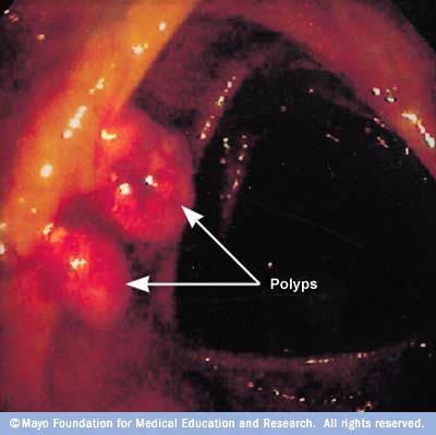
  • Nasal polyps — small noncancerous growths in the nose and sinusesthat block the nasal passage.
  • Injury to the nose and smell nerves from surgery or head trauma.
  • Exposure to toxic chemicals, such as pesticides or solvents.
  • Certain medications, including antibiotics, antidepressants, anti-inflammatory medication, heart medications, and others.
  • Cocaine abuse.
  • Old age. Like vision and hearing, your sense of smell can become weaker as you age. In fact, one’s sense of smell is most keen between the ages of 30 and 60 and begins to decline after age 60.
  • Certain medical conditions, such as Alzheimer’s disease, Parkinson’s disease, multiple sclerosis, nutritional deficiencies, congenital conditions, and hormonal disturbances.
  • Radiation treatment of head and neck cancers.

Anosmia Symptoms

The obvious sign of anosmia is a loss of smell. Some people with anosmia notice a change in the way things smell. For example, familiar things begin to lack odor.

Anosmia Diagnosis

If you experience a loss of smell that you can’t attribute to a cold or allergy or which doesn’t get better after a week or two, tell your doctor. Your doctor can take a look inside your nose with a special instrument to see if a polyp or growth is impairing your ability to smell or if an infection is present.

Further testing by a doctor who specializes in nose and sinus problems an ear, nose, and throat doctor (ENT, or an otolaryngologist) — may be needed to determine the cause of anosmia. A CT scan may be necessary so that the doctor can get a better look of the area.

Anosmia Treatments

If nasal congestion from a cold or allergy is the cause of anosmia, treatment is usually not needed, and the problem will get better on its own. Short-term use of over-the-counter decongestants may open up your nasal passages so that you can breathe easier. However, if the congestion gets worse or does not go away after a few days, see your doctor. You may have an infection and need antibiotics, or another medical condition may be to blame.

Endometriosis: Causes, Symptoms & Treatments

What is endometriosis?

Endometriosis is the abnormal growth of endometrial tissue similar to that which lines the interior of the uterus, but in a location outside of the uterus. Endometrial tissue is shed each month during menstruation. Areas of endometrial tissue found in ectopic locations are called endometrial implants. These lesions are most commonly found on the ovaries, the Fallopian tubes, the surface of the uterus, the bowel, and on the membrane lining of the pelvic cavity (i.e. the peritoneum). They are less commonly found to involve the vagina, cervix, and bladder. Rarely, endometriosis can occur outside the pelvis. Endometriosis has been reported in the liver, brain, lung, and old surgical scars. Endometrial implants, while they may become problematic, are usually benign (i.e. non-cancerous).

What are the stages of endometriosis?

Endometriosis is classified into one of four stages (I-minimal, II-mild, III-moderate, and IV-severe) based upon the exact location, extent, and depth of the endometriosis implants as well as the presence and severity of scar tissue and the presence and size of endometrial implants in the ovaries. Most cases of endometriosis are classified as minimal or mild, which means there are superficial implants and mild scarring. Moderate and severe endometriosis typically result in cysts and more severe scarring. The stage of endometriosis is not related to the degree of symptoms a woman experiences, but infertility is common with stage IV endometriosis.

What are the signs and symptoms endometriosis?

Most women who have endometriosis, in fact, do not have symptoms. Of those who do, the most common include:

  • Pain (usually pelvic) that usually occurs just before menstruation and lessens after menstruation
  • Painful sexual intercourse
  • Cramping during intercourse
  • Cramping or pain during bowel movements or urination
  • Infertility
  • Pain with pelvic examinations

The intensity of the pain can vary from month to month, and can vary greatly among affected individuals. Some women experience progressive worsening of symptoms, while others can have resolution of pain without treatment.

Pelvic pain in women with endometriosis depends partly on where endometrial implants of endometriosis are located.

  • Deeper implants and implants in areas of high nerve density are more apt to produce pain.
  • The implants may also release substances into the bloodstream which are capable of eliciting pain.
  • Pain can result when endometriotic implants incite scarring of surrounding tissues. There appears to be no relationship between severity of pain and the amount of anatomical disease which is present.

Endometriosis can be one of the reasons for infertility for otherwise healthy couples. When laparoscopic examinations are performed during evaluations for infertility, implants are often found in individuals who are totally asymptomatic. The reasons diminished fertility in many patients with endometriosis are not understood. Endometriosis may incite scar tissue formation within the pelvis. If the ovaries and Fallopian tubes are involved, the mechanical processes involved in the transfer of fertilized eggs into the tubes may be altered. Alternatively, the endometriotic lesions may produce inflammatory substances which adversely affect ovulation, fertilization, and implantation.

Other symptoms that can be related to endometriosis include

  • lower abdominal pain,
  • diarrhea and/or constipation,
  • low back pain,
  • chronic fatigue
  • irregular or heavy menstruation,
  • painful urination, or
  • bloody urine (particularly during menstruation).

Rare symptoms of endometriosis include chest pain or coughing blood due to endometriosis in the lungs and headache and/or seizures due to endometriosis in the brain.

Does endometriosis increase a woman’s risk of getting cancer?

Some studies have postulated that women with endometriosis have an increased risk for development of certain types of ovarian cancer, known as epithelial ovarian cancer (EOC). This risk is highest in women with both endometriosis and primary infertility (those who have never conceived a pregnancy). The use of combination oral contraceptive pills (OCPs), which are sometimes used in the treatment of endometriosis, appears to significantly reduce this risk.

The reasons for the association between endometriosis and ovarian epithelial cancer are not clearly understood. One theory is that the endometriosis implants themselves undergo malignanttransformation to cancer. Another possibility is that the presence of endometriosis may be related to other genetic or environmental factors that serve to increase a women’s risk of developing ovarian cancer.

What causes endometriosis?

The cause of endometriosis is unknown. One theory is that the endometrial tissue is deposited in unusual locations by the retrograde flow of menstrual debris through the Fallopian tubes into the pelvic and abdominal cavities. The cause of this retrograde menstruation is not clearly understood. It is clear that retrograde menstruation is not the only cause of endometriosis, as many women who have retrograde menstruation do not develop the condition.

Another possibility is that areas lining the pelvic organs possess primitive cells that are able to develop into other forms of tissue, such as endometrium. (This process is termed coelomic metaplasia.)

It is also likely the direct transfer of endometrial tissues at the time of surgery may be responsible for the endometriosis implants occasionally found in surgical scars (for example, episiotomy or Cesarean section scars). Transfer of endometrial cells via the bloodstream or lymphatic system is the most plausible explanation for the rare cases of endometriosis that are found in the brain and other organs remote from the pelvis.

Finally, there is evidence that some women with endometriosis have an altered immune response in women with endometriosis, which may affect the body’s natural ability to recognize ectopic endometrial tissue.

What medications treat endometriosis?

Nonsteroidal anti-inflammatory drugs (NSAIDs)

Nonsteroidal anti-inflammatory drugs or NSAIDs (such as ibuprofen or naproxen sodium) are commonly prescribed to help relieve pelvic pain and menstrual cramping. These pain-relieving medications have no effect on the endometrial implants or the progression of endometriosis. However, they do decrease prostaglandin production, and prostaglandins are well-known to have a role in the causation of pain. As the diagnosis of endometriosis can only be definitively confirmed with a biopsy, many women with complaints suspected to arise from endometriosis are treated for pain first without a firm diagnosis being established. Under such circumstances, NSAIDs are commonly used as a first line empirical treatment. If they are effective in controlling the pain, no other procedures or medical treatments are needed. If they are ineffective, additional evaluation and treatment will be necessary.

Since endometriosis occurs during the reproductive years, many of the available medical treatments for endometriosis rely on interruption of the normal cyclical hormone production by the ovaries. These medications include GnRH analogs, oral contraceptive pills, and progestins.

Gonadotropin-releasing hormone analogs (GnRH analogs)

Gonadotropin-releasing hormone analogs (GnRH analogs) have been effectively used to relieve pain and reduce the size of endometriosis implants. These drugs suppress estrogen production by the ovaries by inhibiting the secretion of regulatory hormones from the pituitary gland. As a result, menstrual periods stop, mimicking menopause. Nasal and injection forms of GnRH agonists are available.

The side effects are a result of the lack of estrogen, and include:

  • hot flashes,
  • vaginal dryness,
  • irregular vaginal bleeding,
  • mood alterations,
  • fatigue, and
  • loss of bone density (osteoporosis).

Fortunately, by adding back small amounts of progesterone in pill form (similar to treatments sometimes used for symptom relief in menopause), many of the annoying side effects due to estrogen deficiency can be avoided. “Add back therapy” is a term that refers to this modern way of administering GnRH agonists along with progesterone in a way to ensure compliance by eliminating most of the unwanted side effects of GnRH therapy.

Oral contraceptive pills

Oral contraceptive pills (estrogen and progesterone in combination) are also sometimes used to treat endometriosis. The most common combination used is in the form of the oral contraceptive pill (OCP). Sometimes women who have severe menstrual pain are asked to take the OCP continuously, meaning skipping the placebo (hormonally inert) portion of the cycle. Continuous use in this manner will generally stop menstruation altogether. Occasionally, weight gain, breast tenderness, nausea, and irregular bleeding may occur. Oral contraceptive pills are usually well-tolerated in women with endometriosis.

Progestins

Progestins [for example, medroxyprogesterone acetate (Provera, Cycrin, Amen), norethindrone acetate, norgestrel acetate (Ovrette)] are more potent than birth control pills and are recommended for women who do not obtain pain relief from or cannot take a birth control pill. They may be helpful in women who do not respond, or cannot take (for medical reasons) oral contraceptives.

Side effects are more common and include:

  • breast tenderness,
  • bloating,
  • weight gain,
  • irregular uterine bleeding, and
  • depression.

Because the absence of menstruation (amenorrhea) induced by high doses of progestins can last many months following cessation of therapy, these drugs are not recommended for women planning pregnancy immediately following cessation of therapy.

Other drugs used to treat endometriosis

Danazol (Danocrine)

Danazol (Danocrine) is a synthetic drug that creates a high androgen (male type hormone) and low estrogen hormonal environment by interfering with ovulation and ovarian production of estrogen. Eighty percent of women who take this drug will have pain relief and shrinkage of endometriosis implants, but up to 75% of women develop significant side effects from the drug. These include:

  • weight gain,
  • edema (swelling),
  • breast shrinkage,
  • acne,
  • oily skin,
  • hirsutism (male pattern hair growth),
  • deepening of the voice,
  • headache,
  • hot flashes,
  • changes in libido, and
  • mood alterations.

Except for the voice changes, all of these side effects are reversible. In some cases, resolution of the side effects may take many months. Danazol should not be taken by women with certain types of liver, kidney, or heart conditions. This product is rarely used.

Aromatase inhibitors

A more current approach to the treatment of endometriosis has involved the administration of drugs known as aromatase inhibitors (for example, anastrozole [Arimidex] and letrozole [Femara]). These drugs act by interrupting local estrogen formation within the endometriosis implants themselves. They also inhibit estrogen production within the ovary and adipose tissue. Research is ongoing to evaluate the effectiveness of aromatase inhibitors in the management of endometriosis. Aromatase inhibitors can cause significant bone loss with prolonged usage. They must also be employed in combination with other drugs in premenopausal women because of their effects on the ovaries.

Can surgery cure endometriosis?

Surgical treatment for endometriosis can be useful when the symptoms are severe or there has been an inadequate response to medical therapy. Surgery is the preferred treatment when there is anatomic distortion of the pelvic organs or obstruction of the bowel or urinary tract. It may be classified either as conservative, in which the uterus and ovarian tissue are preserved, or definitive, which involves hysterectomy (removal of the uterus), with or without removal of the ovaries.

Conservative surgery is typically performed laparoscopically. Endometrial implants may be excised or destroyed by different sources of energy (e.g. laser, electrical current). If the disease is extensive and anatomy is distorted, laparotomy may be required.

While surgical treatments can be very effective in the reduction of pain, the recurrence rate of endometriosis following conservative surgical treatment has been estimated to be as high as 40%. Many doctors recommend ongoing medical therapy following surgery in an attempt to prevent symptomatic disease recurrence.

Who gets endometriosis?

Endometriosis affects women during their reproductive years. The exact prevalence of endometriosis is not known, since many women who are later identified as having the condition are asymptomatic. Endometriosis is estimated to affect over one million women (estimates range from 3% to 18% of women) in the United States. It is one of the leading causes of pelvic pain and it is responsible for many of the laparoscopic procedures and hysterectomies performed by gynecologists. Estimates suggest that 20% to 50% of women being treated for infertility have endometriosis, and up to 80% of women with chronic pelvic pain may be affected.

While most cases of endometriosis are diagnosed in women aged 25 to 35 years, endometriosis has been reported in girls as young as 11 years of age. Endometriosis is rare in postmenopausal women. Studies further suggest that endometriosis is most common in taller, thin women with a low body mass index (BMI). Delaying pregnancy until an older age, never giving birth, early onset of menses, and late menopause all have been shown to be risk factors for endometriosis. It also is likely that there are genetic factors which predispose a woman to developing endometriosis, since having a first-degree relative with the condition increases the chance that a woman will develop the condition.


Femoral Neck Fracture: Causes, Symptoms & Treatments

What Is Femoral Neck Fracture?

It is a well known fact that bones in the elderly population are quite fragile and are at increased risk for developing various forms of fractures. One such fracture which is quite common in the elderly population is the Femoral Neck Fracture. This fracture may be quite common in the elderly but it may render the individual disabled and virtually dependent on others. It may take many months for people to recuperate from this type of injury and resultant fracture and people require prolonged rehabilitation which may stretch to several months. Since such people are mostly confined to their beds they become prone to other infections and pressure sores from prolonged lying in bed. Studies reflect that more than 100,000 people of which most are elderly sustain Femoral Neck Fracture requiring prolonged hospitalization. They are usually caused due to a fall. To understand Femoral Neck Fracture, it is vital to understand the anatomy of the femoral bone. The femoral bone is attached to the hip bone in a ball and socket fashion. When Femoral Neck Fracture occurs then it is often treated with a procedure called as hemiarthroplasty in which the femoral neck is replaced by prosthesis.

What Causes Femoral Neck Fracture?

The main cause of Femoral Neck Fracture is a medical condition called Osteoporosiswhich causes the bones to become thin and weak due to loss of bone mass. This condition is quite common in the elderly population, specifically females, and hence they are more prone to fractures even due to minor injuries or falls. Talking about the younger generation, Femoral Neck Fracture can be caused due to a motor vehicle crash or a sporting injury like rugby or other contact sports. A spontaneous fracture in this area can occur in cases of carcinomas and is known by the name of pathologic fractures.

What Is The Classification Of A Femoral Neck Fracture?

Femoral Neck Fracture has been classified into four parts which are mentioned below:

Stage I Fracture of Femoral Neck: This type of femoral neck fracture is incomplete and nondisplaced.

Stage II Fracture of Femoral Neck: These types of femoral neck fracture is nondisplaced but are complete.

Stage III Fracture of Femoral Neck: These types of fractures are complete and minimally displaced femoral neck fracture.

Stage IV Fracture of Femoral Neck: These type of fractures are complete fractures which are totally displaced femoral neck fracture.

Symptoms:

  • Severe pain in and around the hip area.
  • Extreme difficulty with ambulation.

How Is Femoral Neck Fracture Treated?

To begin with, the affected leg will be put in such a position so that the leg does not move much causing the fracture to get displaced. Next thing to be done is to give the patient pain medications to control pain. Now, the treating physician will take a decision as to how to approach to treat the fracture. If the patient stays alone and does not have enough support then the patient may have to be sent to extended care facility after treatment since most of the patients are elderly and due to this fracture they would need an aide for support.

The best way to treat Femoral Neck Fracture is with surgical fixation. The procedure done may be a hemiarthroplasty in which femoral neck is replaced by prosthesis. In some cases a total hip replacement may need to be done. In total hip replacement, both the acetabulum and the femur are replaced by prosthesis. Before a surgery is performed, a complete preoperative history and physical is conducted of the patient along with blood tests and EKG to look at the cardiac status and whether the patient will be able to tolerate the procedure.

Postprocedure the patient will need extensive rehabilitation and will take quite a few months before recovery can be anticipated. Just like any other surgery, surgery for Femoral Neck Fracture also has its own inherent complications. Some of the complications of surgery for Femoral Neck Fracture are fracture nonunion, avascular necrosis (AVN), and arthritis. In case if there is development of avascular necrosis then further procedures may be required. If prosthesis is used to correct the fracture then there is always the complication of failure of prosthesis and at times dislocation. Once the patient is through with treatment and rehabilitation and has regained some stability and movement of the joint then the patient is recommended to seek active treatment for osteoporosis which is the main cause of Femoral Neck Fracture. If the patient is taken care of well, then there is every chance that the patient will recover completely from Femoral Neck Fracture

.

Preventative Strategies To Avoid Femoral Neck Fractures

Since Femoral Neck Fracture is mostly seen in the elderly population, it is vital that they follow certain precautions so as to avoid sustaining a Femoral Neck Fracture. The very first step towards this is to prevent development of osteoporosis which can be done by intake of adequate calcium and vitamin D to make the bones strong and indulging in light exercises to stay fit and healthy. Also it should be made sure that such people avoid stairs without handrails to avoid falls which may break the hip and if required use an assistive device for ambulation. It is also vital to stay away from medications that may cause drowsiness during the day which may affect the way a person walks.

It should be noted here that a person who has sustained a Femoral Neck Fracture needs as much as can be given after treatment for complete recovery. The speed of recovery can be made faster by early ambulation and diligent exercises for which support is required. If the patient gets all this then there is no reason why that patient will not be able to recover fully from Femoral Neck Fracture and continue with their normal life.

Whooping Cough: Symptoms & Treatments

  • Whooping cough (also known as pertussis) is a bacterial infection that gets into your nose and throat. It spreads very easily, but vaccines like DTaP and Tdap can help prevent it in children and adults.

Symptoms

At first, whooping cough has the same symptoms as the average cold:

  • Mild coughing
  • Sneezing
  • Runny nose
  • Low fever (below 102 F)

You may also have diarrhea early on.

After about 7-10 days, the cough turns into “coughing spells” that end with a whooping sound as the person tries to breathe in air.

Because the cough is dry and doesn’t produce mucus, these spells can last up to 1 minute. Sometimes it can cause your face to briefly turn red or purple.

Most people with whooping cough have coughing spells, but not everyone does.

Infants may not make the whooping sound or even cough, but they might gasp for air or try to catch their breath during these spells. Some may vomit.

Sometimes adults with the condition just have a cough that won’t go away.

Children and Whooping Cough

Whooping cough is dangerous in babies, especially ones younger than 6 months old. In severe cases, they may need to go to an ER.

If you think your child might have it, see your doctor right away.

Children under the age of 18 months with whooping cough should be watched at all times, because the coughing spells can make them stop breathing. Young babies with bad cases may need hospital care, too.

Help protect your child by making sure he and any adult who’s around him often gets vaccinated.

Treatment

If doctors diagnose whooping cough early on, antibiotics can help cut down coughing and other symptoms. They can also help prevent the infection from spreading to others. Most people are diagnosed too late for antibiotics to work well, though.

Don’t use over-the-counter cough medicines, cough suppressants, or expectorants (medicines that make you cough up mucus) to treat whooping cough. They don’t work.

If your coughing spells are so bad that they keep you from drinking enough fluids, you risk dehydration.

Whooping Cough: What Happens

If a person with whooping cough sneezes, laughs, or coughs, small droplets that contain the bacteria may fly through the air. You might get sick when you breathe the droplets.

When the bacteria get into your airways, they attach to the tiny hairs in the linings of the lungs. The bacteria cause swelling and inflammation, which lead to a dry, long-lasting cough and other cold-like symptoms.

Whooping cough can cause anyone at any age to get sick. It may last 3 to 6 weeks. You can get sick from it even if you’ve already been vaccinated, but that’s not likely.

Hernia: Types, symptoms & repairing procedures

Hernia: A hernia is the protrusion of an organ through the wall of the cavity that normally contains it. The main risk is strangulation, which is a surgical emergency. Asymptomatic hernias can be safely observed, but severe pain is a symptom of strangulation. The most common hernias develop in the abdomen, when a weakness in the abdominal wall evolves into a localized hole, or “defect”, through which adipose tissue, or abdominal organs covered with peritoneum, may protrude. Another common hernia involves the spinal discs and causes sciatica. A hiatus hernia occurs when the stomach protrudes into the mediastinum through the esophageal opening in the diaphragm.

Hernias can be classified according to their anatomical location:

Hernia Examples include:

  • abdominal hernias
  • diaphragmatic hernias and hiatal hernias (for example, paraesophageal hernia of the stomach)
  • pelvic hernias, for example, obturator hernia
  • anal hernias
  • hernias of the nucleus pulposus of the intervertebral discs
  • intracranial hernias

A sportman’s hernia is a syndrome characterized by chronic groin pain in athletes and a dilated superficial ring of the inguinal canal, although a true hernia is not present.

Since many organs or parts of organs can herniate through many orifices, it is very difficult to give an exhaustive list of hernias, with all synonyms and eponyms.

Hernia Symptoms:

  • Constipation – Because a hernia is often created by a portion of the small intestine pressing through the abdominal wall, it can negatively affect the digestive system. By constricting the small intestine, a hernia can cause constipation and other digestive problems.
  • Lumps – While there is not always an external lump or bulge with a hernia, it is the most common indicator that a hernia is present. Often, hernia bulges in women are less visible than in men, especially when lying down, complicating a hernia diagnosis. Other times, a lump is only present in times of stress, like when coughing. If there is a bulge that feels rigid or will not go back into the body, this is referred to as a strangulated hernia, and it requires immediate medical attention.
  • Pain – The earliest warning sign that a patient may be suffering from a hernia is if they feel persistent pain in their lower abdomen or groin. Hernia sufferers usually first experience pain after some sort of singular movement like lifting, coughing, or twisting that puts strain on the abdominal wall. Often this pain will slowly subside and then come back immediately the next time that motion or action is repeated. Persistent pain is the easiest determinant of a chronic problem like a hernia.

Hernia Repairs:

Most physicians believe people avoid treating their hernias because they fear painful surgery. Today, there is little reason to fear. Hernia surgery is usually performed on an outpatient basis and patients are able to return to most normal activities in a matter of a few days.

It is generally advisable to repair hernias quickly in order to prevent complications such as organ dysfunction, gangrene, and multiple organ dysfunction syndrome. Most abdominal hernias can be surgically repaired, and recovery rarely requires long-term changes in lifestyle. Uncomplicated hernias are principally repaired by pushing back, or “reducing”, the herniated tissue, and then mending the weakness in muscle tissue (an operation called herniorrhaphy).

Hernias usually need to be surgically repaired to prevent intestinal damage and further complications. The surgery takes about an hour and is usually performed on an outpatient basis (which means the patient can go home the same day of the procedure). This surgery may be performed by an open repair (small incision over the herniated area) or by laparoscopic surgery (minimally invasive). Your surgeon will determine the best method of repair for your individual situation.

Most patients will be able to go home a few hours after surgery. If needed, a 23-hour extended recovery area is available. Typically, most patients feel fine within a few days after the surgery and resume normal eating habits and activities. Strenuous activity and exercise are restricted for 4 to 6 weeks after surgery.

Bone marrow disorders: Causes, Symptoms & Teatments

Bone marrow is the spongy tissue inside some of your bones, such as your hip and thigh bones. It contains stem cells. The stem cells can develop into the red blood cells that carry oxygen through your body, the white blood cells that fight infections, and the platelets that help with blood clotting.

With bone marrow disease, there are problems with the stem cells or how they develop:

  • In leukemia, a cancer of the blood, the bone marrow makes abnormal white blood cells
  • In aplastic anemia, the bone marrow doesn’t make red blood cells
  • In myeloproliferative disorders, the bone marrow makes too many white blood cells
  • Other diseases, such as lymphoma, can spread into the bone marrow and affect the production of blood cells

Causes of bone marrow diseases include genetics and environmental factors. Tests for bone marrow diseases include blood and bone marrow tests. Treatments depend on the disorder and how severe it is. They might involve medicines, blood transfusions or a bone marrow transplant.

Symptoms of Bone Marrow Diseases

The following features are indicative of Bone Marrow Diseases:
  • feeling tired, weak or short of breath, usually because of anemia
  • pain or fullness below the ribs on the left side, due to an enlarged spleen
  • easy bruising
  • easy bleeding
  • excessive sweating during sleep (night sweats)
  • fever
  • bone pain
Common Causes of Bone Marrow Diseases
The following are the most common causes of Bone Marrow Diseases:
  • genetic mutation in the Janus kinase 2 (JAK2) gene.

Risk Factors of Bone Marrow Diseases

The following factors may increase the likelihood of Bone Marrow Diseases:
  • age more than 50 years
  • another blood cell disorder
  • exposure to toxic chemicals such as toluene and benzene
  • exposure to radiation

Prevention of Bone Marrow Diseases

No, it is not possible to prevent Bone Marrow Diseases.
  • gene mutations in multiple genes such as CEBPA gene

Occurrence of Bone Marrow Diseases.

Degree of Occurrence

The following are number of Bone Marrow Diseases cases seen each year worldwide:
  • Rare between 10K – 50K cases

Common Age Group

Bone Marrow Diseases most commonly occurs in the following age group:
  • Aged – 50 years

Common Gender

Bone Marrow Diseases most commonly occurs in the following gender:
  • Not gender specific
Lab Tests and Procedures for Diagnosis of Bone Marrow Diseases:
The following lab tests and procedures are used to detect Bone Marrow Diseases:
  • Physical Exam: To check the pulse and blood pressure
  • Blood Tests: To check abnormally low levels of the red blood cells
  • Imaging tests: X Rays and MRI used to gather more information about the myelofibrosis
  • Bone marrow examination: To confirm diagnosis of myelofibrosis

Doctor for Diagnosis of Bone Marrow Diseases:

Patients should visit the following specialists if they have symptoms of Bone Marrow Diseases:
  • Myelo-fibrosis Specialist

Complications of Bone Marrow Diseases if Untreated

Yes, Bone Marrow Diseases causes complications if it is not treated. Below is the list of complications and problems that may arise if Bone Marrow Diseases is left untreated:

  • increased pressure on blood flowing into the liver
  • pain
  • growths in other areas of the body
  • bleeding complications
  • painful bones and joints
  • acute leukemia

Procedures for Treatment of Bone Marrow Diseases

The following procedures are used to treat Bone Marrow Diseases:
  • Chemotherapy: Reduce the size of an enlarged spleen and relieve related symptoms
  • Radiation therapy: Reduce the size of the spleen
  • Surgical removal of the spleen (splenectomy): Removes spleen
  • Allogeneic stem cell transplantation: Stem cell transplantation from a suitable donor to cure myelofibrosis

Stage IV Melanoma: Causes, symptoms & Treatments

What Is Metastatic Melanoma?

Melanoma is a type of skin cancer. When it spreads to other places in your body, it’s called metastatic, or advanced. You may also hear your doctor refer to it as stage IV melanoma.

Melanoma often spreads to:

  • Tissue under the skin
  • Lymph nodes
  • Lungs
  • Liver
  • Brain

Although in most cases it can’t be cured, treatments and support can help you live longer and better. Doctors have new therapies that have greatly increased survival rates. And researchers are working to find new medications that can do even more.

Remember: You still have control over the decisions you make about your treatment and your life. It’s important to have people you can talk to about your plans, your fears, and your feelings. So find support and learn about your treatment options. That will help you make the most of your life.


Causes

In most cases, melanoma is caused by exposure to ultraviolet (UV) radiationfrom the sun or tanning beds. It damages the DNA of your skin cells, and they start to grow out of control.

You can get the disease on parts of your body that don’t get sunlight, though, like the palms of your hands and the retinas of your eyes.You’re more likely to get melanoma if you have:

  • Fair skin, along with lighter hair and eye color
  • Many moles or irregular moles (not beauty marks or small brown blemishes)
  • A family history of melanoma

Symptoms

If your melanoma has spread to other areas, you may have:

  • Hardened lumps under your skin
  • Swollen or painful lymph nodes
  • Trouble breathing, or a cough that doesn’t go away
  • Swelling of your liver (under your lower right ribs) or loss of appetite
  • Bone pain or, less often, broken bones
  • Headaches, seizures, or weakness or numbness in your arms or legs
  • Weight loss
  • Fatigue

Treatment

Although metastatic melanoma is not easy to treat, you do have options. Choosing what’s right for you will depend on where and how big the cancer is, what your health is like, and what your wishes are. Since most cases of metastatic melanoma can’t be cured, the goals of treatment are to:

  • Shrink or stop the growth of the disease where it has spread.
  • Stop it from spreading to new areas.
  • Make you more comfortable.

Treatment used to be mainly radiation and chemotherapy. Now there are newer drugs available that can work better, studies show. Your treatment may include:

Surgery. Your doctor may remove tumors or lymph glands. Although surgery alone probably won’t cure the cancer, it can help you live longer and have fewer symptoms. Your doctor will likely also use one or more other treatments.

Radiation and chemotherapy . These can help some people, depending on the size and location of the cancer.

Immunotherapy. These drugs boost your immune system so it can better attack the cancer. You get immunotherapy through an IV or a shot in high doses. It can have serious side effects, but it can also shrink metastatic melanomas and help some people live longer. These drugs include:

  • Interferon-alpha and interleukin-2: These older drugs can help some people live longer.
  • Ipilimumab (Yervoy): There are two uses for this drug. It can be given to individuals who have had surgery to remove melanoma in order to prevent the melanoma from coming back. It can also be used for late-stage melanoma that cannot be removed by surgery. But the drug doesn’t work for everyone, and it can have serious, even life-threatening, side effects.
  • Nivolumab (Opdivo): It works by inhibiting the PD-1 protein on cells, which blocks the body’s immune system from attacking melanoma tumors. It’s been shown to increase overall survival.
  • Pembrolizumab (Keytruda): In people with advanced melanoma who have already been treated with Yervoy, Keytruda may shrink tumors in some. You take it as an IV infusion every 3 weeks.
  • esearchers are studying many other drugs that spur the immune system to fight melanoma. Because metastatic melanoma is so hard to treat, you may want to ask your doctor about taking part in a clinical trial, which uses a treatment that’s still under research.

Targeted therapy. This kind of treatment aims to kill cancer cells without harming healthy ones. They may work for people who have certain changes in genes. Because these treatments target the tumors, they may cause fewer side effects than chemotherapyor radiation.

Some drugs attack a gene called BRAF. About half the people who have melanoma have changes in this gene, which helps cancer cells grow. If you have a tumor with BRAF, these drugs may shrink it and extend your life. They include:

  • Dabrafenib (Tafinlar)
  • Vemurafenib (Zelboraf)

Other drugs block an enzyme called MEK. This enzyme sends abnormal signals to the body, causing cancer. These drugs, working in combination with a BRAF inhibitor to attack cancer cells, seem to shrink tumors for a longer period of time:

  • Cobimetinib (Cotellic)
  • Trametinib (Mekinist)

Rhinoplasty: Details of procedure

Rhinoplasty, also known as nose reshaping plastic surgery or nose job is a very commonly performed plastic surgery in India.

Rhinoplasty or Nose reshaping surgery generally called nose job is a commonly performed plastic surgery procedure. The surgery is performed for two main purposes:

  • Cosmetic: to improve the appearance of the nose i.e. broad nostrils, bulging nose tip, hawk like nose, bump on the bridge etc.
  • Functional: to correct the deviated septum to improve breathing

Nose plastic surgery or Rhinoplasty can accentuate your overall facial appearance by making the nose in proportion with other facial procedures.

Reasons to undergo Rhinoplasty / Nose job
  1. Breathing Difficulties 
    Deviated septum and nostril collapse are the main reasons for difficulty in breathing.
  2. Snoring
    Snoring is mainly caused by deviated septum. It can be congenital or due to trauma.
  3. Shape Change
    People often complain that they have a nose bridge that is either too big or too small or, broad or narrow nostril or, short philtrum etc. These can be re-contoured with a rhinoplasty.
  4. Size Change
    The size of the nose is another complaint most people have to make. A proportionate nose can make a great difference for a beautiful face which can be achieved with rhinoplasty.
  5. Gaining Symmetry
    It is a proven fact that facial symmetry is one of the surest signs of a perfect beauty in all cultures. Since nose occupies the centre of the face there can be substantial disruption in overall symmetry with a crocked or off-centre nose.
Types of Rhinoplasty/ Nose reshaping

There are various treatment options with rhinoplasty which decide the final goal of surgery such as:

  1. Reduction Rhinoplasty
    The commonest type of rhinoplasty to reduce the size of the nose in correlation to rest of the facial features by correcting the bridge, tip and nostrils.
    best nose surgeon in india
  2. Re-constructive Rhinoplasty
    Serious conditions like skin cancer, accidents or other serious diseases can lead to severe deformity and loss of nasal tissue structure. The nose is reconstructed with grafts and other advanced technology. This kind of reconstruction can take more than one surgery, and an extremely skilled surgeon.
  3. Augmentation Rhinoplasty
    Bone or tissue grafts are used to build the tip and/or bridge. Graft can also be cartilage from nasal septum. Both with augmentation and reduction rhinoplasty 2D and 3D models are created for perfect results.
  4. Post-traumatic Rhinoplasty
    Injury can cause serious functional and aesthetic damage. This can be dealt with by correcting the nasal septum which is often broken in trauma cases of nose. This can be done within 10 to 15 days of injury but occasionally takes months to get the surgery done if trauma is extensive.
  5. Ethnic Rhinoplasty
    It follows same method as others but done for African American, Caribbean, Hispanic, Asian and Middle Eastern as they have softer nasal contours and need extra care to conserve these unique features.
  6. Adolescent Rhinoplasty
    Rhinoplasty is usually not done till the growth phase is finished. However in exceptional cases of breathing difficulties or trauma it can be done in teenagers.
  7. Ageing Rhinoplasty
    Ageing can cause constriction and drooping of nasal tissues. These problems can be alleviated by rhinoplasty.
    best nose surgeon in india
Outcome of Rhinoplasty
  • A rhinoplasty surgery done by skilled surgeons will improve the overall appearance of a person’s face.
  • The shape,size,symmetry and other imperfections are smoothened out
  • Breathing ability is improved and certain birth defects are corrected
  • The quality of life of the person gets better leading to a fulfilling life with feelings of anxiety disappearing which were there because of poor shape of the nose.
Candidates

If you are 18 or above, are in good health, and are mentally and emotionally stable, with a dislike for your nose, have realistic expectations from the surgery, you are a good candidate for rhinoplasty or nose reshaping surgery or nose job.

Procedure

Rhinoplasty has 2 primary techniques:

  1. Closed Rhinoplasty: In this all incisions are hidden inside the nose. So the scars cannot be seen and functional or cosmetic purpose is achieved.
  2. Open Rhinoplasty: Here the skin is lifted off the tip to the cartilage between nostrils and the cartilage of the nose is re-contoured precisely. There is a very minor scar below the nose. So the access and precision is better with this method.
  3. Advanced Techniques such as:
  • Bone alterations with high power tools
  • Use of patient’s own blood as adhesive to seal the incisions

These have further improved the rhinoplasty techniques.

The surgery is done under general anaesthesia and starts with incisions on the inside of nostrils to lift a flap and gain access to underlying bone and cartilage. The required addition by grafts or reductions by removing tissues gives the desired result. The flap is then closed and sutured.

Recovery

Nasal packing (if used), stays in the nostrils for one to seven days. The outside stitches, if any, are taken off with the next five to seven days. The inner stitches are however dissolve by themselves. To reduce swelling and to keep the nasal bones in place, the nasal splint is kept in place for seven days. Light activity like returning to work or school can be resumed in the next ten days. It takes about 6 – 8 weeks to resume rigorous activities.

Results

You can expect great results if you have realistic expectations from Rhinoplasty. Final results of nose reshaping cosmetic surgery may not be apparent until 6 months to a year after the surgery.

Time

Rhinoplasty is usually performed under general anaesthesia and takes about 1-2 hours.You would need to stay at some of the best plastic surgery hospitals in India for a maximum of 2 days post-surgery. The stitches are removed within a week’s time. The stitches are removed about a week after the procedure. You would need about 8 to 10 days for getting nose plastic surgery in India.

Face cosmetic surgeries: Types and details

What is a brow lift?

brow lift, also known as a forehead lift:

  • Reduces the wrinkle lines that develop horizontally across the forehead, as well as those that occur on the bridge of the nose, between the eyes
  • Improves frown lines, the vertical creases that develop between the eyebrows
  • Raises sagging brows that are hooding the upper eyelids
  • Places the eyebrows in an alert and youthful position

Other cosmetic procedures that may be performed with a brow lift include:

  • Upper and or lower eyelid surgery (blepharoplasty)
  • Facelifting
  • Skin resurfacing techniques

Eyelid Surgery:

This surgery is usually done for cosmetic reasons. It’s also an effective way to improve sight in older people whose sagging upper eyelids get in the way of their vision.An eye lift will not eliminate dark circles under the eyes, crow’s feet, or other facial wrinkles. It is often done along with other procedures such as laser resurfacing, filler injections, or forehead lifts.

What is facelift surgery:

A facelift is a surgical method that removes excess facial skin to make the face appear younger. However, the aging face not only loses skin elasticity and develops looser skin, but also loses fat and muscle tone. Additional procedures that may be necessary to achieve the best results include: necklift, blepharoplasty (eyelid surgery), liposuction, autologous fat injection, removal of buccal (cheek) fat pad, forehead lift, browlift, chemical or laser peel, and malar (cheek), submalar or chin implants.

How is facelift surgery performed?

A traditional facelift procedure is performed through an incision starting in the hair or hairline above and in front of the ear (the temporal region). The incision is extended downward in front of the ear, comes under the ear and then upward behind the ear ending in the hair or hairline behind the ear. The skin and fatty tissues are then lifted off the underlying muscle and fascia (connective tissue) as far forward as is necessary to correct the loose skin problem. The underlying muscle and fascia can be tightened with sutures if the surgeon feels it is necessary. The skin is pulled back and upward and the excess skin removed. The wound is then closed with sutures and skin staples. Some surgeons leave a drain in the wounds to remove excess blood. Bandages are then applied. There are surgical techniques which go into deeper tissues rather than under the skin and fat. The results are similar.

What are complications of facelift surgery?

Although infrequent, the risks and complications of facelift surgery include:

  • Bleeding, hematoma, bruising
  • Infection
  • Neurological dysfunction (loss of muscle function or sensation), which is usually temporary
  • Widened or thickened scar
  • Loss of hair (around the incision site)
  • Asymmetry (unevenness between two sides)
  • Skin necrosis (loss of skin from tissue death)

Skin Resurfacing:

Laser resurfacing is a treatment to reduce facial wrinkles and skin irregularities, such as blemishes or acne scars.

The technique directs short, concentrated pulsating beams of light at irregular skin, precisely removing skin layer by layer. This popular procedure is also called lasabrasion, laser peel, or laser vaporization.

Who Is a Good Candidate For Laser Resurfacing?

If you have fine lines or wrinkles around your eyes or mouth or on your forehead, shallow scars from acne, or non-responsive skin after a facelift, then you may be a good candidate for laser skin resurfacing.

If you have active acne or if you have very dark skin, you may not be a candidate. This technique is also not recommended for stretch marks. You should discuss whether laser resurfacing is right for you by consulting with the doctor before having the procedure done.

How Does Laser Skin Resurfacing Work?

The two types of lasers most commonly used in laser resurfacing are carbon dioxide (CO2) and erbium. Each laser vaporizes skin cells damaged at the surface-level.

CO2 Laser Resurfacing

This method has been used for years to treat different skin issues, including wrinkles, scars, warts, enlarged oil glands on the nose, and other conditions.

The newest version of CO2 laser resurfacing uses very short pulsed light energy (known as ultrapulse) or continuous light beams that are delivered in a scanning pattern to remove thin layers of skin with minimal heat damage. Recovery takes up to two weeks.

Pages:1...32333435363738...89