Brain swelling, also known as cerebral edema in medical terminology is a serious neurological condition. Swelling can occur in any organ of the body. It is body’s response to any type of injury, infection or an overuse of that particular organ or part of the body. Brain edema can develop as a result of an injury to brain or infection in the brain. It may involve certain part of brain or the whole brain.
Brain swelling produces increase in intracranial pressure. Intracranial pressure prevents proper blood flow to the brain as a result the brain cells are deprived of vital food that is oxygen and glucose.
Causes:
- Brain Injury: An injury on the head can damage the brain. Usually a severe injury to the skull such as in vehicular accidents, falling from a height, etc is responsible for swelling in brain.
The injury can cause swelling of the brain tissue. The broken pieces of skull can injure the blood vessels which can lead to swelling of brain.
- Stroke and high blood pressure: Brain stroke caused due to ischemia of the blood vessel in the brain or hemorrhage inside the brain caused due to trauma or high blood pressure can cause cerebral edema.
- Infections: Bacterial infection or viral infection such as meningitis, encephalitis can cause swelling of brain tissues.
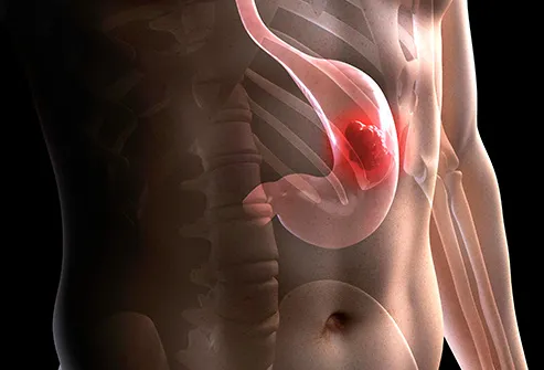
- Brain tumor: Edema of brain can be caused due to brain tumor. The growing brain tumor can compress the adjacent area of brain and block the circulating spinal fluid. These may cause rise in intracranial pressure and brain swelling. The growing blood vessels in and around the tumor can also cause brain edema.
- High altitude: Brain swelling can be one of the symptoms of high altitude sickness although not very common. The swelling is more likely to develop at an altitude 5000 feet and above. The condition is often accompanied with acute mountain sickness.
Symptoms:
- Headache
- Neck pain or stiffness
- Nausea or vomiting
- Dizziness
- Irregular breathing
- Vision loss or changes
- Memory loss
- Inability to walk
- Difficulty speaking
- Stupor
- Seizures
- Loss of consciousness
Treatment for Edema:
Minor cases of brain swelling due to causes such as moderate altitude sickness or a slight concussion often resolve within a few days. In most cases, however, more treatment is needed quickly.
The goal is to assure that the brain receives enough blood and oxygen to remain healthy while the swelling is relieved and any underlying causes are treated. This may require a combination of medical and surgical treatments. Prompt treatment usually results in quicker and more complete recovery. Without it, some damage may remain.
Treatment for brain edema may include any combination of the following:
- Oxygen therapy: Providing oxygen through a respirator or other means helps make sure that the blood has enough oxygen in it. The doctor can adjust the respirator to help reduce the amount of swelling.
- IV fluids: Giving fluids and medicine through an IV can keep blood pressure from dropping too low. This helps to make sure that the body — including the brain — is receiving enough blood. However, some fluids can make swelling worse. Doctors attempt to use the right amounts of the right fluids in someone with brain swelling.
- Lowering body temperature (hypothermia): Lowering the temperature of the body and brain helps relieve swelling and allows the brain to heal. Hypothermia as a treatment for brain swelling is not widely used because it is difficult to perform correctly.
- Medication: In some cases of brain edema, your doctor may start a drug to help relieve the swelling. Medication may also be given for other reasons, such as to slow your body’s response to the swelling or to dissolve any clots. The drugs your doctor gives you depend on the cause and symptoms of brain swelling.
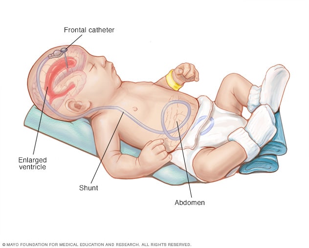
- Ventriculostomy: In this procedure, a surgeon cuts a small hole in the skull and inserts a plastic drain tube. Cerebrospinal fluid is drained from inside the brain, helping to relieve the pressure.
- Surgery: Surgery may have one or more of these goals:
- Removing part of the skull to relieve intracranial pressure; this procedure is called decompressive craniectomy.
- Removing or repairing the source of the swelling, such as repairing a damaged artery or vein or removing a growth