Maternal-fetal medicine (MFM) is a specilaity of Gynecology and Obstetrics which specializes in health of pregnant women foetus.The department provides specialized care of the mother and fetus in complicated, high-risk pregnancies.The speciality is also called perinatology.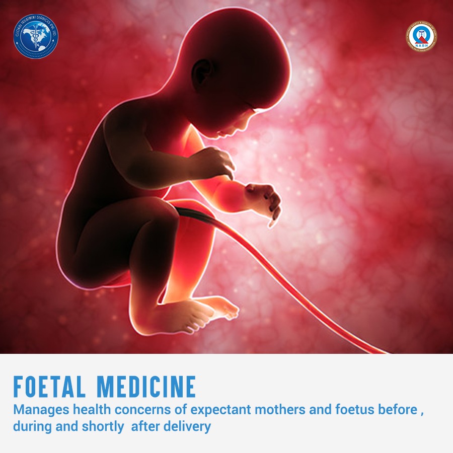
Reasons to visit MFM
Sometimes it is the pregnant women who needs care for unexpected problems that develop during pregnancy such as early labor, bleeding, or high blood pressure. In other cases, it is the baby who faces the non-routine. If birth defects or growth problems are found in the foetus, treatment can be started before birth by providing monitoring, blood transfusions, or surgery to support babies with the best possible care until they are ready for delivery.
Reasons why you may see an MFM physician:
- Pregnant women of advanced maternal age (35 years or older) at the expected time of delivery.
- Pregnant women who have had an abnormal first trimester screening result (for down syndrome and/or Trisomy 18) or an abnormal second trimester quad screen result (for Down Syndrome, Trisomy 18 and/or spina bifida)
- Pregnant women who have had a positive carrier test results for genetic conditions such as cystic fibrosis and sickle cell disease
- Pregnant women experiencing complications such as bleeding, preterm labor, hypertension, diabetes and others
- Pregnant women with a multifetal gestation (twins, triplets, quadruplets)
- Pregnant women using medications, alcohol or other drugs which could be harmful to the unborn baby
- Pregnant women who have an abnormality discovered by ultrasound
- Couples who are pregnant or considering pregnancy who have a family history of birth defects, mental retardation or genetic conditions
- Couples with unexplained infertility, recurrent miscarriages or fetal loss.
- Problems with a previous pregnancy, such as multiple previous miscarriages, premature birth, low birth weight baby, Rh sensitization, prior cesarean delivery or a desire for Trial of Labor after cesarean (TOLAC), prior stillbirth or early neonatal demise
- Cervical insufficiency, also known as incompetent cervix
- Intrauterine growth restriction (IUGR)
- Pre-pregnancy diabetes and gestational diabetes
- Chronic high blood pressure in addition to gestational hypertension, preeclampsia or eclampsia
- Maternal heart disease such as repaired congenital heart malformation or coronary artery disease
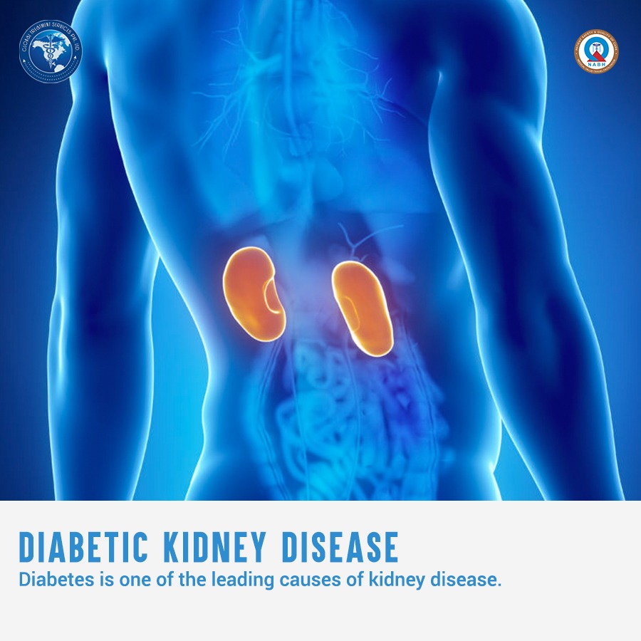
- Kidney disease such as chronic renal failure, nephropathy, kidney stones or lupus nephritis
- Placenta abnormalities such as placenta previa (covering the cervix) or placental abruption (premature separation of the placenta)
- Premature labor threatening to result in early delivery
- Hyperemesis gravidarum (excessive nausea and vomiting during pregnancy)
- Infections that could threaten a pregnancy, such as HIV/AIDS, STDs (sexually transmitted diseases), bacterial vaginosis (BV), cytomegalovirus (CMV), hepatitis B virus (HBV), hepatitis A virus (HAV), listerisosis, parvovirus BI9 infection (also know as Fifth’s disease), toxoplasmosis and urinary tract infections
- Complication from thyroid diseases (Graves disease, Hashimoto’s disease, hypothyroid)
- Complication from liver diseases (intrahepatic cholestasis of pregnancy, hepatitis, acute fatty liver of pregnancy
- Thrombophilias or clotting disorders, such as Factor V Leiden mutation, prothrombin gene mutation, antithrombin III deficiency, protein S and protein C deficiencies
- Autoimmune diseases, such as systemic lupus erythematosus
- Reproductive abnormalities, such as double uterus
- Umbilical cord abnormalities, such as vasa previa, nuchal cord, umbilical cord cysts or knots
- Rh disease and other cases of alloimmunization
- Managing pre-existing conditions that require medications, such as seizure disorders, cancer, interstitial cystitis, Crohn’s disease or ulcerative colitis,
- Management of wide range of obstetrical emergencies, such as maternal hemorrhage
Prenatal Diagnostics
Some fetal problems may be identified during pregnancy through tests.Prenatal diagnosis assists couples in making informed decisions regarding the management of their pregnancy.Genetic counseling assesses the risk of passing an inheritable disease or birth defect to your baby.
Screening tests such as 1st Trimester Screening and Multiple Marker Screening (or Quad Test) provide information regarding the relative risk of having a baby with either a genetic (chromosomal) abnormality such as Down syndrome or a structural anomaly such as spina bifida.
- 1st trimester screening for fetal chromosomal abnormalities is done between about 11 weeks and 13 weeks 6 days of pregnancy. It combines two modalities, ultrasound and a maternal blood specimen. It provides information about a woman’s risk for having a child with Down syndrome and Edwards or Patau’s syndrome
- Multiple Marker Screening or Quad Test assesses the child’s risk for having a chromosomal abnormality or neural tube defect (spina bifida or anencephaly). It is a screening test, not diagnostic. Performed between the 16th and 20th weeks of pregnancy, a sample of the mother’s blood is drawn to help adjust a pregnant woman’s age-related risk.
- Diagnostic tests used to identify conditions are amniocentesis, chorionic villus sampling (CVS), targeted sonogram examination and percutaneous umbilical blood sampling (PUBS).
- Amniocentesis is a technique to obtain amniotic fluid containing fetal cells from the “bag of waters” to analyze the cells for the number of chromosomes. It does require inserting a needle into the “bag of waters” to obtain the fluid containing the cells. It is usually done between 15 and 20 weeks of pregnancy for this purpose.
- CVS is a procedure to obtain a sample of the placenta to detect fetal chromosomal abnormalities. The test is used when information about the chromosomes of the fetus is desired earlier in pregnancy, since it is done between 10 and 13 weeks of pregnancy. This test involves inserting a needle into the developing placenta to obtain the sample.
- High resolution ultrasonography, including 3-D ultrasonography when indicated, helps identify fetal abnormalities.
- Percutaneous umbilical blood sampling (PUBS) detects chromosomal abnormalities and blood abnormalities (fetal hemolytic disease) and may be used to diagnose fetal infection (toxoplasmosis or rubella), abnormal fetal platelet count and Rh incompatibility (alloimmunization). It is rarely needed now but when necessary it typically is done later in pregnancy, at 20 weeks of pregnancy and beyond.
- Maternal blood tests can determine a woman’s status regarding the Rh factor, immunity to rubella (German measles), Parvo virus, toxoplasmosis and chickenpox.
Treatments
Cervical cerclage is a surgical procedure performed when the cervix cannot hold a pregnancy inside the womb against the forces of gravity. A stitch is placed to help the developing baby remain inside the uterus as long as possible or until 37-38 weeks of pregnancy. The procedure is often used if the mother has a history of second-trimester miscarriages without labor, a damaged cervix, a previous cone biopsy or LEEP procedure, or an inherited uterine anomaly.There are currently no treatments for fetal chromosomal abnormalities. When the condition is known before birth, full preparations can be made for the arrival of a child who will have special needs.Alloimmunized pregnancy, either from the Rh factor or other red blood cell factors, may require intrauterine transfusion of blood to the fetus to treat or prevent serious fetal low blood count.