Global Treatment Services Pvt. Ltd.

Global Treatment Services

Endometriosis: Causes, Symptoms & Treatments

Endometriosis is a medical condition that occurs when the lining of the uterus, called the endometrium, grows in other places, such as the fallopian tubes, ovaries or along the pelvis. When that lining breaks down, like the regular lining in the uterus that produces the menstruation, it has nowhere to go. This causes cysts, heavy periods, severe cramps and even infertility.

The endometrial tissue may also grow in the vagina, cervix, bowel or bladder, and in rare cases it may spread to other parts of the body, such as the lungs.

The cause of endometriosis is unknown, but researchers have several theories. One theory is that during menstruation, blood with endometrial cells flows back into the fallopian tubes. The cells are rooted there and grow a new lining. Another theory is that the bloodstream carries endometrial cells throughout the body.

It’s also possible that problems with the immune system contribute to the development of endometriosis, when the immune system does not properly detect and destroy endometrial tissue outside of the uterus.

Yet another theory is that abdominal cells that were present since a woman’s embryonic state retain their ability to become endometrial cells. Because endometriosis sometimes run in families, it’s theorized that a person’s genetics can contribute to the development of the condition.

The most common symptom of endometriosis is cramping during menstruation, which can get worse over time, and can be debilitating. The pain is due to internal bleeding from the lining being shed inside the body — in a place where it doesn’t belong — and can also lead to scar tissue formation, blocked fallopian tubes, and bowel problems.

Although many women experience some pain during their periods, women with endometriosis often say their menstrual pain is much worse than usual, and increases over time.

Women should see their doctor if they experience pelvic pain that persists. At first, some women with endometriosis experience pain just before the start of their period, but as the condition progresses, the pain may start in mid-cycle (during ovulation), and continue until after the woman’s period ends.

Women with the condition may also experience pain while having sex, fatigue, painful urination or bowel movements during menstruation and gastrointestinal upset. Infertility and bleeding between periods are also symptoms of endometriosis.

Some women don’t experience any symptoms at all, but realize they have the condition when they are unable to get pregnant.

Diagnosis

The only way for endometriosis to be diagnosed for certain is through laparoscopy, which is a minor surgical procedure that involves your doctor putting a thin scope into your abdomen to view your pelvic organs. If the doctor sees the extra endometrial tissue during the laparoscopy, he or she can also remove it right there to treat the condition.

However, other basic medical tests are typically performed first, before a laparoscopy. These include a pelvic exam, where the doctor manually feels for abnormalities such as cysts, or an abdominal or vaginal ultrasound, which uses sound waves to create an image of the uterus and reproductive organs. Vaginal and abdominal ultrasounds can’t definitively diagnosis endometriosis, but they can test for cysts that may be caused by the condition.

If all other causes of pelvic pain can be ruled out, the doctor can choose to treat the condition surgically or with medicine.

There are several risk factors for endometriosis none of which can really be helped. They include: never having given birth, having a mother with endometriosis, having menstrual cycles that are shorter than 27 days and bleeding that lasts longer than eight days,  having a medical condition that makes the passage of menstrual flow irregular, and having previous damage to cells that line the pelvis.

The biggest complication with endometriosis is fertility problems. About a third to a half of women with endometriosis have difficulties getting pregnant. Infertility can occur because the condition can create adhesions that trap the egg near the ovary, making it difficult for it to travel down the fallopian tube to be fertilized by sperm.

However, that doesn’t mean all women with endometriosis can’t get pregnant – it just might take them a little longer.

Treatment for endometriosis can involve pain medication (for the severe cramping), hormone therapy to slow growth of the endometrial tissue and surgery to remove the tissue. A number of factors go into determining a woman’s treatment, including her age, the severity of her symptoms, and whether the patient wants to become pregnant.

For women who are not trying to become pregnant, hormonal birth control pills are usually the first step in treatment.

For women who are trying to become pregnant, treatment may involve gonadotropin-releasing hormone agonist, which stops ovulation. This treatment is usually recommended for only six months at a time, and pregnancy is possible once treatment is stopped.

For women whose symptoms are not relieved by medication, surgery is another treatment option. Laparoscopy and laparotomy are common forms of surgery used to treat endometriosis. They require going in through the abdominal region to remove the endometrial tissue.

Most women experience relief from pain after surgery, but the pain may come back within two years. Birth-control pills and other medications may be used along with surgery to help extend the pain-free period.

However, if symptoms keep coming back after these surgical procedures, a hysterectomy, or total removal of the uterus, may be a “last resort” option. The condition is less likely to come back after this procedure.


Mini-thoracotomy: Causes, Symptoms & Treatments

What is an atrial septal defect?

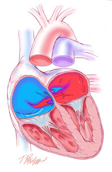
An atrial septal defect or ASD is an opening between the upper two chambers of the heart, known as the right atrium and the left atrium. A congenital heart defect, an ASD permits mixing of deoxygenated blood returning to the heart from the body (right atrium) and freshly oxygenated blood coming from the lungs (left atrium). The degree of mixing is largely related to the size of the defect and the relative pressures in each atrium. Early in life, blood from the left atrium preferentially moves into the right atrium, causing excessive blood to flow through the lungs. If this flow, otherwise known as “shunting” is significant enough, resistance to flow develops in the lung resulting in a gradual reversal of shunting of deoxygenated blood from the right atrium into the left atrium and subsequently through the body. This latter condition can lead to a condition called “cyanosis” whereby inadequately oxygenated blood is delivered to the body causing early fatigue and congestive heart failure.

Atrial Septal DefectAtrial Septal Defect

Causes:

Blood can flow between the two upper heart chambers through the ASD.

When blood flows between the two heart chambers. This is called a shunt. Pressure in the lungs may build up. Over time, there will be less oxygen in the blood that goes to the body.

Small atrial septal defects often cause very few problems, and may be discovered much later in life. Many problems can occur if the opening is large, or if there is more than one opening.  ASD is not very common.

Symptoms:

A person with no other heart defect, or a small defect (less than 5 millimeters) may not have symptoms, or the symptoms may not occur until middle age or later.

Symptoms that do occur may begin at any time after birth through childhood. They can include:

  • Difficulty breathing (dyspnea)
  • Frequent respiratory infections in children
  • Feeling the heart beat (palpitations) in adults
  • Shortness of breath with activity

How is an atrial septal defect treated?

Small atrial septal defects can often be followed conservatively without surgery, due to minimal shunting. Larger ASDs should be closed to prevent the late irreversible consequences of excessive left-to-right shunting. ASDs can be closed surgically by simply sewing them closed or, in the case of larger ASDs, placing a patch of the patient’s own tissue or synthetic material (e.g., Dacron) over it.

How is an atrial septal defect repaired?

ASD repairs require the use of cardiopulmonary bypass, otherwise known as “the heart-lung machine.” This permits the surgeon to safely open the right atrium and access the ASD in a relatively bloodless field. In some cases, the heart is also stopped for 1 to 2 hours to facilitate the repair. Repairs range from relatively simple operations to more complex procedures depending on the location, size, and characteristics of the ASD. The total duration of the operation ranges from 2 to 3 hours.

What are minimally invasive approaches to an atrial septal defect?

The most common surgical approach requires the surgeon to saw open the breastbone and spread the edges apart to gain direct access to the heart. Although this approach provides excellent access to the heart, the resulting wound requires several months to heal completely, an extended recovery period with substantial activity restrictions, and can be subject to serious complications including infection, breakdown, and even death.

Mini-ASD Repair

Minimally Invasive ApproachMinimally Invasive Approach to ASD Closure

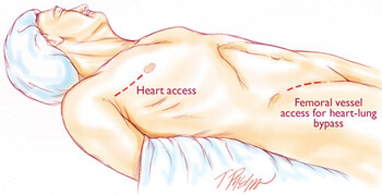
Currently at The Johns Hopkins Hospital, the most commonly employed minimally invasive approach to ASDs is a “mini-thoracotomy” which consists of a 3 inch incision made through the right side of the chest between the ribs. Heart-lung bypass is instituted with small tubes placed in the main artery and vein of the right leg through a 1 to 2 inch incision placed in the right groin crease. The heart is then stopped and the right atrium is opened to expose the ASD. At this point, specialized hand-held “chopstick” like instruments are inserted through this small incision by the surgeon to repair the defect. After the defect is repaired, the heart is then closed and restarted. Finally, heart-lung bypass is discontinued and the incisions are closed.


Systemic Sclerosis: Features and Treatments

Systemic sclerosis (SSc) is a multisystem autoimmune disease in which there is increased fibroblast activity resulting in abnormal growth of connective tissue. This causes vascular damage and fibrosis. Fibrosis occurs in skin, the gastrointestinal (GI) tract and other internal organs.

The name scleroderma is derived from the Greek for ‘hard skin’ and emphasises the dermatological component of the disease. It was described by Hippocrates. There is a localised form of scleroderma, also known as morphoea

General features

  • Fatigue.
  • Weight loss.

Skin features

  • Signs in the hand:
    • Swelling (non-pitting oedema) of fingers and toes – a common early sign; digits may look sausage-like; hand movement may be limited.
    • Skin becomes hard and thickened – this may limit joint movement or cause joint contractures; in the fingers, this is known as sclerodactyly.
    • Swelling and sclerosis reduce hand movements, so patients may be unable to make a fist, or to place the palmar surfaces together – the ‘prayer sign’.
    • Fingertips may have pitting, ulcers or loss of bulk from finger pads.
    • Raynaud’s phenomenon. This is the most common symptom and is present at some point in 90% of cases. Raynaud’s phenomenon with puffy fingers is thought to be a cardinal sign of likely SSc.
  • Calcinosis – nodules or lumps of chalky material which may break through the skin.
  • Face and mouth:
    • Tightening of facial skin.
    • Tight lips (microstomia) – can make dental hygiene difficult.
  • Telangiectasia.
  • ‘Salt and pepper’ appearance of skin, due to areas of hypopigmentation and hyperpigmentation.
  • Dry or itchy skin; reduced hair over affected skin areas.

Musculoskeletal features

  • Joint pain and swelling.
  • Myalgia (due to inflammatory myopathy).
  • Restriction of joint movement, contractures and muscle atrophy due to skin sclerosis.
  • Tendon friction rubs – palpable/audible over the flexor/extensor tendons of the hands, knees and ankles.

GI features

  • Heartburn and reflux oesophagitis.
  • Oesophageal scarring and dysphagia.
  • Delayed gastric emptying – eg, fullness after meals.
  • Reduced small bowel motility – can cause bacterial overgrowth, with bloating, malabsorption, diarrhoea and malnutrition.
  • Constipation due to reduced colonic motility.

Pulmonary features

  • Pulmonary fibrosis (interstitial lung disease):
    • Occurs in as many as 75% of scleroderma patients, but only a few develop end-stage disease.
    • Causes restrictive lung disease.
    • Symptoms and signs: exertional dyspnoea, cough, coarse basal crackles.
  • Pulmonary arterial hypertension (PAH):
    • Occurs in about 10-15% of patients with scleroderma.
    • A leading cause of death in SSc. The presence of PAH drastically reduces survival rate.
    • Symptoms and signs: exertional dyspnoea, syncope, right ventricular strain features.
    • Recent research has attempted to define predictive screening tools. These include monitoring lung function, ECG, urate levels, and N-terminal prohormone of brain natriuretic peptide (NT-proBNP); and by taking into account anti-centromere antibody (ACA) presence and history or presence of telangiectasia.

Management of GI symptoms

For upper GI symptoms:

  • Maintain upright posture after meals; raise the head of the bed; limit alcohol.
  • Proton pump inhibitors.
  • May also need H2-receptor antagonists and pro-motility agents (metoclopramide or domperidone).
  • Dilatation of oesophageal strictures if required.

For intestinal bacterial overgrowth and malabsorption:

  • Cyclical antibiotics.
  • Nutritional advice and nutritional supplements; rarely, parenteral nutrition is required.

For constipation:

  • Dietary fibre and good fluid intake.
  • Softening laxatives (such as lactulose) and/or soluble fibre (such as ispaghula).

Management of pulmonary disease

Pulmonary fibrosis (interstitial lung disease)

  • Benefits of cyclophosphamide seem clear, but must be weighed against side-effects.
  • Supportive treatment: prompt treatment of chest infections – oxygen if needed.

Pulmonary arterial hypertension (PAH)

  • Drug treatment of PAH has improved recently and includes:
    • Endothelin receptor antagonists – eg, bosentan or sitaxsentan.
    • Vasodilators – eg, sildenafil.
    • Prostaglandin derivatives – eg, iloprost (nebulised or intravenous) or epoprostenol (infusion).
  • Supportive treatment – eg, oxygen.

GI complications

See also specific GI sections under ‘Clinical features’ and ‘Management’ headings.

  • Malnutrition due to swallowing problems and other digestive issues.
  • ‘Watermelon stomach’ (gastric antral vascular ectasia):
    • May cause anaemia and GI bleeding.
    • May need endoscopic laser coagulation to prevent bleeding.
  • Obstruction and pseudo-obstruction:
    • Can occur due to reduced motility and bacterial overgrowth.
    • Can be complicated by perforation and peritonitis.
    • Pseudo-obstruction is treated initially by bowel rest and antibiotics.
    • Laparotomy may be needed.
  • Anorectal dysfunction:
    • In some cases, the rectum and anus are involved, causing faecal incontinence.
    • This may require surgery.

Scleroderma renal crisis

  • A serious complication with features of accelerated hypertension.
  • Can lead to renal failure if not treated promptly.
  • Occurs in 5-10% of patients with scleroderma and is more common in those with diffuse or rapidly progressive disease.
  • Presentation:
    • Usually presents as accelerated hypertension with oliguria, headache, fatigue, oedema, rapidly rising serum creatinine levels, proteinuria and microscopic haematuria.
    • Scleroderma renal crises can occur with apparently normal blood pressure, but the blood pressure is higher than baseline values – hence the importance of regular blood pressure monitoring.
  • Treatment is with angiotensin-converting enzyme (ACE) inhibitors, plus dialysis if necessary.
  • Testing patients with scleroderma for anti-RNA polymerase III antibodies may help identify at-risk patients.

Pulmonary complications

  • Aspiration pneumonia from severe reflux.
  • Respiratory muscle weakness if there is severe myositis or extensive skin disease involving the chest.
  • Pneumothorax

Cardiac complications

  • Many different cardiac abnormalities can be associated with SSc. These include:
    • Microvascular coronary artery disease (with resultant myocardial ischemia).
    • Myocardial fibrosis.
    • Left ventricular (LV) systolic dysfunction, LV diastolic dysfunction.
    • Pericarditis or pericardial effusion; these may cause cardiac impairment or congestive cardiac failure.
    • Arrhythmias and conduction defects (including bradyarrhythmias and tachyarrhythmias).
  • The wide variety of abnormalities makes it difficult to assess prevalence. It is likely that the subclinical cardiac involvement rates are very high.
  • Treatment is according to the clinical features.

Other complications

  • Erectile dysfunction.
  • Depression.
  • Osteoporosis – due to reduced blood flow.
  • Hypothyroidism may be associated.


Treatments:

There is no cure for SSc, and management consists of controlling symptoms and preventing complications.

Monitoring

Regular monitoring and reviews are aimed at early detection and treatment of complications. Monitoring includes:

  • Regular review of symptoms.
  • Blood pressure monitoring.
  • Renal function monitoring.
  • Lung function tests and chest CT scan.
  • ECG and echocardiography.

Non-drug treatments

  • Patient involvement and education:
    • ‘Expert patient’ programmes and the Scleroderma Society.
    • Awareness of urgent problems such as renal crisis or intestinal obstruction symptoms.
  • Physiotherapy to promote joint mobility and muscle strength.
  • Home exercises to maintain range of motion (such as gentle mouth, face and hand stretches).
  • Avoid tobacco and maintain healthy weight.
  • Nutritional advice, and supplements if needed.
  • For Raynaud’s phenomenon:
    • Prevention – avoid cold and trauma; use warm clothing or heated clothing.
    • For an attack – warm the body, hands and feet gently (the skin may be numb and unable to feel if the heat source is too hot); use gentle arm movements or gentle massage to help restore circulation.
  • Occupational therapists – for adaptations to assist in daily living.
  • Camouflage products – for cosmetic help with skin changes.


Bone Cancer: Causes, Symptoms and Treatments

Bone cancer can be distinguished into primary bone cancer or secondary bone cancer. Primary bone cancer starts in the bone, where the cancer spreads within the cells of the bone. Secondary cancer, on the other hand, starts elsewhere in the body and spreads to the bones. Examples of primary bone cancer include steosarcoma, Ewing sarcoma, malignant fibrous histiocytoma, and chondrosarcoma.

Causes:

Although bone cancer does not have a clearly defined cause, researchers have identified several factors that increase the likelihood of developing these tumours.

Osteosarcoma occurs more frequently in people who have had high-dose external radiation therapy or treatment with certain anticancer drugs; children seem to be particularly susceptible.

A small number of bone cancers are due to heredity. For example, children who have had hereditary retinoblastoma (an uncommon cancer of the eye) are at a higher risk of developing osteosarcoma, particularly if they are treated with radiation.

Additionally, people who have hereditary defects of bones and people with metal implants, which doctors sometimes use to repair fractures, are more likely to develop osteosarcoma.

The following groups of people may be at a higher risk of developing bone cancer:

  • Being a child or very young adult – most cases of bone cancer occurs in children or young adults aged up to 20
  • Patients who have received radiation therapy (radiotherapy)
  • People with a history of Paget’s disease
  • People with a close relative (parent or sibling) who has/had bone cancer
  • People with Li-Fraumeni syndrome – a rare genetic condition
  • Babies born with an umbilical hernia

Symptoms:

The most common symptom of bone tumours is pain, which gradually increases over time. A person may go weeks, months, and sometimes years before seeking help; the pain increases with the growth of the tumour.

Additional symptoms may include:

  • Fatigue
  • Fever
  • Anemia
  • Unexplained bone fractures

Many patients will not experience any symptoms, except for a painless mass. Some bone tumours may weaken the structure of the bone, causing pathologic fractures.

Treatments:

The type of treatment for bone cancer depends on several factors, including what type of bone cancer it is, where it is located, how aggressive it is, and whether it is localized or has spread. There are three approaches to bone cancer:

  • Surgery
  • Radiotherapy (radiation therapy)
  • Chemotherapy

Surgery – the aim is to remove the tumour, all of it if possible, and some of the bone tissue that surrounds it. If some of the cancer is left behind after surgically removing the tumour it may continue to grow and eventually spread. Limb sparing surgery, also known as limb salvage surgery means that surgical intervention occurs without having to amputate the limb. The surgeon may take some bone from another part of the body to replace lost bone (bone graft), or an artificial bone may be put in. In some cases, however, amputation of a limb may be necessary.

Radiation therapy is also known as radiotherapy, radiation oncology and XRT. Approximately 40% of patients of all types of cancer undergo some kind of radiotherapy. It involves the use of beams of high-energy X-rays or particles (radiation) to destroy cancer cells. Radiotherapy works by damaging the DNA inside the tumour cells, destroying their ability to reproduce. Radiotherapy can be used for different reasons:

  • Total Cure – to cure the patient by completely destroying the tumour
  • To alleviate symptoms – radiotherapy is often used to relieve pain in more advanced cancers
  • Neo-adjuvant radiotherapy (before surgery) – if a tumour is large, radiotherapy can shrink it, making it easier and less harmful to then surgically remove it
  • Adjuvant radiotherapy – given after surgery. The aim is to eliminate the cancer cells that remained behind
  • Combination therapy (radiotherapy combined with another type of therapy) – in some cases, chemo radiation – radiotherapy combined with chemotherapy – is more effective

Chemotherapy is the use of chemicals (medication) to treat disease – more specifically, it usually refers to the destruction of cancer cells. Cytotoxic medication prevents cancer cells from dividing and growing. In general, chemotherapy has 5 possible goals:

  • Total remission – to cure the patient completely. In some cases chemotherapy alone can get rid of the cancer completely
  • Combination therapy – chemotherapy can help other therapies, such as radiotherapy or surgery have more effective results
  • Delay/Prevent recurrence – chemotherapy, when used to prevent the return of a cancer, is most often used after a tumour is removed surgically
  • Slow down cancer progression – used mainly when the cancer is in its advanced stages and a cure is unlikely. Chemotherapy can slow down the advancement of the cancer
  • To relieve symptoms – also more frequently used for patients with advanced cancer

Idiopathic Thrombocytopenic Purpura (ITP): Causes, Symptoms and Treatments

What is Idiopathic Thrombocytopenic Purpura (ITP)?

Idiopathic thrombocytopenic purpura (ITP) is a platelet disorder that occurs in people who have an abnormally low number of platelets in the blood. Platelets are essential in forming blood clots, which consist of a mass of fibers and blood cells. Platelets travel to a damaged or cut area of the body and stick together to form a clot. Having fewer platelets can result in easy bruising, bleeding gums and internal bleeding.

Idiopathic means the cause is unknown.
Thrombocytopenia means a decreased number of platelets in the blood.
Purpura refers to the purple discoloring of the skin, as with a bruise.

Acute thrombocytopenic purpura is most commonly seen in young children (2 to 6 years old). The symptoms may follow a viral illness, such as chicken pox. Acute ITP usually has a very sudden onset, and the symptoms usually disappear in less than six months (often within a few weeks). The disorder usually does not recur. Acute ITP is the most common form of the disorder.
Chronic thrombocytopenic purpura can happen at any age, and the symptoms can last a minimum of six months or several years. It is more common in adults than in children, but it does affect adolescents. Two to three times more females have ITP than males. Chronic ITP can recur often and requires continual follow-up care with a hematologist.
What are the causes of ITP?

In most cases, the cause of ITP is unknown. It is not contagious, meaning a child cannot “catch it” from playing with another child with ITP. It also is important to know that nothing the parents or the child did caused the disorder.

Often, the child may have had a virus or viral infection approximately three weeks before developing ITP. It is believed that the body’s immune system, when making antibodies to fight against a virus, “accidentally” also made an antibody that can stick to the platelet cells. The body recognizes any cells with antibodies as foreign cells and destroys them. Doctors think that in people who have ITP, platelets are being destroyed because they have antibodies. That is why ITP is also referred to as immune thrombocytopenic purpura.

Although there has been research looking at whether certain medications can cause ITP, no direct link has been made with any specific medication that may cause ITP.

What are the symptoms of ITP?

A normal platelet count is 150,000 to 450,000. With ITP, the platelet count is less than 100,000. By the time significant bleeding occurs, the child may have a platelet count of less than 10,000. The lower the platelet count, the greater the risk of bleeding.

Because platelets help stop bleeding, the symptoms of ITP are related to increased bleeding. However, each child may experience symptoms differently. Symptoms can include:

  1. Purpura, which is the purple color of the skin after blood has “leaked” under it.
  2. A bruise is blood under the skin.
  3. Children with ITP can have large bruises from no known trauma. Bruises can appear at the joints of elbows Petechia, which are tiny red dots under the skin caused by very small bleeds.
  4. Nosebleeds.
  5. Bleeding in the mouth and/or in and around the gums.
  6. Blood in the vomit, urine or stool.
  7. Bleeding in the head, which is the most dangerous symptom of ITP.

Any head trauma that occurs when there are not enough platelets to stop the bleeding can be life-threatening.
The symptoms of ITP can resemble other blood disorders or medical problems. Always consult your child’s physician for a diagnosis.

How is ITP diagnosed?

In addition to a complete medical history and physical examination, diagnostic procedures for ITP can include:

Complete blood count (CBC), which is a measurement of size, number, and maturity of different blood cells in a specific volume of blood.
Additional blood and urine tests to measure bleeding time and detect possible infections.
Careful review of the child’s medications.
A bone marrow aspiration, which involves taking a small amount of bone marrow fluid, to look at the production of platelets and to rule out that any abnormal cells the marrow may be producing could lower platelet counts.
Treatment

A child’s specific treatment plan, including the frequency of visits, the length of treatment, side effects and long-term effects are determined by the:

Child’s age, overall health, and medical history
Extent of the disease
Child’s tolerance for specific medications, procedures, or therapies
Expectations for the course of the disease
The patient and family’s opinion or preference
Not all children with ITP require treatment. Close monitoring of a child’s platelets and prevention of serious bleeding complications may be the course of action chosen until the body is able to correct the disorder on its own.

When treatment is necessary, the two most common forms of treatment are:

Steroids, which can help prevent bleeding by decreasing the rate of platelet destruction. Steroids, if effective, will increase platelet counts within two to three weeks.
Intravenous gamma globulin (IVGG), a protein that contains many antibodies and also slows the destruction of platelets. It works more quickly than steroids (within 24 to 48 hours).
Other treatments for ITP can include:

Immune globulin, which is a medication that temporarily stops the spleen from destroying platelets. A child must be Rh positive and have a spleen for this medication to be effective.
Medication changes, because when a medication is the suspected cause, stopping the use of or changing the medication may be necessary.
Infection treatment to increase platelet counts, if infection is the cause for ITP.
Splenectomy to remove a child’s spleen if the spleen is the site of platelet destruction. This is considered more often in older children with chronic ITP.
Hormone therapy in teenage girls to stop their menstrual cycle when their platelets are low if excessive bleeding occurs.
Preventing bleeding

Parents of a child with ITP need to be aware of how to prevent injuries and bleeding. Consider the following:

Making the environment as safe as possible for young children by padding a crib, having the child wear a helmet, and providing protective clothing when platelet counts are low.
Restricting contact sports, riding bicycles, and rough play may be necessary.
Avoiding medications that contain aspirin, ibuprofen, naprosen, or other non-steroidal anti-inflammatory drugs because they may interfere with the body’s ability to control bleeding.
Discuss prevention measures with the child’s physician.
Myocardinal Infarction: Causes, Symptoms and Treatments

“Heart Attack”, technically termed Myocardial Infarction, is a result of total blockage of the blood vessel (coronary artery) which supplies blood to the heart. The blockage is due to the formation of plug composed of cholesterol, blood cells, fibrin and calcium. The heart attack may manifest with a sudden onset of severe central chest pain described as heavy, compressing pain, which can radiate to the left hand, jaw or to the back. It is often associated with profuse sweating and weakness. The presentation can be so sudden that the patient suddenly collapses and becomes unresponsive with profuse sweating and cold peripheries.

A patient with the above mentioned symptoms has to be rushed to the nearest hospital equipped with ECG facilities and a physician / cardiologists on call. Emergency measures include supplemental oxygen through a face mask, pain relief with strong analgesics, intravenous access and initial dose of aspirin and statins (lipid lowering drug). Once the diagnosis of heart attack is confirmed, the treatment is initiated. The important aim of early institution of treatment is to save the heart muscle from damage following lack of blood supply. Blood thinning medications, drugs that improve demand-supply imbalance (beta blockers), drugs that reduce blood cholesterol (statins) and drugs that improve the remodeling of the heart after the attack (ACE inhibitors) form the mainstay of management of heart attacks.

However, the most important component of the treatment consists of Conventional Thrombolytic Therapy -Using clot dissolving medications like Streptokinase, Urokinase or Recombinant TPA to improve blood flow.

Primary Percutaneous Coronary Intervention or Primary Angioplasty (PCI)-An emergency coronary angiogram (to visualize the blockade) followed by balloon Angioplasty with stenting thereby improving the blood flow.

Both methods have got their own merits and demerits. Advantages of primary PCI are that the blockage is visualized, opened and stented, ensuring normal blood flow. The chances of recurrent heart attack, recurrent block and death are less when compared to the clot dissolving medications. Disadvantages include inadequate availability of cathlabs with trained professionals who could perform PCI, and the high cost of the procedure.

Thrombolysis with clot dissolving medications is a time-tested method of treatment. The chief advantage is its universal availability at affordable cost. The disadvantages are the lesser degree of blood flow improvement (54% success rate when compared to 93-98% success with primary PCI), bleeding complications and allergic reactions.

Whatever may be the modality of treatment, the earlier it is instituted the better will be the outcome for the patient. The first one hour is called “The Golden hour” and if treatment as described above is given, the complications and mortality are significantly reduced.

Leg veins: Causes, symptoms and treatments

Leg veins come in many shapes and sizes. Spider veins are generally characterized as smaller blue, red, and purple veins. Varicose veins are characterized as large, ropy, bulging, and sometimes painful veins.

Where did these visible leg veins come from?

There are many different factors that contribute to spider veins and varicose veins including age, occupations that involve long periods of sitting and standing, childbirth, hormones, obesity, history of blood clots, and heredity.

How do I know if I have Varicose Veins?

Visible bulging or ropy veins are usually an indication of varicose veins. Other indications are pain in the legs, ulcers, swelling, leg cramps or pinching sensations, as well as no response to cosmetic vein treatments. An ultrasound is used to determine if varicose veins are present.

What exactly are varicose veins and how are they treated?

The legs are made up of a series of one way valves that carry blood to the heart. If there is reflux present the blood is coming back down instead of going one way. The good news is that varicose veins are treatable and with minimal downtime. Endovenous Laser Therapy is a minimally invasive surgical procedure that allows patients to return to their normal activities directly post treatment. If you don’t have varicose veins and are unhappy with leg vein appearance there is a treatment called sclerotherapy that diminishes the appearance of the veins with just a few injections.

Minimal Access Breast Surgery : Causes, Symptoms & Treatments

Breast cancer develops when one malignant cell starts dividing in an uncontrolled manner from the lining of the breast ducts. As the cells divide, they create a lump which can be felt by the patient if the lump happens to be superficial, if the lump is small or deep, then it is hard to detect with your fingers and must be discovered early with periodic mammograms or ultrasounds.

Causes:

There is no one single factor that causes breast cancer. Breast cancer is multi-factorial disease caused by a combination of stress, abdominal obesity, smoking, lack of exercise, alcohol, pollution, birth control pills, delayed breast feeding, large breasts, advancing age and genetic factors. You do not have to have all these factors get breast cancer but the more factors you have the more likely that you get breast cancer.

Symptoms:

  • A lump in the breast
  • Thickening of the breast
  • Change in shape or look of the breast
  • Skin changes – dimpling or puckering of skin over the breast
  • Swelling, redness or warmth that does not go away
  • Pain in the breast: particularly in one spot: that does not vary with the monthly cycle.
  • Blood or any other discharge from the nipple that starts suddenly and appears in one breast.
  • Any itchy, sore or scaling area on one nipple.

What are the factors that place women at risk?

  • Age: This is the single most important factor. The older a woman gets, the higher the risk, as 80% of all breast cancer occurs in post-menopausal women.
  • Family History: Women with a strong family history of either breast/ovarian cancer, especially among relatives, are more susceptible to breast cancer and they account for 5-10% of all breast cancer patients.
  • Not Having Children: Studies show that women who have borne children are lesser prone to breast cancer.
  • Late First Pregnancy: A woman who has her first child after the age of 30 is more prone to breast cancer.
  • Early Start of Periods: Women who have had their periods at the age of 11 years or so are more prone to breast cancer.
  • Late Menopause: Women, in whom the menopause has started after the age of 55 years.

If you have any of the above signs, be alerted and not alarmed. Consult your doctor.

It is not possible to prevent breast cancer yet, but one can reduce the risk of suffering from the disease.

How can you reduce the risk of breast cancer?

  • Be Breast Aware: Early detection is the aim and therefore one should be aware of the signs of breast cancer and act immediately on noticing any abnormality.
  • Self-Breast Examination: The technique of self-breast examination should be learnt from the doctor and it should be carried out monthly after the age of 20 years. (Technique of self-breast examination is also given in this leaflet.)
  • Clinical Breast Examination: Clinical breast examination is done by a trained doctor and should be done at least every 3 years beginning at the age of 20 years and annually after the age of 40.
  • Mammography: Also known as x-ray of the breast should be done should be done after the age of age of 40.
  • Healthy Well Balanced Diet & Regular Exercise: A low-fat, high fiber diet with plenty of fresh fruit and vegetables, combined with regular exercise is known to enhance overall health and maintain healthy body weight. Remember that post-menopausal and overweight women have an increased risk of breast cancer.

 Prevention & easy detection:

There are now major health programs involved in research and development into prevention, early detection, and new treatments with the aim of reducing the morbidity and mortality from breast cancer

The diagnosis is made by a combination of mammography, breast ultra-sound and increasingly core biopsy and histopathology of the breast lesion rather than the previously used fine needle aspiration and cytological analysis.


Fibroids in females:Causes, symptoms, Treatments

What are Fibroids and How Common are they?

​Fibroids are considered noncancerous tumors of the uterus, and are found in over 80% of women. Although 97% benign, there does exist a malignancy rate of approximately 1-3%.

​Fibroid tumors are the most common uterine pathology and are the primary reason for approximately 80% of the hysterectomies in this country today. Also known as myomas, leiyomomas, fibromas, and fibromyomas, these tumors can have an extensive blood supply from within the uterus.

                            Fibroid Tumors

Fibroid Types

Fibroid tumors usually present in four different types:pedunculated, subserosal,, intramural, submucosal.

  • Pedunculated fibroids are those extending from the uterus on a stalk.This type of tumor may be found within the endometrial cavity or extending outside the uterus into the pelvis. These are the most easily removed tumors, and often the surgical removal can be performed through laparoscopy or hysteroscopy. The symptoms include pain or pressure in a specific area, or bleeding depending on where the tumor is located.
  • Subserosal are those fibroids located just beneath the outer layer of the uterus. These are the tumors that are most easily accessible and are often removed via a simple or laparoscopic myomectomy. If allowed to increase in size, symptoms can include pelvic pain, back pain, urinary frequency, constipation, bloating and indigestion.
  • Intramural tumors are those located deep in the uterus in the main body of the organ. These are the most common and the most difficult to remove, and are responsible for the highest percentage of hysterectomies. These tumors can produce bleeding problems, abdominal pressure, and painful intercourse, in addition to all the symptoms listed above
  • Finally, submucosal fibroids are those directly adjacent to the endometrial lining. These are the tumors primarily responsible for heavy menstrual bleeding. These tumors impinge on the endometrial cavity and can produce long heavy periods, cramps, clots, and cervical
  • pressure.

What Causes Fibroids?

The true etiology of fibroid tumors is unknown. Research has shown significant genetic correlation, as well as a strong cultural component: fibroids are 4-6 times more likely in the African American populations, and yet are rarely seen in the Asian culture. With the introduction of hormones into our general food supply, the incidence of fibroid tumor disease is increasing across the general population regardless
of race or culture.​

We do know that these tumors are directly responsive to estrogen and exhibit their most significant growth spurts just prior to the monthly menses.Women that have a documented estrogen dominance have a much higher rate of tumor
growth, which is also witnessed during pregnancy. Conversely, women in the menopausal phase of life, demonstrate a reduction in the growth of tumors, with some fibroids even shrinking slightly in size.

The Signs and Symptoms of Fibroid Tumors

Signs and symptoms of fibroids are dependent on the size, number and location of the tumors. Many patients complain of menstrual problems like heavy periods, with extended days of flow, cramps and clots.​

However, many women have absolutely no symptoms at all. The following symptoms are known (but not limited)to be associated with fibroids and will increase in severity based on size and number of tumors:

  • heavy bleeding or increased flow
  • clotting
  • cramps
  • abdominal pressure
  • irregular and painful periods
  • bloating
  • indigestion
  • urinary frequency
  • constipation
  • painful intercourse
  • back pain
  • pressure in the legs
  • PMS
  • fatigue

Fibroid Tumor Treatments

For any long term relief, once symptommatic, most fibroids need to be surgically removed. Delaying removal of tumors usually allows the fibroids to grow in size and number. There are multiple treatments available from herbal supplements, birth control pills, IUDs, minimally invasive procedures like UAE to laparoscopic, Robotic and open surgeries.

The type of treatment recommended is often based on the size, number and location of the fibroids and the skill and philosophy of your physician. Please refer to our section on Types of Treatments for more indepth and comparative information and analysis.

 Fertility & Fibroids:

Fertility outcomes with fibroids are dependent on the size, number and location of the tumors. In general, only -12% of fibroids are associated with primary infertilty so most women can have uncomplicated pregnancies with these tumors, and research has shown that if conception takes place only 10-15% of women will have complications with pregnancy.

Submucosal and intracavity tumors are an example of those that can produce complications and even pregnancy loss. Intracavity fibroids are in direct competition for space within the endometrial cavity with a developing fetus. This can create obstacles to development and miscarriage can occur. Submucosal tumors by nature of their location near the lining can also contribute to infertility by inhibiting implanation of the fertilitzed egg. Fundal fibroids are known to block fallopian tubes and are therefore a primary source of infertility by preventing conception.

No matter what type, the longer fibroid tumors are left untreated, the greater the chance of infertility.


Laparoscopic Appendectomy: Symptoms, causes & Treatments

What is the appendix?

If the appendix becomes infected (appendicitis), the infected appendix must be surgically removed (emergency appendectomy) before a hole develops in the appendix (perforation) and spreads the infection to the entire abdominal space (peritonitis).

The appendix is a narrow, small, finger-shaped portion of the large intestine that generally hangs down from the lower right side of the abdomen.

Laparoscopic Appendectomy Surgery India offers info on Cost Appendectomy Hospital India, Laparoscopic Abdominal Surgery India

What is appendicitis and what causes it?

Appendicitis is a sudden inflammation of the appendix. Although the appendix does not seem to serve any purpose, it can become diseased and, if untreated, can burst, causing infection and even death.

The cause of appendicitis is usually unknown. Appendicitis may occur after a viral infection in the digestive tract or when the tube connecting the large intestine and appendix is blocked or trapped by stool. It is thought that blockage of the opening of the appendix into the bowel by a hard, small stool fragment causes inflammation and infection of the appendix (appendicitis). The inflammation can cause infection, a blood clot, or rupture of the appendix.

The infected appendix then must be surgically removed (emergency appendectomy) before a hole develops in the appendix and spreads the infection to the entire abdominal space.

What are the symptoms of appendicitis?

  • Abdominal pain – pain may begin in the upper-middle abdomen then develop to sharp localized pain
  • Abdominal pain may be worse when walking or coughing
  • Fever usually occurs within several hours
  • Loss of appetite
  • Nausea
  • Vomiting
  • Constipation
  • Rectal tenderness
  • Chills and shaking

If you have these symptoms, see a doctor immediately! Because of the risk of rupture, which may happen as soon as 48 to 72 hours after symptoms begin, appendicitis is considered an emergency.

How is appendicitis treated?

The best treatment of appendicitis is its surgical removal. Mild appendicitis may sometimes be cured with antibiotics. More serious cases are treated with surgery to remove the appendix, called an appendectomy. Doctors either use an “open” technique or a minimally invasive approach to remove the appendix.

Appendicitis is considered an emergency and anyone with symptoms needs to see a doctor immediately!

What is a laparoscopic appendectomy?

The laparoscopic (minimally invasive) surgical technique involves making several tiny cuts in the abdomen and inserting a miniature camera and surgical instruments. As many as three or four incisions are made. The surgeon then removes the appendix with the instruments, so there is usually no need to make a large incision in the abdomen. The camera projects a magnified image of the area onto a television monitor which helps guide the surgeons as they remove the appendix.
What is the benefit of laparoscopic appendectomy?

Most cases of acute appendicitis can be treated laparoscopically. The main advantages are : -

  • Less post-operative pain
  • Faster recovery and return to normal activity
  • Shorter hospital stay
  • Less post-operative complications
  • Minimally sized incisions/scars

In most cases, patients can be discharged within 24 to 36 hours. By contrast, the hospital stay is typically two to five days for an open procedure.

Can every patient have a laparoscopic appendectomy?

No. Patients with cardiac diseases and COPD would not be good candidates for laparoscopic appendectomy. In addition, laparoscopic appendectomy is not recommended for those with pre-existing disease conditions.

Laparoscopic appendectomy may also be more difficult in patients who have had previous lower abdominal surgery and for obese patients. The elderly may also be at increased risk for complications with general anesthesia. We evaluate every patient to determine the appropriate type of surgery to perform.

Treatment Options:

The appendix is then removed using a camera through the belly button as well as two other small incisions. Depending on how advanced the infection is, the patient is kept on antibiotics. If the infection is very advanced, the operation may be delayed while the patient receives antibiotics for several weeks. After the infection has improved, a laparoscopic appendectomy is performed.

Laparoscopic appendectomy is a safe and effective treatment for appendicitis. However, in the presence of infection, adhesions, or variations in anatomy, this method becomes dangerous and your surgeon may need to make the prudent decision to continue by making the traditional incision to safely complete the operation. This should not be seen as a failure, but as a wise decision by your surgeon to prevent dangerous complications. Other complications, although rare, include bleeding and infection. It is extremely uncommon to require a blood transfusion for this operation.

This operation is generally performed with general anesthesia. An IV line will be placed in your arm for fluids and you will be brought into the operation room. The anesthesiologist and nurses will use monitors to check your heart rate and breathing during the procedure. These may include EKG leads, a blood pressure cuff, an oxygen mask and sleeves on your legs to prevent clots from forming.

Once you are asleep, the operating room team will work together to perform your operation. When your operation is complete, you will be awakened from anesthesia in the operating room but you may not remember this. After a few hours in the recovery room, the nurses will help you out of bed and give you something to drink. It is common to feel groggy and nauseated soon after surgery and medication is available to help with these discomforts. This information is not intended to replace a visit with your physician.

Picturised Procedure of Laparoscopic Appendectomy:

Laparoscopic Appendectomy

Laparoscopic Abdominal Surgery India, Laparoscopic Surgery Training India

This is a normal appendix in a female patient found to have an infection of the reproductive organs. Since the appendix serves no useful function, it will be removed to save her from ever getting appendicitis in the future

Laparoscopic Appendectomy Surgery India offers info on Cost Appendectomy Hospital India

The first step is to free up the appendix down to where it is attached to the large bowel. This requires dividing the mesentery which contains the blood vessels that supply the appendix. Bipolar forceps apply an electric current which seals the blood vessels and prevents bleeding.

Laparoscopic Surgery Training India, Laparoscopic Abdominal Surgery Gurgaon India

Next, scissors are used to divide the mesentery. Alternating application of the bipolar forceps and scissors allow complete mobilization of the appendix down to its base.


Laparoscopic Appendectomy Surgery India offers info on Cost Appendectomy Hospital India, Laparoscopic Abdominal Surgery India

Now the base of the appendix is tied off using a pretied suture which is now being positioned.

Laparoscopic Surgery Training India, Laparoscopic Abdominal Surgery Gurgaon India

The suture has now been tightened and appears secure. The knot is a fisherman’s knot, which can be tightened, but will not loosen on its own.

Laparoscopic Surgery Training India, Laparoscopic Abdominal Surgery Gurgaon India, Laparoscopic Cancer Surgery India

As shown in this picture, the suture is now cut with the scissors.

Laparoscopic Appendectomy Surgery India offers info on Cost Appendectomy Hospital India, Laparoscopic Abdominal Surgery India, Laparoscopic Cancer Surgery India

Finally, the appendix is cut free and is ready to be removed.

Laparoscopic Appendectomy Surgery India offers info on Cost Appendectomy Hospital India, Laparoscopic Abdominal Surgery Gurgaon India, Laparoscopic Abdominal Surgery India
The operation is complete, and the area is inspected a final time to make sure there is no bleeding.


Pages:1...53545556575859...81