Global Treatment Services Pvt. Ltd.

Global Treatment Services

Hemicrania continua : Causes, Symptoms & Treatments

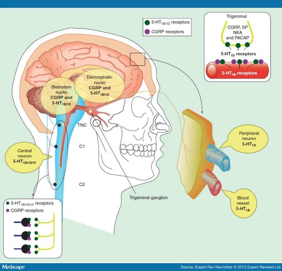
This rare type of headache doesn’t stop. It causes pain on one side of your face or head.

Doctors don’t know what causes this “continuous headache.” But women seem to get it more often than men.

With the right treatment, though, most people can get nearly complete relief from the pain.

Related image

Symptoms

People with hemicrania continua describe a dull ache or throb that’s interrupted by pain that is:

  • Jolting
  • Sharp
  • Stabbing

These attacks usually happen three to five times a day.

Some people will have these headaches steadily for months or years. For others, the pain will go away for weeks or months, then come back.

The headaches often have some of the same symptoms as other kinds. This overlap can make them tricky for doctors to diagnose.

Like migraines, they can cause:

  • Nausea or vomiting
  • Sensitivity to noise or light
  • Throbbing pain

Hemicrania continua also shares features of cluster headaches. For example, people who have it may have problems with how part of their nervous system works. That causes symptoms that happen on the painful side of the face and head, including:

  • Stuffy or runny nose
  • Nosebleeds (which are rare)
  • Tearing, redness, or irritation of the eyes
  • Drooping eyelids
  • Sweating

Some things tend to make symptoms worse, such as:

  • Stress
  • Changes in sleep patterns
  • Bright lights
  • Fatigue
  • Overdoing exercise
  • Alcohol

About 10% of people have symptoms when they:

  • Feel pressure on their neck
  • Flex or rotate their neck

Doctors can make a diagnosis of hemicrania continua if you’ve had pain consistently, without it switching sides or disappearing even briefly, for at least 3 months.

Related image

Treatments

Some anti-inflammatory medications ease hemicrania continua headaches. Indomethacin, a nonsteroidal anti-inflammatory drug (NSAID), often gives fast relief. One way doctors know that you have hemicrania continua is if your headaches go away after a dose of the drug. But some people may need more testing — such as an MRI — to look into their symptoms.

Daily doses of indomethacin for hemicrania continua typically range from 25 to 200 milligrams. A common side effect of the drug is irritation of the lining of the stomach and digestive tract. So, people who take it may also need medication to help their stomach make less acid.

If the side effects of indomethacin are too much for you, another NSAID, celecoxib, may also help.

Tricyclic antidepressants, like amitriptyline, can also prevent these headaches.

Amyotrophic Lateral Sclerosis: Causes, Symptoms & Treatments

What is ALS?

Amyotrophic lateral sclerosis (ALS) is a rare group of neurological diseases that mainly involve the nerve cells (neurons) responsible for controlling voluntary muscle movement. Voluntary muscles produce movements like chewing, walking, breathing and talking. The disease is progressive, meaning the symptoms get worse over time. Currently, there is no cure for ALS and no effective treatment to halt, or reverse, the progression of the disease.

ALS belongs to a wider group of disorders known as motor neuron diseases, which are caused by gradual deterioration (degeneration) and death of motor neurons. Motor neurons are nerve cells that extend from the brain to the spinal cord and to muscles throughout the body. These motor neurons initiate and provide vital communication links between the brain and the voluntary muscles.

Messages from motor neurons in the brain (called upper motor neurons) are transmitted to motor neurons in the spinal cord and to motor nuclei of brain (called lower motor neurons) and from the spinal cord and motor nuclei of brain to a particular muscle or muscles.

In ALS, both the upper motor neurons and the lower motor neurons degenerate or die, and stop sending messages to the muscles. Unable to function, the muscles gradually weaken, start to twitch (called fasciculations), and waste away (atrophy). Eventually, the brain loses its ability to initiate and control voluntary movements.

Early symptoms of ALS usually include muscle weakness or stiffness. Gradually all muscles under voluntary control are affected, and individuals lose their strength and the ability to speak, eat, move, and even breathe.

Most people with ALS die from respiratory failure, usually within 3 to 5 years from when the symptoms first appear. However, about 10 percent of people with ALS survive for 10 or more years.

Related image

Causes and types

It is unclear exactly what causes ALS. There are different types, according to their signs and symptoms and whether or not there is a clear genetic association.

ALS can be sporadic or familial.

Sporadic ALS occurs randomly, and it accounts for 90 to 95 percent of cases. There is no clear risk factor or cause.

Familial ALS is inherited. Around 5 to 10 percent of cases are familial. The child of a person with ALS will have a 50 percent chance of developing the condition. Rarely, it can affect a person in their teens. Researchers are investigating which genes are involved.

Other possible causes of ALS include:

  • Disorganized immune response: The immune system may attack some of the body’s cells, possibly killing nerve cells.
  • Chemical imbalance: People with ALS often have higher levels of glutamate, a chemical messenger in the brain, near the motor neurons. Glutamate in high quantities is known to be toxic to nerve cells.
  • Mishandling of proteins: If proteins are not processed correctly by nerve cells, abnormal proteins could potentially accumulate and cause the nerve cells to die.

Possible environmental factors

Environmental factors may also play a role.

One study has reported that military personnel deployed in the Gulf region during the 1991 war were more likely to develop ALS than military personnel deployed elsewhere.

Some possible links have been found between ALS and exposure to:

  • mechanical or electrical trauma
  • military service
  • high levels of exercise
  • high levels of agricultural chemicals
  • high levels of a variety of heavy metals

However, there is no conclusive evidence that specific lifestyle changes can decrease the risk.

Signs and symptoms

The symptoms of ALS usually appear when a person is in their late 50s or early 60s, but it can happen at other ages.

Progression varies between individuals. In the early stages, signs and symptoms may be barely noticeable, but the weakness becomes more visible over time.

Common symptoms include:

  • difficulty carrying out daily activities, including walking
  • increased clumsiness
  • weakness in the feet, hands, legs, and ankles
  • cramping and twitching in the arms, shoulders, or tongue
  • difficulty maintaining good posture and holding the head up
  • uncontrolled outbursts of laughing or crying, known as emotional lability
  • cognitive changes
  • slurring of speech and difficulty with voice projection
  • pain
  • fatigue
  • problems with saliva, and mucus
  • difficulty breathing and swallowing, in the later stages

Related image

Progressive muscle weakness occurs in all cases of ALS, but this may not be the first indication of the condition.

Early symptoms often include clumsiness, abnormal limb fatigue, muscle cramps and twitches, and slurred speech. Symptoms will spread to all parts of the body as ALS progresses.

Some people may have problems with decision-making and memory, eventually leading to a form of dementia called frontotemporal dementia.

Emotional lability can cause fluctuations in mood and emotional response.

Treatment and prevention

There is no cure for ALS, so treatment aims to alleviate symptoms, prevent unnecessary complications, and slow the rate of disease progression.

ALS can cause a range of physical, mental, and social changes, so a team of specialists will often help patients manage their symptoms and care, improve their qualify of life, and prolong survival.

Riluzole (Rilutek) was approved for ALS treatment by the Food and Drug Administration (FDA) in 1995, and it appears to slow the progression of the disease. It may work by reducing the body’s levels of glutamate, an excitotoxin that has been linked to neuronal damage.

In May 2017, Radicava (Edaravone) was approved to treat ALS. It may slow the decline in physical function by one third.

A number of research projects are looking at ways to use new and existing drugs to treat different aspects of ALS. Doctors can also prescribe medications to treat the different symptoms.

Therapy

Physical therapy can help people with ALS manage pain and address mobility issues.

A physical therapist can provide help and information with:

  • low-impact exercises to enhance cardiovascular fitness and overall well-being
  • mobility aids, such as walkers and wheelchairs
  • devices to make life easier, such as ramps

Occupational therapy can help a patient maintain their independence for longer by:

  • helping patients choose adaptive equipment and assistive technologies to help them keep up their daily routines
  • train them in ways to compensate for hand and arm weaknesses

Breathing therapy may be needed in time, as the respiratory muscles get weaker.

Breathing devices can help the patient breathe better at night. Some patients may need mechanical ventilation. One end of a tube is connected to a respirator, while the other end is inserted into the windpipe through a surgically-created hole in the neck, or tracheostomy.

Speech therapy is useful when ALS begins to make it harder to talk. Speech therapists can help by teaching adaptive techniques. Other methods of communication include writing and computer-based communications equipment.

Nutritional support is important, as difficulty with swallowing can make it hard to get enough nutrients. Nutritionists can advise on preparing nutritious meals that are easier to swallow. Suction devices and feeding tubes may help.

Mastoiditis: Causes, Symptoms & Treatments

Mastoiditis is a bacterial infectionof the mastoid  air cells surrounding the inner and middle ear. The mastoid bone, which is full of these air cells, is part of the temporal bone of the skull. The mastoid air cells are thought to protect the delicate structures of the ear, regulate ear pressure and possibly protect the temporal bone during trauma.

When the mastoid cells become infected or inflamed, often as a result of an unresolved middle ear infection (otitis media), mastoiditis can develop. Because so many vital structures pass through the mastoid, infection may spread outside of the mastoid bone and cause serious health complications.

Acute mastoiditis typically affects children, but adults can also be affected.

Some people have chronic mastoiditis, an ongoing infection of the middle ear and mastoid that causes persistent drainage from the ear.

Related image

Mastoiditis Causes

As mentioned above, mastoiditis most often develops as a result of a middle ear infection. Bacteria from the middle ear can travel into the air cells of the mastoid bone. Less commonly, a growing collection of skin cells called a cholesteatoma, may block drainage of the ear, leading to mastoiditis.

Mastoiditis Symptoms :

Mastoiditis symptoms may include:

  • Fever, irritability, and lethargy
  • Swelling of the ear lobe
  • Redness and tenderness behind the ear
  • Drainage from the ear
  • Bulging and drooping of the ear

Mastoiditis Complications :

Mastoiditis complications may include:

  • Facial paralysis
  • Nausea, vomiting, vertigo (labyrinthitis)
  • Hearing loss
  • Brain abscess or meningitis
  • Vision changes or headaches (blood clots in the brain)

Mastoiditis Treatments :

Antibiotic therapy is the mainstay of treatment for both acute and chronic mastoiditis.

If you or your child is diagnosed with acute mastoiditis, you may be put in the hospital to receive treatment and care by an otolaryngologist, a doctor who specializes in ear, nose, and throat disorders. Antibiotics will be given through an IV (intravenous line) to treat the infection.

Surgery may also be needed to drain the fluid from the middle ear, called a myringotomy. During a myringotomy, the doctor makes a small hole in the eardrum to drain the fluid and relieve pressure from the middle ear. A small tube may be inserted into the middle ear to to keep the hole from closing so as to allow for continued drainage. Typically, the tube will fall out on its own after six to 12 months.


If the symptoms don’t quickly improve on IV antibiotics, or complications, like an abscess or bone erosion are present, surgery to remove the infected mastoid bone may be recommended. This is called a mastoidectomy.

Chronic mastoiditis is treated with oral antibiotics, eardrops, and regular ear cleanings by a doctor. If these treatments do not work, surgery may be necessary to prevent further complications.

If left untreated, mastoiditis can cause serious, even life-threatening, health complications, including hearing loss, blood clot, meningitis, or a brainabscess. But with early and appropriate antibiotic treatment and drainage, these complications can usually be avoided and you can recover completely.

If you have an earache, discharge or trouble hearing, see your doctor.  He will decide if you need antibiotics to help prevent mastoiditis and other serious health complications.

Spinal Fractures : Causes, Symptoms & Treatments

Spinal fractures caused by trauma represent a serious orthopedic injury. Fractures that occur as a result from a high velocity accident are most commonly in the mid to low back. High velocity accidents are associated with trauma from motor vehicle accidents, a fall from height, or sporting accidents. Patients with high energy trauma or who lose consciousness require urgent emergency evaluation and treatment. The most common symptom of a spinal fracture is moderate to severe pain in the back that is increased with movement.

Fractures of the neck usually occur as a result of high energy trauma and are uncommon in other situations. There are seven bones in the neck and a break in any of those bones may be referred to as a “broken neck.” Any fracture in the neck has serious consequences because the spinal cord runs through the center of the cervical vertebrae. Damage or injury to the spinal cord can result in paralysis or death. All patients with trauma who develop cervical fractures should be evaluated in an emergency room setting.

FRACTURES  :

Low Energy Fractures

People can develop spinal fractures unrelated to trauma; they usually have weakened bones from osteoporosis, tumors, or other medical conditions. Fractures that occur during daily activities are most commonly associated with conditions that may weaken the bone including osteoporosis, spinal tumors or spinal infections. For patients with low energy fractures due to osteoporosis these fractures are called a compression fracture. Treatment options include observation, bracing, and pain management. For some patients a minimally invasive procedure including vertebroplasty and kyphoplasty may improve pain and help to prevent further compression.

Types of Spinal Fractures

There are several types of spinal fractures based on pattern of injury and the extent of spinal cord injury. Below are the more common fractures.

Compression Fracture: This is often caused by osteoporosis or a tumor and rarely has spinal cord or nerve involvement. The front of the vertebra fractures and loses height however the back, posterior, part remains stable. It can be painful or asymptomatic and is usually a stable fracture.

Axial Burst Fraction: This is usually caused from a fall from a significant height, landing on the feet. The vertebra loses height on both the front and back side; depending on the stability of the fracture this may require surgery.

Chance Fracture: This fracture is known as a “seat-belt injury” and is caused by a violent forward flexed injury.  The vertebra is pulled apart, often from a car accident where the upper body is pulled forward while the pelvis is stabilized by a lap only seat belt

Image result for Spinal fracture

Image result for Spinal fracture

PREVENTION

Prevention from high impact accidents can save your life. Always wear a seat belt when riding in a car. Always wear protective equipment when playing sports, riding bicycles, motorcycles or even horses. Obey all regulations in place for your safety. Never dive head first into shallow pool or swimming area.

TREATMENT

Treatment for spinal fractures will depend on the location and type of fracture as well as other the amount of neurologic compression. Minor fractures can be treated with cervical bracing. More complex fractures or fractures with spinal cord compression may require traction, surgery, or some combination of the above. For patients with neurologic compromise or unstable fractures, surgical intervention may be appropriate in order to stabilize the fracture, remove any nerve compression and assist with healing. For most patients, non-operative treatment is appropriate. Rehabilitation is an essential part to the treatment plan once the spinal fracture has healed. Rehabilitation allows patients to reduce pain and minimize disability with a hopeful full return to their functional level before the injury.


Strabismus : Causes, Symptoms & Treatments

Strabismus is a failure of the two eyes to maintain proper alignment and work together as a team.

If you have strabismus, one eye looks directly at the object you are viewing, while the other eye is misaligned inward (esotropia, “crossed eyes” or “cross-eyed”), outward (exotropia or “wall-eyed”), upward (hypertropia) or downward (hypotropia).

Strabismus can be constant or intermittent. The misalignment also might always affect the same eye (unilateral strabismus), or the two eyes may take turns being misaligned (alternating strabismus).

To prevent double vision from congenital and early childhood strabismus, the brain ignores the visual input from the misaligned eye, which typically leads to amblyopia or “lazy eye” in that eye.

According to the American Association for Pediatric Ophthalmology and Strabismus, approximately 4 percent of the U.S. population has crossed eyes or some other type of strabismus.

Related image

Strabismus Symptoms And Signs

The primary sign of strabismus is a visible misalignment of the eyes, with one eye turning in, out, up, down or at an oblique angle.

Corneal light reflex (Hirschberg) test: A screening test for strabismus that evaluates eye alignment based on the location of reflections of light shined at the eyes.

When the misalignment of the eyes is large and obvious, the strabismus is called “large-angle,” referring to the angle of deviation between the line of sight of the straight eye and that of the misaligned eye. Less obvious eye turns are called small-angle strabismus.

Typically, constant large-angle strabismus does not cause symptoms such as eye strain and headaches because there is virtually no attempt by the brain to straighten the eyes. Because of this, large-angle strabismus usually causes severe amblyopia in the turned eye if left untreated.

Less noticeable cases of small-angle strabismus are more likely to cause disruptive visual symptoms, especially if the strabismus is intermittent or alternating. In addition to headaches and eye strain, symptoms may include an inability to read comfortably, fatigue when reading and unstable or “jittery” vision. If small-angle strabismus is constant and unilateral, it can lead to significant amblyopia in the misaligned eye.

Both large-angle and small-angle strabismus can be psychologically damaging and affect the self-esteem of children and adults with the condition, as it interferes with normal eye contact with others, often causing embarrassment and awkwardness.

Newborns often have intermittent crossed eyes due to incomplete vision development, but this frequently disappears as the infant grows and the visual system continues to mature. Most types of strabismus, however, do not disappear as a child grows.

Routine children’s eye exams are the best way to detect strabismus. Generally, the earlier strabismus is detected and treated following a child’s eye exam, the more successful the outcome. Without treatment, your child may develop double vision, amblyopia or visual symptoms that could interfere with reading and classroom learning.

What Causes Strabismus?

Each eye has six external muscles (called the extraocular muscles) that control eye position and movement. For normal binocular vision, the position, neurological control and functioning of these muscles for both eyes must be coordinated perfectly.

Strabismus occurs when there are neurological or anatomical problems that interfere with the control and function of the extraocular muscles. The problem may originate in the muscles themselves, or in the nerves or vision centers in the brain that control binocular vision.

Genetics also may play a role: If you or your spouse has strabismus, your children have a greater risk of developing strabismus as well.

Accommodative Esotropia

Occasionally, when a farsighted child tries to focus to compensate for uncorrected farsightedness, he or she will develop a type of strabismus called accommodative esotropia, where the eyes cross due to excessive focusing effort. This condition usually appears before 2 years of age but also can occur later in childhood. Often, accommodative esotropia can be fully corrected with eyeglasses or contact lenses.

Strabismus Surgery

In most cases, the only effective treatment for a constant eye turn is strabismus surgery. If your general eye doctor finds that your child has strabismus, he or she can refer you to an ophthalmologist who specializes in strabismus surgery.

In most cases, the only effective treatment for a constant eye turn is strabismus surgery.

The success of strabismus surgery depends on many factors, including the direction and magnitude of the eye turn. In some cases, more than one surgery may be required. The strabismus surgeon can give you more information about this during a pre-surgical consultation.

Strabismus surgery also can effectively align the eyes of adults with long-standing strabismus. In many cases of adult strabismus, however, a significant degree of amblyopia may remain even after the affected eye is properly aligned. This is why early treatment of strabismus is so important.

The earlier strabismus is treated surgically, the more likely it is that the affected eye will develop normal visual acuity and the two eyes will function together properly as a team.

Non-Surgical Strabismus Treatment

In some cases of intermittent and small-angle strabismus, it may be possible to improve eye alignment non-surgically with vision therapy.


Esotropia (crossed eyes) needs to be treated early in life to prevent amblyopia.

For example, convergence insufficiency (CI) is a specific type of intermittent exotropia in which the eyes usually align properly when viewing a distant object, but fail to achieve or maintain proper alignment when looking at close object, such as when reading, resulting in one eye drifting outward. Convergence insufficiency can interfere with comfortable reading, causing eye strain, blurred vision, double vision and headaches.

There also is some evidence that suggests CI can cause attention problems and affect academic performance in children. A recent study conducted by Mayo Clinic researchers found that children with exotropia (including convergence insufficiency) at an early age were significantly more likely to develop attention deficit hyperactivity disorder (ADHD), adjustment disorder and learning disabilities by early adulthood.

Certain types of strabismus also have been associated with an increased risk of myopia. Another Mayo Clinic study published in 2010 followed 135 children with intermittent exotropia over a 20-year period and found that more than 90 percent of these children became nearsighted by the time they reached their 20s.

On the bright side, it appears non-surgical vision therapy can be an effective treatment for convergence insufficiency. In a study published in Archives of Ophthalmology, 73 percent of 221 children with symptomatic convergence insufficiency had a successful or improved outcome following a 12-week program of office-based vision therapy combined with eye exercises performed at home.

Sometimes, a strabismus surgeon may recommend a program of vision therapy for a period of time after strabismus surgery to treat amblyopia and minor binocular vision problems that might remain after surgery. In these cases, the term “orthoptics” (“ortho” = straight; “optics” = eyes) rather than “vision therapy” might be used to describe this treatment, which may be provided by an orthoptist working closely with the surgeon rather than by an optometrist.

Cornesal Ulcer : Causes, Symptoms & Treatments

A corneal ulcer is an open sore on the cornea, the thin clear structure overlying the iris (the colored part of the eye).

Corneal Ulcer Causes

Most corneal ulcers are caused by infections.

Bacterial infectionscause corneal ulcers and are common in people who wear contact lenses.


Viral infections are also possible causes of corneal ulcers. Such viruses include the herpes simplex virus(the virus that causes cold sores) or the varicella virus (the virus that causes chickenpoxand shingles).


Fungal infections are an unusual cause of corneal ulcers and may happen after injury with organic material such as branches or twigs. People who contract this type of infection have been treated with steroid eyedrops or are wearing contact lenses which are not properly disinfected.

Tiny tears to the cornea may also cause corneal ulcers. These tears can come from direct trauma; scratches; or particles, such as sand, glass, or small pieces of steel. Such injuries damage the cornea and make it easier for bacteria to invade and cause a serious ulcer.

  • Disorders that cause dry eyes can leave your eye without the germ-fighting protection of tears and cause ulcers.
  • Disorders that affect the eyelid and prevent your eye from closing completely, such as Bell’s palsy, can dry your cornea and make it more vulnerable to ulcers.
  • Chemical burns or other caustic (damaging) solution splashes can injure the cornea.
  • People who wear contact lenses are at an increased risk of corneal ulcers. In fact, your risk of corneal ulcerations increases tenfold when using extended-wear (overnight) soft contact lenses. Extended-wear contact lenses are those contact lenses that are worn for several days without removing them at night. Contact lenses can damage your cornea in many ways.
  • Scratches on the edge of your contact lens can scrape the cornea’s surface and make it more open to bacterial infections.
  • Similarly, tiny particles of dirt trapped underneath the contact lens can scratch the cornea.
  • Bacteria may be on the lens or in your cleaning solutions and, thus, get trapped on the undersurface of the lens. If your lenses are left in your eyes for long periods of time, these bacteria can multiply and cause damage to the cornea.
  • Wearing lenses for extended periods of time can also block oxygen to the cornea, making it more susceptible to infections.

Corneal Ulcer Symptoms

  • Red eye
  • Severe pain
  • Feeling that something is in your eye
  • Tears
  • Pus or thick discharge draining from your eye
  • Blurry vision
  • Pain when looking at bright lights
  • Swollen eyelids
  • A white round spot on the cornea that is visible with the naked eye if the ulcer is very large

Corneal Ulcer Treatment – Self-Care at Home

  • If you wear contact lenses, remove them immediately.
  • Apply cool compresses to the affected eye.
  • Do not touch or rub your eye with your fingers.
  • Limit spread of infection by washing your hands often and drying them with a clean towel.
  • Take over-the-counter pain medications, such as acetaminophen(Tylenol) or ibuprofen (Motrin).

Medical Treatment

  • Your ophthalmologist will remove your contact lenses if you are wearing them.
  • Your ophthalmologist will generally not place a patch over your eye if he or she suspects that you have a bacterial infection. Patching creates a warm dark environment that allows bacterial growth.
  • Hospitalization may be required if the ulcer is severe.
  • Because infection is a common occurrence in corneal ulcers, your ophthalmologist will prescribe antibiotic eyedrops. If the infection appears very large, you may need to use these drops as often as 1 drop an hour.
  • Oral pain medications will be prescribed to control the pain. Pain can also be controlled with special eyedrops that keep your pupil dilated.

Surgery

If the ulcer cannot be controlled with medications or if it threatens to perforate the cornea, you may require an emergency surgical procedure known as corneal transplant.

Aboulia : Causes, Symptoms & Treatments

Abulia, also known as aboulia, is a state of diminished motivation. In general, abulia refers to a type of apathy that develops as a result of disease, specifically disease that affects the brain. Some people who have abulia are aware of the change in level of motivation, but it is particularly noticeable and upsetting for friends and loved ones.

An individual who experiences abulia may show symptoms that range from subtle to overwhelming in levels of severity, with mutism (not speaking) being the most extreme form.

Abulia is most commonly associated with neurologic or psychiatric conditions. It is estimated that about 20-30% of stroke survivors experience apathy or abulia.

When abulia results from a sudden event, such as a stroke, it may become apparent during or after stroke recovery. When abulia results from a progressive disease, such as Alzheimer’s disease or Parkinson’s disease, it may worsen over time.

Image result for abulia causes, symptoms & treatments

Image result for abulia causes, symptoms & treatments

Symptoms

There are a number of symptoms of abulia.

Typical symptoms may include any combination of the following:

  • Apathy- lack of interest or motivation in getting things started or in joining activities. Not enthusiastic about getting things done. Difficulty initiating or completing projects or important tasks.
  • Disinterest in things that are going on or in participating in activities or events.
  • Passivity
  • Lack of opinion or lack of expression of opinion about preferences
  • Asocial- lack of interest in other people, lack of interaction with others, limited communication with others, avoiding others.
  • Extreme quiet or muteness
  • Physical slowness of movement
  • Lack of spontaneity
  • Emotionally remote
  • Difficulty in making decisions or plans, indecisiveness
  • Chewing food for long periods of time without swallowing

Causes

Brain damage or dementia can interfere with normal functioning in the brain, resulting in abulia.

Damage to the nerve circuits of the frontal lobes of the brain that impact the dopaminergic system, is thought to be the cause of abulia. The dopaminergic system describes the system of chemicals produced in the brain that provide positive feelings of happiness and satisfaction. Thus, it is possible that once positive feedback is disrupted, there is little incentive to try to attain things that make a person ‘happy.’

Most people who experience and show symptoms of abulia have experienced one of the following conditions:

  • Stroke
  • Huntington’s disease
  • Schizophrenia
  • Acquired brain injury- from trauma, cancer or infection
  • Alzheimer’s disease
  • Vascular dementia
  • Lewy body dementia
  • Parkinson’s disease
  • Frontotemporal dementia

Difference From Depression

While they share some features in common, abulia and depression are not the same. Abulia is clinically distinct from depression in that people living with abulia typically do not exhibit signs of sadness or negative thoughts. Instead, they exhibit a lack of concern about their condition or about the world around them.

Establishing the difference between the two conditions and getting an accurate diagnosis is important because abulia does not improve with antidepressant medications, while people who have depression should generally expect to experience improvement of symptoms with antidepressants.

Diagnosis

You might be wondering whether you have abulia or depression or whether your friend, coworker or loved one has abulia or depression. You should start by seeking medical attention so that the symptoms and signs can be carefully evaluated.

The diagnosis of abulia may take some time. Abulia is diagnosed through a medical history that details the feelings and actions of the person who might have abulia, as well as a history of the observations make by friends and loved ones. Your doctor may ask further questions and make clinical observations during the office visit. In some cases of abulia, diagnostic imaging with a Brain CT scan or Brain MRI scan can reveal localized brain lesions such as stroke, one of the main causes of abulia.

Treatment

There are some medical treatments available for the treatment and management of abulia. Medications that have an effect on the body’s dopaminergic system, which is disrupted in abulia, have shown positive results when it comes to objective symptoms of abulia.

Cognitive rehabilitation can help in optimizing thinking skills, which may improve abulia. Physical therapy can help with initiating physical activity, possibly increasing motivation in people who are affected with this condition. Counseling and family counseling can improve communication and help in dealing with abulia.

Whiplash: Causes, Symptoms & Treatments

Whiplash occurs when a person’s head moves backward and then forward suddenly with great force. This injury is most common following a rear-end car collision. It can also result from physical abuse, sports injuries, or amusement park rides.

Whiplash results when the soft tissues (the muscles and ligaments) of your neck extend beyond their typical range of motion. Your symptoms might not appear for a while, so it’s important to pay attention to any physical changes for a few days following any accident.

Whiplash is thought of as a relatively mild condition, but it can cause long-term pain and discomfort.

  • Most mild to moderate cases of whiplash can be treated at home using over-the-counter drugs, ice, and other remedies.
  • Common symptoms of whiplash include neck pain and stiffness, headaches, and dizziness.
  • Average recovery time is anywhere from a few days to several weeks. Most people fully recover within three months.

CAUSES:

Whiplash occurs when the muscles in your neck suffer a strain because of a rapid movement backward and then forward. The sudden motion causes your neck’s tendons and ligaments to stretch and tear, resulting in whiplash.

Some things that can cause whiplash include:

  • car accidents
  • physical abuse, such as being punched or shaken
  • contact sports such as football, boxing, and karate
  • horseback riding
  • cycling accidents
  • falls in which the head violently jerks backward
  • blows to the head with a heavy object
 Image result for Neck pain

Symptoms:

What does whiplash feel like?

Symptoms usually appear within 24 hours after the incident that caused the whiplash. Sometimes, symptoms may develop after a few days. They can last for several weeks.

Common symptoms include:

  • neck pain and stiffness
  • headaches, specifically at the base of the skull
  • dizziness
  • blurred vision
  • constant weariness

Less common symptoms associated with chronic whiplash include:

  • problems with concentration and memory
  • ringing in the ears
  • inability to sleep well
  • irritability
  • chronic pain in the neck, shoulders, or head

You should follow up with your doctor immediately if:

  • your symptoms spread to your shoulders or arms
  • moving your head is painful
  • you have numbness or weakness in your arms

How is whiplash diagnosed?

Most mild to moderate cases of whiplash can be treated at home using over-the-counter (OTC) drugs, ice, and other remedies. However, you should seek medical help if you have the following symptoms:

  • pain or stiffness in the neck that goes away and then returns
  • severe neck pain
  • pain, numbness, or tingling in your shoulders, arms, or legs
  • any issues with your bladder or bowels
  • localized weakness in an arm or leg

Your doctor will normally ask you questions about your injury, such as how it occurred, where you feel pain, and whether the pain is dull, shooting, or sharp. They may also do a physical exam to check your range of motion and look for areas of tenderness.

Your doctor might order an X-ray to ensure your pain isn’t connected to any other type of injury or degenerative disease such as arthritis.

Other tests, such as CT scans and MRI, will allow your doctor to assess any damage or inflammation in the soft tissues, spinal cord, or nerves. Certain imaging studies, such as diffuse tensor imaging (DTI) or positron emission tomography (PET scan), may be helpful, especially when there may be a brain injury. These tests will help localize and measure the extent of an injury to the brain or other areas.

Treatment for whiplash:

The treatments for whiplash are relatively simple. Doctors will often prescribe an OTC pain medication like Tylenol or aspirin. More severe injuries may require prescription painkillers and muscle relaxants to reduce muscle spasms.

In addition to medication, physical therapy plays a crucial role in recovery. You may want to apply ice or heat to the injured area and practice simple exercises to build strength and flexibility in your neck. Practice good posture and learn relaxation techniques to keep your neck muscles from straining and to help with recovery.

You might also be given a foam collar to keep your neck stable. Collars should not be worn for more than three hours at a time. They should only be used the first couple of days after your injury, as well.


Alternative remedies

You may also want to try alternative remedies to treat pain. Some include:

  • chiropractic care
  • massage, which may relieve some of the tension in the neck muscles
  • electronic nerve stimulation, which involves a gentle electric current that may help reduce neck pain
  • acupuncture, though no studies confirm acupuncture’s ability to ease neck pain

Complications associated with whiplash

Some people with whiplash do experience chronic pain or headaches for years following their accident. Doctors may be able to trace this pain to damaged neck joints, disks, and ligaments. But chronic pain following a whiplash injury typically has no medical explanation.

However, very few people have any long-term complications from whiplash. Usually, recovery time is anywhere from a few days to several weeks.  most people recover fully within three months.

Spine Surgery: Types, Benefits & Risks

By the time you sign up for back surgery, your doctor will probably have tried a number of treatments to ease your back pain. While there are no guarantees the operation will provide relief, there are lots of options. Learn as much as you can about back surgery ahead of time. The more you know, the better the choice you’ll make.

What Are the Benefits?

Often, the result is more than just a drop in pain. You may find:

  • You can move around better.
  • You’re more physically fit.
  • Your mood improves.
  • You don’t need to take as much pain medicine.
  • You can go back to work.
  • You’re more productive at work

Risks:

That said, any operation will have some degree of risk, including:

  • Reaction to anesthesia or other drugs
  • Bleeding
  • Infection
  • Blood clots, for instance in your legs or lungs
  • Heart attack
  • Stroke
  • Herniated disk
  • Nerve damage, which can lead to weakness, paralysis, pain, sexual dysfunction, or loss of bowel or bladder control

The odds that something could go wrong go up for people with certain health conditions. They also vary by type of surgery. Part of your surgeon’s job is to identify potential problems. So talk it over before you head to the OR.

Each type comes with its own risks and benefits.

Spinal fusion. This is the most common surgery for back pain. The doctor will join spinal bones, called vertebrae, together. This limits the motion between them and how far your nerves can stretch. But it probably won’t limit your activity. It’s rare, but the bones don’t always fuse completely. Smoking can make this more likely. If it happens, you may need another operation to fix it.

Related image

Laminectomy. In this procedure, a surgeon removes parts of the bone, bone spurs, or ligaments in your back. This relieves pressure on spinal nerves and can ease pain or weakness. It can make your spine less stable. If that happens, you’ll probably need a spinal fusion. Doctors sometimes do the two procedures together.

Related image

Foraminotomy. The surgeon cuts away bone at the sides of your vertebrae to widen the space where nerves exit your spine. The extra room may relieve pressure on the nerves and ease your pain. Like a laminectomy, this procedure can also make your spine less stable. So the surgeon may do a spinal fusion at the same time. That’ll increase the amount of time you need for recovery.

Diskectomy. Sometimes a disk, the cushion that separates your vertebrae, can slip out of place, press on a spinal nerve, and cause back pain. In a diskectomy, the surgeon removes all or part of the disk. He may have to make a big cut in your back, or he may be able to do it through a small one. Sometimes it’s part of a larger surgery that includes laminectomy, foraminotomy, or spinal fusion.

Disk replacement. A surgeon removes the damaged spinal disk and inserts an artificial one between your vertebrae. Unlike fusion, this lets you continue to move your spine. Recovery time may be shorter than for a spinal fusion, too. But there is a slight chance the new disk could slip or fall out of place and require repair.

Interlaminar implant. The surgeon implants a U-shaped device between two vertebrae in your lower back. It helps keep the space between them open and eases pressure on your spinal nerves. It can be done at the same time as a laminectomy. Unlike spinal fusion, the implant provides stability and lets you move your back almost like normal. You may not be able to bend backward as easily in that area.

Pages:1...32333435363738...81