Global Treatment Services Pvt. Ltd.

Global Treatment Services

Varicose Veins: Types & Treatments

Varicose Veins

Varicose (VAR-i-kos) veins are swollen, twisted veins that you can see just under the surface of the skin. These veins usually occur in the legs. However, they also can form in other parts of your body. Varicose veins are a common condition. They usually cause few signs or symptoms. In some cases, varicose veins may cause complications, such as mild to moderate pain, blood clots, or skin ulcers.

Veins are blood vessels that carry blood from your body’s tissues to your heart. The heart pumps the blood to your lungs to pick up oxygen. The oxygen-rich blood is then pumped out to your body through your arteries.  From your arteries, the blood flows through tiny blood vessels called capillaries, where it gives up its oxygen to the body’s tissues. Your blood then returns to your heart through your veins to pick up more oxygen.

Veins have one-way valves that help keep blood flowing toward your heart. If your valves are weak or damaged, blood can back up and pool in your veins. This causes the veins to swell and can lead to varicose veins. A number of factors may increase your risk for varicose veins. These include family history, age, gender, pregnancy, overweight or obesity, and lack of movement.

Varicose veins are treated with lifestyle changes and medical procedures. The goals of treatment are to relieve symptoms, prevent complications, and improve appearance.

Outlook

Varicose veins usually don’t cause medical problems. If your varicose veins cause few signs and symptoms, your doctor may suggest simply making lifestyle changes.

In some cases, varicose veins can cause complications, such as pain, blood clots, or skin ulcers. If your condition is more severe, your doctor may recommend one or more medical procedures. Some people choose to have these procedures to improve the appearance of their varicose veins or to relieve pain.

A number of treatments are available for varicose veins that are quick and easy and don’t require a long recovery time.

Vein Problems Related to Varicose Veins

A number of vein problems are related to varicose veins, such as telangiectasias, spider veins, varicoceles, and other vein problems.

Telangiectasias

Telangiectasias are small clusters of blood vessels. They’re usually found on the upper body, including the face.

These blood vessels appear red. They may form during pregnancy and often are found in people who have certain genetic disorders, viral infections, or other medical conditions, such as liver disease.

Because telangiectasias can be a sign of a more serious condition, see your doctor if you think you have them.

Spider Veins

Spider veins are a smaller version of varicose veins and a less serious type of telangiectasias. Spider veins involve the capillaries, the smallest blood vessels in the body.

Spider veins often show up on the legs and face. They usually look like a spider web or tree branch and can be red or blue. They usually aren’t a medical concern.

Varicoceles

Varicoceles are varicose veins in the scrotum (the skin over the testicles). Varicoceles may be linked to male infertility. If you think you have varicoceles, see your doctor.

Other Related Vein Problems

Other types of varicose veins include venous lakes, reticular veins, and hemorrhoids. Venous lakes are varicose veins that appear on the face and neck.

Reticular veins are flat blue veins often seen behind the knees. Hemorrhoids are varicose veins in and around the anus.

What Causes Varicose Veins?

Weak or damaged valves in the veins can cause varicose veins. After your arteries deliver oxygen-rich blood to your body, your veins return the blood to your heart. The veins in your legs must work against gravity to do this.

One-way valves inside the veins open to let blood flow through and then shut to keep blood from flowing backward. If the valves are weak or damaged, blood can back up and pool in your veins. This causes the veins to swell.

Weak valves may be due to weak vein walls. When the walls of the veins are weak, they lose their normal elasticity. They become like an overstretched rubber band. This makes the walls of the veins longer and wider and causes the flaps of the valves to separate.

When the valve flaps separate, blood can flow backward through the valves. The backflow of blood fills the veins and stretches the walls even more. As a result, the veins get bigger, swell, and often get twisted as they try to squeeze into their normal space. These are varicose veins.

Normal Vein and Varicose Vein
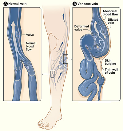
varicose veins

You may be at higher risk for weak vein walls due to increasing age or a family history of varicose veins. You also may be at higher risk if you have increased pressure in your veins due to overweight or obesity or pregnancy.

Signs and Symptoms

The signs and symptoms of varicose veins include:

  • Large veins that you can see on your skin.
  • Mild swelling of your ankles and feet.
  • Painful, achy, or “heavy” legs.
  • Throbbing or cramping in your legs.
  • Itchy legs, especially on the lower leg and ankle. This is sometimes incorrectly diagnosed as dry skin.
  • Discolored skin in the area around the varicose vein.

Signs of telangiectasias are red clusters of veins that you can see on your skin. They’re usually found on the upper body, including the face. Signs of spider veins are red or blue veins in a web pattern that often show up on the legs and face.

See your doctor if you have these signs and symptoms. They also may be signs of other, sometimes more serious conditions.

Complications

Sometimes varicose veins can lead to dermatitis (der-ma-TI-tis), an itchy rash. If you have varicose veins in your legs, dermatitis may affect your lower leg or ankle. Dermatitis can cause bleeding or skin ulcers if the skin is scratched or irritated.

Varicose veins also may lead to a condition called superficial thrombophlebitis. Thrombophlebitis is a blood clot in a vein. Superficial thrombophlebitis means that the blood clot occurs in a vein close to the surface of the skin. This type of blood clot may cause pain and other problems in the affected area.

Treatment Overview

Varicose veins are treated with lifestyle changes and medical procedures. The goals of treatment are to relieve symptoms, prevent complications, and improve appearance.

If your varicose veins cause few symptoms, your doctor may suggest simply making lifestyle changes. If your symptoms are more severe, your doctor may recommend one or more medical procedures. For example, you may need a medical procedure if you have significant pain, blood clots, or skin disorders as a result of your varicose veins.

Some people who have varicose veins choose to have procedures to improve the appearance of their varicose veins.

Although treatment can help existing varicose veins, it can’t keep new varicose veins from forming.

Lifestyle Changes

Lifestyle changes often are the first treatment for varicose veins. These changes can prevent varicose veins from getting worse, reduce pain, and delay other varicose veins from forming. Lifestyle changes include the following:

  • Avoid standing or sitting for long periods without taking a break. When sitting, avoid crossing your legs. Raise your legs when sitting, resting, or sleeping. When you can, raise your legs above the level of your heart.
  • Do physical activities to get your legs moving and improve muscle tone. This helps blood move through your veins.
  • If you’re overweight or obese, lose weight. This will improve blood flow and ease the pressure on your veins.
  • Avoid wearing tight clothes, especially those that are tight around your waist, groin (upper thighs), and legs. Tight clothes can make varicose veins worse.
  • Avoid wearing high heels for long periods. Lower heeled shoes can help tone your calf muscles. Toned muscles help blood move through the veins.

Your doctor may recommend compression stockings. These stockings create gentle pressure up the leg. This pressure keeps blood from pooling and decreases swelling in the legs.

There are three types of compression stockings. One type is support pantyhose. These offer the least amount of pressure. A second type is over-the-counter compression hose. These stockings give a little more pressure than support pantyhose. Over-the-counter compression hose are sold in medical supply stores and pharmacies.

Prescription-strength compression hose are the third type of compression stockings. These stockings offer the greatest amount of pressure. They also are sold in medical supply stores and pharmacies. However, you need to be fitted for them in the store by a specially trained person.

Procedures & Surgical Interventions

Medical procedures are done either to remove varicose veins or to close them. Removing or closing varicose veins usually doesn’t cause problems with blood flow because the blood starts moving through other veins.

You may be treated with one or more of the procedures listed below. Common side effects right after most of these procedures include bruising, swelling, skin discoloration, and slight pain.

The side effects are most severe with vein stripping and ligation (li-GA-shun). Although rare, this procedure can cause severe pain, infection, blood clots, and scarring.

Sclerotherapy

Sclerotherapy uses a liquid chemical to close off a varicose vein. The chemical is injected into the vein to cause irritation and scarring inside the vein. The irritation and scarring cause the vein to close off, and it fades away.

This procedure often is used to treat smaller varicose veins and spider veins. It can be done in your doctor’s office, while you stand. You may need several treatments to completely close off a vein.

Treatments are typically done every 4 to 6 weeks. Following treatments, your legs will be wrapped in elastic bandaging to help healing and decrease swelling.

Microsclerotherapy

Microsclerotherapy is used to treat spider veins and other very small varicose veins. A small amount of liquid chemical is injected into a vein using a very fine needle. The chemical scars the inner lining of the vein, causing it to close off.

Laser Surgery

This procedure applies light energy from a laser onto a varicose vein. The laser light makes the vein fade away. Laser surgery is mostly used to treat smaller varicose veins. No cutting or injection of chemicals is involved.

Endovenous Ablation Therapy

Endovenous ablation (ab-LA-shun) therapy uses lasers or radiowaves to create heat to close off a varicose vein. Your doctor makes a tiny cut in your skin near the varicose vein. He or she then inserts a small tube called a catheter into the vein. A device at the tip of the tube heats up the inside of the vein and closes it off.

You will be awake during this procedure, but your doctor will numb the area around the vein. You usually can go home the same day as the procedure.

Endoscopic Vein Surgery

For endoscopic (en-do-SKOP-ik) vein surgery, your doctor will make a small cut in your skin near a varicose vein. He or she then uses a tiny camera at the end of a thin tube to move through the vein. A surgical device at the end of the camera is used to close the vein. Endoscopic vein surgery usually is used only in severe cases when varicose veins are causing skin ulcers. After the procedure, you usually can return to your normal activities within a few weeks.

Ambulatory Phlebectomy

For ambulatory phlebectomy (fle-BEK-to-me), your doctor will make small cuts in your skin to remove small varicose veins. This procedure usually is done to remove the varicose veins closest to the surface of your skin. You will be awake during the procedure, but your doctor will numb the area around the vein. Usually, you can go home the same day that the procedure is done.

Vein Stripping and Ligation

Vein stripping and ligation typically is done only for severe cases of varicose veins. The procedure involves tying shut and removing the veins through small cuts in your skin. You will be given medicine to temporarily put you to sleep so you don’t feel any pain during the procedure. Vein stripping and ligation usually is done as an outpatient procedure. The recovery time from the procedure is about 1 to 4 weeks.

Limiting the Effects of Varicose Veins

You can’t prevent varicose veins from forming. However, you can prevent the ones you have from getting worse. You also can take steps to delay other varicose veins from forming:

  • Avoid standing or sitting for long periods without taking a break.
  • When sitting, avoid crossing your legs. Raise your legs when sitting, resting, or sleeping. When you can, raise your legs above the level of your heart.
  • Do physical activities to get your legs moving and improve muscle tone. This helps blood move through your veins.
  • If you’re overweight or obese, lose weight. This will improve blood flow and ease the pressure on your veins.
  • Avoid wearing tight clothes, especially those that are tight around your waist, groin (upper thighs), and legs. Tight clothes can make varicose veins worse.
  • Avoid wearing high heels for long periods. Lower heeled shoes can help tone your calf muscles. Toned muscles help blood move through the veins.
  • Wear compression stockings if your doctor advises you to. These stockings create gentle pressure up the leg. This pressure keeps blood from pooling in the veins and decreases swelling in the legs.


Urinary Tract Infection: Causes, Symptoms & Treatments

Urinary Tract Infection (UTI) Facts

  • A urinary tract infection (UTI) is an infection that occurs when bacteria enters into any part of the urinary tract, including the kidneys, ureters, bladder, or urethra.
  • Risk factors for urinary tract infections include being female, menopause, wiping from back to front after a bowel movement, sexual intercourse, some types of birth control, douches, diabetes, urinary catheters, kidney stones, genitourinary surgery, or structural abnormalities of the urinary tract.
  • UTI symptoms and signs include
    • pain or burning when urinating,
    • frequent urination,
    • sudden urge to urinate,
    • frequent urge to urinate without much urine passing, and
    • urine that is milky/cloudy/bloody/foul smelling.
  • See a health-care provider for diagnosis because some types of UTIs can be serious to life-threatening conditions.
  • UTIs are usually treated with antibiotics.
  • Most cases of UTIs go away with treatment, but in some cases, people may have recurrent urinary tract infections.
  • Serious UTIs may lead to scarring of the urinary tract or pyelonephritis (kidney infection).

What Is the Definition of a Urinary Tract Infection (UTI)?

 A urinary tract infection is an infection that can occur in any area of the urinary tract, including the ureters, bladder, kidneys, or urethra. Bladder infections (cystitis) and urethra infections (urethritis) are most common.

Picture of the urinary tract

Urinary tract infections can be categorized as either simple or complicated.

  • Simple UTIs occur in healthy people with normal urinary tracts. This is the type of UTI that occurs most frequently in women.
  • Complicated UTIs occur in individuals with abnormal urinary tracts or when underlying medical conditions make treatment failure more likely. Men and children are more likely to have this type of UTI.


What Are Causes of Urinary Tract Infections?

When bacteria enter into the urinary tract, this can result in an infection. Escherichia coli (E. coli) is the bacteria that causes the vast majority of UTIs. However, other bacterial pathogens can also cause UTIs. A urine culture can help isolate the bacteria responsible for a particular UTI.

Urinary tract infections are not considered to be contagious, and you can’t acquire a UTI from someone else.

What Are Urinary Tract Infection Risk Factors?

Risk factors for developing urinary tract infections include the following:

  • Wiping from back to front following a bowel movement, particularly in women, can introduce bacteria into the urethra.
  • Sexual intercourse can push bacteria from the vaginal area into the urethra.
  • Holding the urine too long: When someone holds it in, more bacteria have a chance to multiply, which can cause or worsen a UTI.
  • Kidney stones can make it hard to empty the bladder completely, which can also lead to urine remaining in the bladder too long.
  • Certain types of birth control devices (contraceptives), including diaphragms or condoms with spermicides
  • Hormonal changes and changes in the vagina following menopause
  • Using urinary catheters, which are small tubes inserted into the bladder to drain urine, can predispose someone to UTIs.
  • Surgery of the genitourinary tract may introduce bacteria into the urinary tract, resulting in a UTI.
  • Women tend to get UTIs more frequently than men because the urethra in women is shorter and located closer to the rectum.
  • Use of douches
  • Older adults
  • Taking oral antibiotics
  • Diabetes or other illness that compromise the immune system
  • Urinary incontinence
  • Spinal cord injuries
  • Multiple sclerosis
  • Parkinson’s disease
  • Previous urinary tract infection
  • Enlarged prostate (benign prostatic hyperplasia [BPH])
  • Structural abnormalities of the genitourinary tract
  • Uncircumcised men

What Are Symptoms and Signs of a Urinary Tract Infection?

Symptoms and signs of a urinary tract infection include

  • pain or burning when urinating (dysuria);
  • frequent urination;
  • sudden urge to urinate (bladder spasm);
  • frequent or persistent urge to urinate without much urine passing when you go;
  • sense of incomplete emptying of the bladder;
  • loss of bladder control (urinary incontinence);
  • a feeling of pressure or pain in the lower abdomen or pelvis;
  • foul odor to the urine;
  • urine that is milky, cloudy, reddish, or dark in color;
  • blood in the urine;
  • back pain, flank (side) pain, or groin pain;
  • fever or chills;
  • pain during sexual intercourse;
  • fatigue;
  • general feeling of being unwell (malaise);
  • vaginal irritation; and
  • in elderly patients, subtle symptoms such as altered mental status (confusion) or decreased activity may be signs of a UTI.

Vaginal itching is not a typical symptom of a UTI. It may be a sign of bacterial vaginosis or a vaginal yeast infection.

If one is experiencing fever or back pain, this may be a sign of a kidney infection (pyelonephritis), which can be a serious medical issue. Seek medical attention immediately.

What Are Medications and Treatments for Urinary Tract Infections?

Antibiotics are the most commonly used treatment for urinary tract infections. The duration of treatment with antibiotics for UTIs varies according to the part of the urinary tract that is infected.

  • If one has a bladder infection, antibiotics are usually taken for three to seven days.
  • If one has a kidney infection, antibiotics may be taken for up to two weeks. In certain cases, one may also require hospitalization and intravenous antibiotics.


ME/CFS: Causes, Symptoms & Treatments

Myalgic encephalomyelitis/chronic fatigue syndrome (ME/CFS) is a disabling and complex illness.

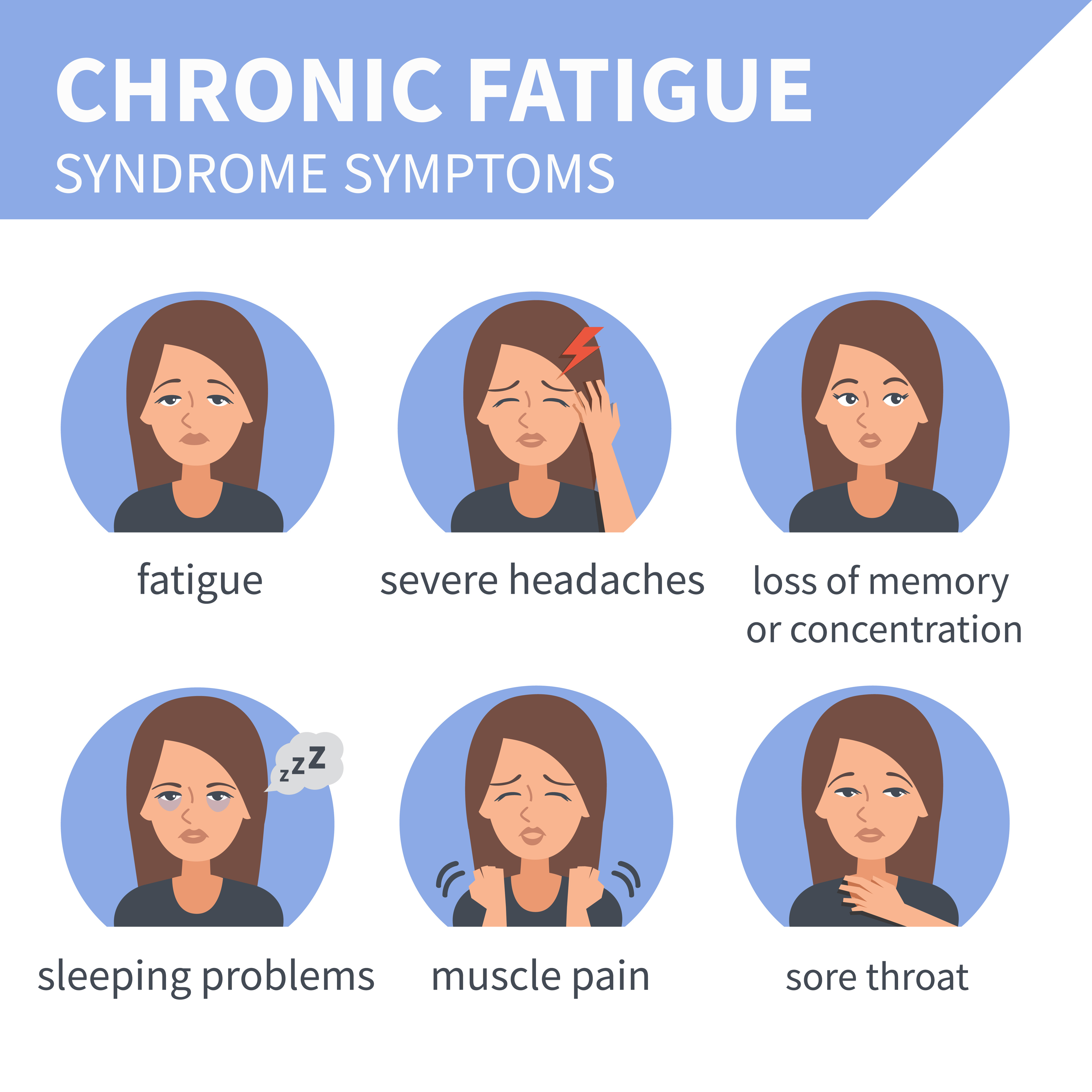
People with ME/CFS are often not able to do their usual activities. At times, ME/CFS may confine them to bed. People with ME/CFS have overwhelming fatigue that is not improved by rest. ME/CFS may get worse after any activity, whether it’s physical or mental. This symptom is known as post-exertional malaise (PEM). Other symptoms can include problems with sleep, thinking and concentrating, pain, and dizziness. People with ME/CFS may not look ill. However,

  • People with ME/CFS are not able to function the same way they did before they became ill.
  • ME/CFS changes people’s ability to do daily tasks, like taking a shower or preparing a meal.
  • ME/CFS often makes it hard to keep a job, go to school, and take part in family and social life.
  • ME/CFS can last for years and sometimes leads to serious disability.
  • At least one in four ME/CFS patients is bed- or house-bound for long periods during their illness.

Anyone can get ME/CFS. While most common in people between 40 and 60 years old, the illness affects children, adolescents, and adults of all ages. Among adults, women are affected more often than men. Whites are diagnosed more than other races and ethnicities. But many people with ME/CFS have not been diagnosed, especially among minorities.

Scientists have not yet identified what causes myalgic encephalomyelitis/chronic fatigue syndrome (ME/CFS). It is possible that ME/CFS has more than one cause, meaning that patients with ME/CFS could have illness resulting from different causes (see below). In addition, it is possible that two or more triggers might work together to cause the illness.

Some of the areas that are being studied as possible causes of ME/CFS are:

  • Infections collapsed
  • Immune System Changes collapsed
  • Stress Affecting Body Chemistry collapsed
  • Changes in Energy Production collapsed
  • Possible Genetic Link

Primary Symptoms:

Also called “core” symptoms, these occur in most patients with ME/CFS. The three primary symptoms required for diagnosis are:

  • Greatly lowered ability to do activities that were usual before the illness. This drop in activity level occurs along with fatigue and must last six months or longer. People with ME/CFS have fatigue that is very different from just being tired. The fatigue of ME/CFS:
    • Can be severe.
    • Is not a result of unusually difficult activity.
    • Is not relieved by sleep or rest.
    • Was not a problem before becoming ill (not life-long).
  • Worsening of ME/CFS symptoms after physical or mental activity that would not have caused a problem before illness. This is known as post-exertional malaise (PEM). People with ME/CFS often describe this experience as a “crash,” “relapse,” or “collapse.” It may take days, weeks, or longer to recover from a crash. Sometimes patients may be house-bound or even completely bed-bound during crashes. People with ME/CFS may not be able to predict what will cause a crash or how long it will last. As examples:
    • Attending a child’s school event may leave someone house-bound for a couple of days and not able to do needed tasks, like laundry.
    • Shopping at the grocery store may cause a physical crash that requires a nap in the car before driving home or a call for a ride home.
    • Taking a shower may leave someone with severe ME/CFS bed-bound and unable to do anything for days.
  • Sleep problems. People with ME/CFS may not feel better or less tired, even after a full night of sleep. Some people with ME/CFS may have problems falling asleep or staying asleep.

In addition to these core symptoms, one of the following two symptoms is required for diagnosis:

  • Problems with thinking and memory. Most people with ME/CFS have trouble thinking quickly, remembering things, and paying attention to details. Patients often say they have “brain fog” to describe this problem because they feel “stuck in a fog” and not able to think clearly.
  • Worsening of symptoms while standing or sitting upright. This is called orthostatic intolerance. People with ME/CFS may be lightheaded, dizzy, weak, or faint while standing or sitting up. They may have vision changes like blurring or seeing spots.

Other Common Symptoms:

Many but not all people with ME/CFS have other symptoms.

Pain is very common in people with ME/CFS. The type of pain, where it occurs, and how bad it is varies a lot. The pain people with ME/CFS feel is not caused by an injury. The most common types of pain in ME/CFS are:

  • Muscle pain and aches
  • Joint pain without swelling or redness
  • Headaches, either new or worsening

Some people with ME/CFS may also have:

  • Tender lymph nodes in the neck or armpits
  • A sore throat that happens often
  • Digestive issues, like irritable bowel syndrome
  • Chills and night sweats
  • Allergies and sensitivities to foods, odors, chemicals, or noise

Treatments:

There is no cure or approved treatment for myalgic encephalomyelitis/chronic fatigue syndrome (ME/CFS). However, some symptoms can be treated or managed. Treating these symptoms might provide relief for some patients with ME/CFS but not others. Other strategies, like learning new ways to manage activity, can also be helpful.

1.Depression, Stress, and Anxiety

Adjusting to a chronic, debilitating illness sometimes leads to other problems, including depression, stress, and anxiety. Many patients with ME/CFS develop depression during their illness. When present, depression or anxiety should be treated. Although treating depression or anxiety can be helpful, it is not a cure for ME/CFS.

Some people with ME/CFS might benefit from antidepressants and anti-anxiety medications. However, doctors should use caution in prescribing these medications. Some drugs used to treat depression have other effects that might worsen other ME/CFS symptoms and cause side effects. When healthcare providers are concerned about patient’s psychological condition, they may recommend seeing a mental health professional.

Some people with ME/CFS might benefit from trying techniques like deep breathing and muscle relaxation, massage, and movement therapies (such as stretching, yoga, and tai chi). These can reduce stress and anxiety, and promote a sense of well-being.

2.Dizziness and Lightheadedness (Orthostatic Intolerance)

Some people with ME/CFS might also have symptoms of orthostatic intolerance that are triggered when-or made worse by-standing or sitting upright. These symptoms can include:

  • Frequent dizziness and lightheadedness
  • Changes in vision (blurred vision, seeing white or black spots)
  • Weakness
  • Feeling like your heart is beating too fast or too hard, fluttering, or skipping a beat

For patients with these symptoms, their doctor will check their heart rate and blood pressure, and may recommend they see a specialist, like a cardiologist or neurologist.

For people with ME/CFS who do not have heart or blood vessel disease, doctor might suggest patients increase daily fluid and salt intake and use support stockings. If symptoms do not improve, prescription medication can be considered.

3. Sleep Problems

Patients with ME/CFS often feel less refreshed and restored after sleep than they did before they became ill. Common sleep complaints include difficulty falling or staying asleep, extreme sleepiness, intense and vivid dreaming, restless legs, and nighttime muscle spasms.

Good sleep habits are important for all people, including those with ME/CFS. Some common tips for good sleep are:

  • Start a regular bedtime routine with a long, calming wind-down period.
  • Go to bed at same time each night and wake up at same time each morning.
  • Limit daytime naps to 30 minutes in total during the day.
  • Remove all TVs, computers, phones, and gadgets from bedroom.
  • Use the bed only for sleep and sex and not for other activities (avoid reading, watching TV, listening to music, or using phones).
  • Control noise, light, and temperature.
  • Avoid caffeine, alcohol, and large meals before bedtime.
  • Avoid exercise right before going to bed. Light exercise and stretching earlier in the day, at least four hours before bedtime, might improve sleep.

When people try these techniques but are still unable to sleep, their doctor might recommend taking medicine to help with sleep. First, people should try over-the-counter sleep products. If this does not help, doctors can offer a prescription sleep medicine, starting at the smallest dose and using for the shortest possible time.

People might continue to feel unrefreshed even after the medications help them to get a full night of sleep. If so, they should consider seeing a sleep specialist. Most people with sleep disorders, like sleep apnea (brief pause in breathing during sleep) and narcolepsy (uncontrollable sleeping), respond to therapy. However, for people with ME/CFS, not all symptoms may go away.

Pulmonary Hypertension: Symptoms, Causes & Treatments

Pulmonary hypertension is a type of high blood pressure that affects the arteries in the lungs and the right side of your heart.

Pulmonary hypertension begins when tiny arteries in your lungs, called pulmonary arteries, and capillaries become narrowed, blocked or destroyed. This makes it harder for blood to flow through your lungs, and raises pressure within your lungs’ arteries. As the pressure builds, your heart’s lower right chamber (right ventricle) must work harder to pump blood through your lungs, eventually causing your heart muscle to weaken and eventually fail.

Pulmonary hypertension is a serious illness that becomes progressively worse and is sometimes fatal. Although pulmonary hypertension isn’t curable, treatments are available that can help lessen symptoms and improve your quality of life.

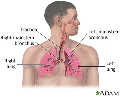
Respiratory system

Symptoms:

The signs and symptoms of pulmonary hypertension in its early stages may not be noticeable for months or even years. As the disease progresses, symptoms become worse.

Pulmonary hypertension symptoms include:

  • Shortness of breath (dyspnea), initially while exercising and eventually while at rest
  • Fatigue
  • Dizziness or fainting spells (syncope)
  • Chest pressure or pain
  • Swelling (edema) in your ankles, legs and eventually in your abdomen (ascites)
  • Bluish color to your lips and skin (cyanosis)
  • Racing pulse or heart palpitations

Causes:

Your heart has two upper and two lower chambers. Each time blood passes through your heart, the lower right chamber (right ventricle) pumps blood to your lungs through a large blood vessel (pulmonary artery). In your lungs, the blood releases carbon dioxide and picks up oxygen. The oxygen-rich blood then flows through blood vessels in your lungs (pulmonary arteries, capillaries and veins) to the left side of your heart.

Ordinarily, the blood flows easily through the vessels in your lungs, so blood pressure is usually much lower in your lungs. With pulmonary hypertension, the rise in blood pressure is caused by changes in the cells that line your pulmonary arteries. These changes cause extra tissue to form, eventually narrowing or completely blocking the blood vessels, making the arteries stiff and narrow. This makes it harder for blood to flow, raising the blood pressure in the pulmonary arteries.

Idiopathic pulmonary hypertension

When an underlying cause for high blood pressure in the lungs can’t be found, the condition is called idiopathic pulmonary hypertension (IPH).

Some people with IPH may have a gene that’s a risk factor for developing pulmonary hypertension. But in most people with idiopathic pulmonary hypertension, there is no recognized cause of their pulmonary hypertension.

Secondary pulmonary hypertension

Pulmonary hypertension that’s caused by another medical problem is called secondary pulmonary hypertension. This type of pulmonary hypertension is more common than is idiopathic pulmonary hypertension. Causes of secondary pulmonary hypertension include:

  • Blood clots in the lungs (pulmonary emboli)
  • Chronic obstructive pulmonary diseases, such as emphysema
  • Connective tissue disorders, such as scleroderma or lupus
  • Sleep apnea and other sleep disorders
  • Heart abnormalities you’re born with (congenital heart defects)
  • Sickle cell anemia
  • Chronic liver disease (cirrhosis)
  • AIDS
  • Lung diseases such as pulmonary fibrosis, a condition that causes scarring in the tissue between the lungs’ air sacs (interstitium)
  • Left-sided heart failure
  • Living at altitudes higher than 8,000 feet (2,438 meters)
  • Climbing or hiking to altitudes higher than 8,000 feet (2,438 meters) without acclimating first
  • Use of certain stimulant drugs, such as cocaine

Eisenmenger syndrome and pulmonary hypertension

Eisenmenger syndrome, a type of congenital heart defect, causes pulmonary hypertension. It is most commonly caused by a large hole in your heart between the two lower heart chambers (ventricles), called a ventricular septal defect (VSD). This hole in your heart causes blood to circulate abnormally in your heart. Oxygen-carrying blood (red blood) mixes with oxygen-poor blood (blue blood). The blood then returns to your lungs instead of going to the rest of your body, increasing the pressure in the pulmonary arteries and causing pulmonary hypertension.


 Treatments:

Medications:


  • Blood vessel dilators (vasodilators). Vasodilators open narrowed blood vessels. One of the most commonly prescribed vasodilators for pulmonary hypertension is epoprostenol (Flolan). The drawback to epoprostenol is that its effects last only a few minutes. This drug is continuously injected through an intravenous (IV) catheter via a small pump that you wear in a pack on your belt or shoulder. This means that you’ll learn to prepare your own medication mixture, operate the pump and care for the IV catheter. You’ll need comprehensive follow-up care. Potential side effects of epoprostenol include jaw pain, nausea, diarrhea, leg cramps, as well as pain and infection at the IV site.Another form of the drug, iloprost (Ventavis), avoids many of these problems. Iloprost can be inhaled every three hours through a nebulizer, a machine that vaporizes your medication, making it far more convenient and less painful to use. And because it’s inhaled, it goes directly to the lungs. Side effects associated with iloprost include chest pain — often accompanied by headache and nausea — and breathlessness.
  • Endothelin receptor antagonists. These medications reverse the effect of endothelin, a substance in the walls of blood vessels that causes them to narrow. One of these medications, bosentan (Tracleer), may improve your energy level and symptoms. The drug isn’t for pregnant women. If you take bosentan, you’ll need monthly liver monitoring, because the drug can damage your liver. Ambrisentan (Letairis) is another medication that stops the narrowing of your blood vessels. This drug can cause serious liver damage if not taken appropriately, and it shouldn’t be taken by pregnant women. Before taking the drug, tell your doctor about any liver condition you have.
  • Sildenafil and tadalafil. Sildenafil (Revatio, Viagra) and tadalafil (Cialis, Adcirca) are sometimes used to treat pulmonary hypertension. These drugs work by opening the blood vessels in the lungs to allow blood to flow through more easily. Side effects include upset stomach, dizziness and vision problems.
  • High-dose calcium channel blockers. These drugs help relax the muscles in the walls of your blood vessels. They include medications such as amlodipine (Norvasc), diltiazem (Cardizem, Tiazac, others) and nifedipine (Adalat, Procardia). Although calcium channel blockers can be effective, only a small number of people with pulmonary hypertension respond to them.
  • Anticoagulants. Your doctor is likely to prescribe the anticoagulant warfarin (Coumadin, Jantoven) to help prevent the formation of blood clots within the small pulmonary arteries. Because anticoagulants prevent normal blood coagulation, they increase your risk of bleeding complications. Take warfarin exactly as prescribed, because warfarin can cause severe side effects if taken incorrectly. If you’re taking warfarin, your doctor will ask you to have periodic blood tests to check how well the drug is working. Many other drugs, herbal supplements and foods can interact with warfarin, so be sure your doctor knows all of the medications you’re taking.
  • Diuretics. Commonly known as water pills, these medications help eliminate excess fluid from your body. This reduces the amount of work your heart has to do. They also may be used to limit fluid buildup in your lungs.
  • Oxygen. Your doctor may suggest that you sometimes breathe pure oxygen, a treatment known as oxygen therapy, to help treat pulmonary hypertension, especially if you live at a high altitude or have sleep apnea. Some people with pulmonary hypertension eventually require constant oxygen therapy.

Surgeries

  • Atrial septostomy. If medications don’t control your pulmonary hypertension, this open-heart surgery may be an option. In an atrial septostomy, a surgeon will create an opening between the left and right chambers of your heart to relieve the pressure on the right side of your heart. Atrial septostomy can have serious complications, including heart rhythm abnormalities (arrhythmias).
  • Transplantation. In some cases, a lung or heart-lung transplant may be an option, especially for younger people who have idiopathic pulmonary hypertension. Major risks of any type of transplantation include rejection of the transplanted organ and serious infection, and you must take immunosuppressant drugs for life to help reduce the chance of rejection.

Anosmia: Causes, Symptoms & Treatments

Fortunately, for most people, anosmia is a temporary nuisance caused by a severely stuffy nose from a cold. Once the cold runs its course, a person’s sense of smell returns.But for some people, including many elderly, the loss of a sense of smell may persist. In addition, anosmia can be a sign of a more serious medical condition. Any ongoing problems with smell should be checked out by a doctor.

The Basics of Smell

A person’s sense of smell is driven by certain processes. First, a molecule released from a substance (such as fragrance from a flower) must stimulate special nerve cells (called olfactory cells) found high up in the nose. These nerve cells then send information to the brain, where the specific smell is identified. Anything that interferes with these processes, such as nasal congestion, nasal blockage, or damage to the nerve cells themselves, can lead to loss of smell.

The ability to smell also affects our ability to taste. Without the sense of smell, our taste buds can only detect a few flavors, and this can affect your quality of life.

Anosmia Causes

Nasal congestion from a cold, allergy, sinus infection, or poor air quality is the most common cause of anosmia. Other anosmia causes include:

  • Nasal polyps — small noncancerous growths in the nose and sinusesthat block the nasal passage.
  • Injury to the nose and smell nerves from surgery or head trauma.
  • Exposure to toxic chemicals, such as pesticides or solvents.
  • Certain medications, including antibiotics, antidepressants, anti-inflammatory medication, heart medications, and others.
  • Cocaine abuse.
  • Old age. Like vision and hearing, your sense of smell can become weaker as you age. In fact, one’s sense of smell is most keen between the ages of 30 and 60 and begins to decline after age 60.
  • Certain medical conditions, such as Alzheimer’s disease, Parkinson’s disease, multiple sclerosis, nutritional deficiencies, congenital conditions, and hormonal disturbances.
  • Radiation treatment of head and neck cancers.

Anosmia Symptoms

The obvious sign of anosmia is a loss of smell. Some people with anosmia notice a change in the way things smell. For example, familiar things begin to lack odor.

Anosmia Diagnosis

If you experience a loss of smell that you can’t attribute to a cold or allergy or which doesn’t get better after a week or two, tell your doctor. Your doctor can take a look inside your nose with a special instrument to see if a polyp or growth is impairing your ability to smell or if an infection is present.

Further testing by a doctor who specializes in nose and sinus problems an ear, nose, and throat doctor (ENT, or an otolaryngologist) — may be needed to determine the cause of anosmia. A CT scan may be necessary so that the doctor can get a better look of the area.

Anosmia Treatments

If nasal congestion from a cold or allergy is the cause of anosmia, treatment is usually not needed, and the problem will get better on its own. Short-term use of over-the-counter decongestants may open up your nasal passages so that you can breathe easier. However, if the congestion gets worse or does not go away after a few days, see your doctor. You may have an infection and need antibiotics, or another medical condition may be to blame.

Endometriosis: Causes, Symptoms & Treatments

What is endometriosis?

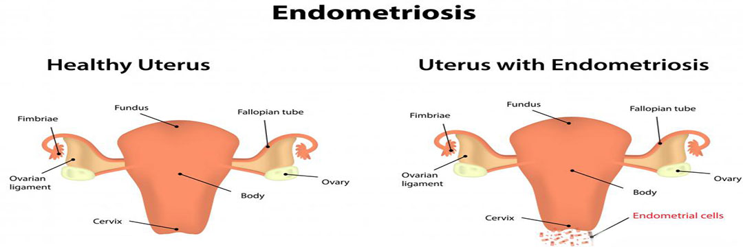
Endometriosis is the abnormal growth of endometrial tissue similar to that which lines the interior of the uterus, but in a location outside of the uterus. Endometrial tissue is shed each month during menstruation. Areas of endometrial tissue found in ectopic locations are called endometrial implants. These lesions are most commonly found on the ovaries, the Fallopian tubes, the surface of the uterus, the bowel, and on the membrane lining of the pelvic cavity (i.e. the peritoneum). They are less commonly found to involve the vagina, cervix, and bladder. Rarely, endometriosis can occur outside the pelvis. Endometriosis has been reported in the liver, brain, lung, and old surgical scars. Endometrial implants, while they may become problematic, are usually benign (i.e. non-cancerous).

What are the stages of endometriosis?

Endometriosis is classified into one of four stages (I-minimal, II-mild, III-moderate, and IV-severe) based upon the exact location, extent, and depth of the endometriosis implants as well as the presence and severity of scar tissue and the presence and size of endometrial implants in the ovaries. Most cases of endometriosis are classified as minimal or mild, which means there are superficial implants and mild scarring. Moderate and severe endometriosis typically result in cysts and more severe scarring. The stage of endometriosis is not related to the degree of symptoms a woman experiences, but infertility is common with stage IV endometriosis.

What are the signs and symptoms endometriosis?

Most women who have endometriosis, in fact, do not have symptoms. Of those who do, the most common include:

  • Pain (usually pelvic) that usually occurs just before menstruation and lessens after menstruation
  • Painful sexual intercourse
  • Cramping during intercourse
  • Cramping or pain during bowel movements or urination
  • Infertility
  • Pain with pelvic examinations

The intensity of the pain can vary from month to month, and can vary greatly among affected individuals. Some women experience progressive worsening of symptoms, while others can have resolution of pain without treatment.

Pelvic pain in women with endometriosis depends partly on where endometrial implants of endometriosis are located.

  • Deeper implants and implants in areas of high nerve density are more apt to produce pain.
  • The implants may also release substances into the bloodstream which are capable of eliciting pain.
  • Pain can result when endometriotic implants incite scarring of surrounding tissues. There appears to be no relationship between severity of pain and the amount of anatomical disease which is present.

Endometriosis can be one of the reasons for infertility for otherwise healthy couples. When laparoscopic examinations are performed during evaluations for infertility, implants are often found in individuals who are totally asymptomatic. The reasons diminished fertility in many patients with endometriosis are not understood. Endometriosis may incite scar tissue formation within the pelvis. If the ovaries and Fallopian tubes are involved, the mechanical processes involved in the transfer of fertilized eggs into the tubes may be altered. Alternatively, the endometriotic lesions may produce inflammatory substances which adversely affect ovulation, fertilization, and implantation.

Other symptoms that can be related to endometriosis include

  • lower abdominal pain,
  • diarrhea and/or constipation,
  • low back pain,
  • chronic fatigue
  • irregular or heavy menstruation,
  • painful urination, or
  • bloody urine (particularly during menstruation).

Rare symptoms of endometriosis include chest pain or coughing blood due to endometriosis in the lungs and headache and/or seizures due to endometriosis in the brain.

Does endometriosis increase a woman’s risk of getting cancer?

Some studies have postulated that women with endometriosis have an increased risk for development of certain types of ovarian cancer, known as epithelial ovarian cancer (EOC). This risk is highest in women with both endometriosis and primary infertility (those who have never conceived a pregnancy). The use of combination oral contraceptive pills (OCPs), which are sometimes used in the treatment of endometriosis, appears to significantly reduce this risk.

The reasons for the association between endometriosis and ovarian epithelial cancer are not clearly understood. One theory is that the endometriosis implants themselves undergo malignanttransformation to cancer. Another possibility is that the presence of endometriosis may be related to other genetic or environmental factors that serve to increase a women’s risk of developing ovarian cancer.

What causes endometriosis?

The cause of endometriosis is unknown. One theory is that the endometrial tissue is deposited in unusual locations by the retrograde flow of menstrual debris through the Fallopian tubes into the pelvic and abdominal cavities. The cause of this retrograde menstruation is not clearly understood. It is clear that retrograde menstruation is not the only cause of endometriosis, as many women who have retrograde menstruation do not develop the condition.

Another possibility is that areas lining the pelvic organs possess primitive cells that are able to develop into other forms of tissue, such as endometrium. (This process is termed coelomic metaplasia.)

It is also likely the direct transfer of endometrial tissues at the time of surgery may be responsible for the endometriosis implants occasionally found in surgical scars (for example, episiotomy or Cesarean section scars). Transfer of endometrial cells via the bloodstream or lymphatic system is the most plausible explanation for the rare cases of endometriosis that are found in the brain and other organs remote from the pelvis.

Finally, there is evidence that some women with endometriosis have an altered immune response in women with endometriosis, which may affect the body’s natural ability to recognize ectopic endometrial tissue.

What medications treat endometriosis?

Nonsteroidal anti-inflammatory drugs (NSAIDs)

Nonsteroidal anti-inflammatory drugs or NSAIDs (such as ibuprofen or naproxen sodium) are commonly prescribed to help relieve pelvic pain and menstrual cramping. These pain-relieving medications have no effect on the endometrial implants or the progression of endometriosis. However, they do decrease prostaglandin production, and prostaglandins are well-known to have a role in the causation of pain. As the diagnosis of endometriosis can only be definitively confirmed with a biopsy, many women with complaints suspected to arise from endometriosis are treated for pain first without a firm diagnosis being established. Under such circumstances, NSAIDs are commonly used as a first line empirical treatment. If they are effective in controlling the pain, no other procedures or medical treatments are needed. If they are ineffective, additional evaluation and treatment will be necessary.

Since endometriosis occurs during the reproductive years, many of the available medical treatments for endometriosis rely on interruption of the normal cyclical hormone production by the ovaries. These medications include GnRH analogs, oral contraceptive pills, and progestins.

Gonadotropin-releasing hormone analogs (GnRH analogs)

Gonadotropin-releasing hormone analogs (GnRH analogs) have been effectively used to relieve pain and reduce the size of endometriosis implants. These drugs suppress estrogen production by the ovaries by inhibiting the secretion of regulatory hormones from the pituitary gland. As a result, menstrual periods stop, mimicking menopause. Nasal and injection forms of GnRH agonists are available.

The side effects are a result of the lack of estrogen, and include:

  • hot flashes,
  • vaginal dryness,
  • irregular vaginal bleeding,
  • mood alterations,
  • fatigue, and
  • loss of bone density (osteoporosis).

Fortunately, by adding back small amounts of progesterone in pill form (similar to treatments sometimes used for symptom relief in menopause), many of the annoying side effects due to estrogen deficiency can be avoided. “Add back therapy” is a term that refers to this modern way of administering GnRH agonists along with progesterone in a way to ensure compliance by eliminating most of the unwanted side effects of GnRH therapy.

Oral contraceptive pills

Oral contraceptive pills (estrogen and progesterone in combination) are also sometimes used to treat endometriosis. The most common combination used is in the form of the oral contraceptive pill (OCP). Sometimes women who have severe menstrual pain are asked to take the OCP continuously, meaning skipping the placebo (hormonally inert) portion of the cycle. Continuous use in this manner will generally stop menstruation altogether. Occasionally, weight gain, breast tenderness, nausea, and irregular bleeding may occur. Oral contraceptive pills are usually well-tolerated in women with endometriosis.

Progestins

Progestins [for example, medroxyprogesterone acetate (Provera, Cycrin, Amen), norethindrone acetate, norgestrel acetate (Ovrette)] are more potent than birth control pills and are recommended for women who do not obtain pain relief from or cannot take a birth control pill. They may be helpful in women who do not respond, or cannot take (for medical reasons) oral contraceptives.

Side effects are more common and include:

  • breast tenderness,
  • bloating,
  • weight gain,
  • irregular uterine bleeding, and
  • depression.

Because the absence of menstruation (amenorrhea) induced by high doses of progestins can last many months following cessation of therapy, these drugs are not recommended for women planning pregnancy immediately following cessation of therapy.

Other drugs used to treat endometriosis

Danazol (Danocrine)

Danazol (Danocrine) is a synthetic drug that creates a high androgen (male type hormone) and low estrogen hormonal environment by interfering with ovulation and ovarian production of estrogen. Eighty percent of women who take this drug will have pain relief and shrinkage of endometriosis implants, but up to 75% of women develop significant side effects from the drug. These include:

  • weight gain,
  • edema (swelling),
  • breast shrinkage,
  • acne,
  • oily skin,
  • hirsutism (male pattern hair growth),
  • deepening of the voice,
  • headache,
  • hot flashes,
  • changes in libido, and
  • mood alterations.

Except for the voice changes, all of these side effects are reversible. In some cases, resolution of the side effects may take many months. Danazol should not be taken by women with certain types of liver, kidney, or heart conditions. This product is rarely used.

Aromatase inhibitors

A more current approach to the treatment of endometriosis has involved the administration of drugs known as aromatase inhibitors (for example, anastrozole [Arimidex] and letrozole [Femara]). These drugs act by interrupting local estrogen formation within the endometriosis implants themselves. They also inhibit estrogen production within the ovary and adipose tissue. Research is ongoing to evaluate the effectiveness of aromatase inhibitors in the management of endometriosis. Aromatase inhibitors can cause significant bone loss with prolonged usage. They must also be employed in combination with other drugs in premenopausal women because of their effects on the ovaries.

Can surgery cure endometriosis?

Surgical treatment for endometriosis can be useful when the symptoms are severe or there has been an inadequate response to medical therapy. Surgery is the preferred treatment when there is anatomic distortion of the pelvic organs or obstruction of the bowel or urinary tract. It may be classified either as conservative, in which the uterus and ovarian tissue are preserved, or definitive, which involves hysterectomy (removal of the uterus), with or without removal of the ovaries.

Conservative surgery is typically performed laparoscopically. Endometrial implants may be excised or destroyed by different sources of energy (e.g. laser, electrical current). If the disease is extensive and anatomy is distorted, laparotomy may be required.

While surgical treatments can be very effective in the reduction of pain, the recurrence rate of endometriosis following conservative surgical treatment has been estimated to be as high as 40%. Many doctors recommend ongoing medical therapy following surgery in an attempt to prevent symptomatic disease recurrence.

Who gets endometriosis?

Endometriosis affects women during their reproductive years. The exact prevalence of endometriosis is not known, since many women who are later identified as having the condition are asymptomatic. Endometriosis is estimated to affect over one million women (estimates range from 3% to 18% of women) in the United States. It is one of the leading causes of pelvic pain and it is responsible for many of the laparoscopic procedures and hysterectomies performed by gynecologists. Estimates suggest that 20% to 50% of women being treated for infertility have endometriosis, and up to 80% of women with chronic pelvic pain may be affected.

While most cases of endometriosis are diagnosed in women aged 25 to 35 years, endometriosis has been reported in girls as young as 11 years of age. Endometriosis is rare in postmenopausal women. Studies further suggest that endometriosis is most common in taller, thin women with a low body mass index (BMI). Delaying pregnancy until an older age, never giving birth, early onset of menses, and late menopause all have been shown to be risk factors for endometriosis. It also is likely that there are genetic factors which predispose a woman to developing endometriosis, since having a first-degree relative with the condition increases the chance that a woman will develop the condition.


Femoral Neck Fracture: Causes, Symptoms & Treatments

What Is Femoral Neck Fracture?

It is a well known fact that bones in the elderly population are quite fragile and are at increased risk for developing various forms of fractures. One such fracture which is quite common in the elderly population is the Femoral Neck Fracture. This fracture may be quite common in the elderly but it may render the individual disabled and virtually dependent on others. It may take many months for people to recuperate from this type of injury and resultant fracture and people require prolonged rehabilitation which may stretch to several months. Since such people are mostly confined to their beds they become prone to other infections and pressure sores from prolonged lying in bed. Studies reflect that more than 100,000 people of which most are elderly sustain Femoral Neck Fracture requiring prolonged hospitalization. They are usually caused due to a fall. To understand Femoral Neck Fracture, it is vital to understand the anatomy of the femoral bone. The femoral bone is attached to the hip bone in a ball and socket fashion. When Femoral Neck Fracture occurs then it is often treated with a procedure called as hemiarthroplasty in which the femoral neck is replaced by prosthesis.

What Causes Femoral Neck Fracture?

The main cause of Femoral Neck Fracture is a medical condition called Osteoporosiswhich causes the bones to become thin and weak due to loss of bone mass. This condition is quite common in the elderly population, specifically females, and hence they are more prone to fractures even due to minor injuries or falls. Talking about the younger generation, Femoral Neck Fracture can be caused due to a motor vehicle crash or a sporting injury like rugby or other contact sports. A spontaneous fracture in this area can occur in cases of carcinomas and is known by the name of pathologic fractures.

What Is The Classification Of A Femoral Neck Fracture?

Femoral Neck Fracture has been classified into four parts which are mentioned below:

Stage I Fracture of Femoral Neck: This type of femoral neck fracture is incomplete and nondisplaced.

Stage II Fracture of Femoral Neck: These types of femoral neck fracture is nondisplaced but are complete.

Stage III Fracture of Femoral Neck: These types of fractures are complete and minimally displaced femoral neck fracture.

Stage IV Fracture of Femoral Neck: These type of fractures are complete fractures which are totally displaced femoral neck fracture.

Symptoms:

  • Severe pain in and around the hip area.
  • Extreme difficulty with ambulation.

How Is Femoral Neck Fracture Treated?

To begin with, the affected leg will be put in such a position so that the leg does not move much causing the fracture to get displaced. Next thing to be done is to give the patient pain medications to control pain. Now, the treating physician will take a decision as to how to approach to treat the fracture. If the patient stays alone and does not have enough support then the patient may have to be sent to extended care facility after treatment since most of the patients are elderly and due to this fracture they would need an aide for support.

The best way to treat Femoral Neck Fracture is with surgical fixation. The procedure done may be a hemiarthroplasty in which femoral neck is replaced by prosthesis. In some cases a total hip replacement may need to be done. In total hip replacement, both the acetabulum and the femur are replaced by prosthesis. Before a surgery is performed, a complete preoperative history and physical is conducted of the patient along with blood tests and EKG to look at the cardiac status and whether the patient will be able to tolerate the procedure.

Postprocedure the patient will need extensive rehabilitation and will take quite a few months before recovery can be anticipated. Just like any other surgery, surgery for Femoral Neck Fracture also has its own inherent complications. Some of the complications of surgery for Femoral Neck Fracture are fracture nonunion, avascular necrosis (AVN), and arthritis. In case if there is development of avascular necrosis then further procedures may be required. If prosthesis is used to correct the fracture then there is always the complication of failure of prosthesis and at times dislocation. Once the patient is through with treatment and rehabilitation and has regained some stability and movement of the joint then the patient is recommended to seek active treatment for osteoporosis which is the main cause of Femoral Neck Fracture. If the patient is taken care of well, then there is every chance that the patient will recover completely from Femoral Neck Fracture

.

Preventative Strategies To Avoid Femoral Neck Fractures

Since Femoral Neck Fracture is mostly seen in the elderly population, it is vital that they follow certain precautions so as to avoid sustaining a Femoral Neck Fracture. The very first step towards this is to prevent development of osteoporosis which can be done by intake of adequate calcium and vitamin D to make the bones strong and indulging in light exercises to stay fit and healthy. Also it should be made sure that such people avoid stairs without handrails to avoid falls which may break the hip and if required use an assistive device for ambulation. It is also vital to stay away from medications that may cause drowsiness during the day which may affect the way a person walks.

It should be noted here that a person who has sustained a Femoral Neck Fracture needs as much as can be given after treatment for complete recovery. The speed of recovery can be made faster by early ambulation and diligent exercises for which support is required. If the patient gets all this then there is no reason why that patient will not be able to recover fully from Femoral Neck Fracture and continue with their normal life.

Whooping Cough: Symptoms & Treatments

  • Whooping cough (also known as pertussis) is a bacterial infection that gets into your nose and throat. It spreads very easily, but vaccines like DTaP and Tdap can help prevent it in children and adults.

Symptoms

At first, whooping cough has the same symptoms as the average cold:

  • Mild coughing
  • Sneezing
  • Runny nose
  • Low fever (below 102 F)

You may also have diarrhea early on.

After about 7-10 days, the cough turns into “coughing spells” that end with a whooping sound as the person tries to breathe in air.

Because the cough is dry and doesn’t produce mucus, these spells can last up to 1 minute. Sometimes it can cause your face to briefly turn red or purple.

Most people with whooping cough have coughing spells, but not everyone does.

Infants may not make the whooping sound or even cough, but they might gasp for air or try to catch their breath during these spells. Some may vomit.

Sometimes adults with the condition just have a cough that won’t go away.

Children and Whooping Cough

Whooping cough is dangerous in babies, especially ones younger than 6 months old. In severe cases, they may need to go to an ER.

If you think your child might have it, see your doctor right away.

Children under the age of 18 months with whooping cough should be watched at all times, because the coughing spells can make them stop breathing. Young babies with bad cases may need hospital care, too.

Help protect your child by making sure he and any adult who’s around him often gets vaccinated.

Treatment

If doctors diagnose whooping cough early on, antibiotics can help cut down coughing and other symptoms. They can also help prevent the infection from spreading to others. Most people are diagnosed too late for antibiotics to work well, though.

Don’t use over-the-counter cough medicines, cough suppressants, or expectorants (medicines that make you cough up mucus) to treat whooping cough. They don’t work.

If your coughing spells are so bad that they keep you from drinking enough fluids, you risk dehydration.

Whooping Cough: What Happens

If a person with whooping cough sneezes, laughs, or coughs, small droplets that contain the bacteria may fly through the air. You might get sick when you breathe the droplets.

When the bacteria get into your airways, they attach to the tiny hairs in the linings of the lungs. The bacteria cause swelling and inflammation, which lead to a dry, long-lasting cough and other cold-like symptoms.

Whooping cough can cause anyone at any age to get sick. It may last 3 to 6 weeks. You can get sick from it even if you’ve already been vaccinated, but that’s not likely.

Hernia: Types, symptoms & repairing procedures

Hernia: A hernia is the protrusion of an organ through the wall of the cavity that normally contains it. The main risk is strangulation, which is a surgical emergency. Asymptomatic hernias can be safely observed, but severe pain is a symptom of strangulation. The most common hernias develop in the abdomen, when a weakness in the abdominal wall evolves into a localized hole, or “defect”, through which adipose tissue, or abdominal organs covered with peritoneum, may protrude. Another common hernia involves the spinal discs and causes sciatica. A hiatus hernia occurs when the stomach protrudes into the mediastinum through the esophageal opening in the diaphragm.

Hernias can be classified according to their anatomical location:

Hernia Examples include:

  • abdominal hernias
  • diaphragmatic hernias and hiatal hernias (for example, paraesophageal hernia of the stomach)
  • pelvic hernias, for example, obturator hernia
  • anal hernias
  • hernias of the nucleus pulposus of the intervertebral discs
  • intracranial hernias

A sportman’s hernia is a syndrome characterized by chronic groin pain in athletes and a dilated superficial ring of the inguinal canal, although a true hernia is not present.

Since many organs or parts of organs can herniate through many orifices, it is very difficult to give an exhaustive list of hernias, with all synonyms and eponyms.

Hernia Symptoms:

  • Constipation – Because a hernia is often created by a portion of the small intestine pressing through the abdominal wall, it can negatively affect the digestive system. By constricting the small intestine, a hernia can cause constipation and other digestive problems.
  • Lumps – While there is not always an external lump or bulge with a hernia, it is the most common indicator that a hernia is present. Often, hernia bulges in women are less visible than in men, especially when lying down, complicating a hernia diagnosis. Other times, a lump is only present in times of stress, like when coughing. If there is a bulge that feels rigid or will not go back into the body, this is referred to as a strangulated hernia, and it requires immediate medical attention.
  • Pain – The earliest warning sign that a patient may be suffering from a hernia is if they feel persistent pain in their lower abdomen or groin. Hernia sufferers usually first experience pain after some sort of singular movement like lifting, coughing, or twisting that puts strain on the abdominal wall. Often this pain will slowly subside and then come back immediately the next time that motion or action is repeated. Persistent pain is the easiest determinant of a chronic problem like a hernia.

Hernia Repairs:

Most physicians believe people avoid treating their hernias because they fear painful surgery. Today, there is little reason to fear. Hernia surgery is usually performed on an outpatient basis and patients are able to return to most normal activities in a matter of a few days.

It is generally advisable to repair hernias quickly in order to prevent complications such as organ dysfunction, gangrene, and multiple organ dysfunction syndrome. Most abdominal hernias can be surgically repaired, and recovery rarely requires long-term changes in lifestyle. Uncomplicated hernias are principally repaired by pushing back, or “reducing”, the herniated tissue, and then mending the weakness in muscle tissue (an operation called herniorrhaphy).

Hernias usually need to be surgically repaired to prevent intestinal damage and further complications. The surgery takes about an hour and is usually performed on an outpatient basis (which means the patient can go home the same day of the procedure). This surgery may be performed by an open repair (small incision over the herniated area) or by laparoscopic surgery (minimally invasive). Your surgeon will determine the best method of repair for your individual situation.

Most patients will be able to go home a few hours after surgery. If needed, a 23-hour extended recovery area is available. Typically, most patients feel fine within a few days after the surgery and resume normal eating habits and activities. Strenuous activity and exercise are restricted for 4 to 6 weeks after surgery.

Bone marrow disorders: Causes, Symptoms & Teatments

Bone marrow is the spongy tissue inside some of your bones, such as your hip and thigh bones. It contains stem cells. The stem cells can develop into the red blood cells that carry oxygen through your body, the white blood cells that fight infections, and the platelets that help with blood clotting.

With bone marrow disease, there are problems with the stem cells or how they develop:

  • In leukemia, a cancer of the blood, the bone marrow makes abnormal white blood cells
  • In aplastic anemia, the bone marrow doesn’t make red blood cells
  • In myeloproliferative disorders, the bone marrow makes too many white blood cells
  • Other diseases, such as lymphoma, can spread into the bone marrow and affect the production of blood cells

Causes of bone marrow diseases include genetics and environmental factors. Tests for bone marrow diseases include blood and bone marrow tests. Treatments depend on the disorder and how severe it is. They might involve medicines, blood transfusions or a bone marrow transplant.

Symptoms of Bone Marrow Diseases

The following features are indicative of Bone Marrow Diseases:
  • feeling tired, weak or short of breath, usually because of anemia
  • pain or fullness below the ribs on the left side, due to an enlarged spleen
  • easy bruising
  • easy bleeding
  • excessive sweating during sleep (night sweats)
  • fever
  • bone pain
Common Causes of Bone Marrow Diseases
The following are the most common causes of Bone Marrow Diseases:
  • genetic mutation in the Janus kinase 2 (JAK2) gene.

Risk Factors of Bone Marrow Diseases

The following factors may increase the likelihood of Bone Marrow Diseases:
  • age more than 50 years
  • another blood cell disorder
  • exposure to toxic chemicals such as toluene and benzene
  • exposure to radiation

Prevention of Bone Marrow Diseases

No, it is not possible to prevent Bone Marrow Diseases.
  • gene mutations in multiple genes such as CEBPA gene

Occurrence of Bone Marrow Diseases.

Degree of Occurrence

The following are number of Bone Marrow Diseases cases seen each year worldwide:
  • Rare between 10K – 50K cases

Common Age Group

Bone Marrow Diseases most commonly occurs in the following age group:
  • Aged – 50 years

Common Gender

Bone Marrow Diseases most commonly occurs in the following gender:
  • Not gender specific
Lab Tests and Procedures for Diagnosis of Bone Marrow Diseases:
The following lab tests and procedures are used to detect Bone Marrow Diseases:
  • Physical Exam: To check the pulse and blood pressure
  • Blood Tests: To check abnormally low levels of the red blood cells
  • Imaging tests: X Rays and MRI used to gather more information about the myelofibrosis
  • Bone marrow examination: To confirm diagnosis of myelofibrosis

Doctor for Diagnosis of Bone Marrow Diseases:

Patients should visit the following specialists if they have symptoms of Bone Marrow Diseases:
  • Myelo-fibrosis Specialist

Complications of Bone Marrow Diseases if Untreated

Yes, Bone Marrow Diseases causes complications if it is not treated. Below is the list of complications and problems that may arise if Bone Marrow Diseases is left untreated:

  • increased pressure on blood flowing into the liver
  • pain
  • growths in other areas of the body
  • bleeding complications
  • painful bones and joints
  • acute leukemia

Procedures for Treatment of Bone Marrow Diseases

The following procedures are used to treat Bone Marrow Diseases:
  • Chemotherapy: Reduce the size of an enlarged spleen and relieve related symptoms
  • Radiation therapy: Reduce the size of the spleen
  • Surgical removal of the spleen (splenectomy): Removes spleen
  • Allogeneic stem cell transplantation: Stem cell transplantation from a suitable donor to cure myelofibrosis
Pages:1...23242526272829...81