Global Treatment Services Pvt. Ltd.

Global Treatment Services

Uterine Transplant: Reasons & Risks

The uterine transplant is the surgical procedure whereby a healthy uterus is transplanted into an organism of which the uterus is absent or diseased. As part of normal mammalian sexual reproduction, a diseased or absent uterus does not allow normal embryonic implantation, effectively rendering the female infertile. This phenomenon is known as absolute uterine factor infertility (AUFI). Uterine transplant is a potential treatment for this form of infertility.

Image result for uterus transplant reasons

The first uterine transplant performed in India took place on 18 May 2017 at the Galaxy Care Hospital in Pune, Maharashtra. The 26-year-old patient had been born without a uterus, and received her mother’s womb in the transplant. India’s first uterine transplant baby, weighing 1.45 kg, was delivered through a Caesarean section at Galaxy Care Hospital in Pune on Thursday. The surgery was performed by a team of doctors at Pune’s Galaxy Care Hospital and led by the hospital’s medical director, Dr. Shailesh Puntambekar.

The transplant is intended to be temporary – the recipient will undergo a hysterectomy after one or two successful pregnancies. This is to avoid the need for her to take immunosuppressive drugs for life with a consequent increased risk of infection.

The procedure remains the last resort: it is a relatively new and somewhat experimental procedure, performed only by certain specialist surgeons in select centres, it is expensive and unlikely to be covered by insurance, and it involves risk of infection and organ rejection. Some ethics specialists consider the risks to a live donor too great, and some find the entire procedure ethically questionable, especially since the transplant is not a life-saving procedure

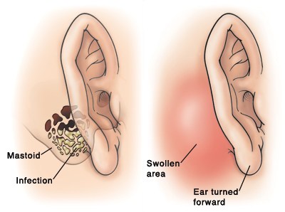
Mastoiditis: Symptoms & Treatments

One of the most important structures in your inner ear is the mastoid bone. Although it’s called a bone, the mastoid doesn’t have the typical structure associated with other bones in the human body. It’s made of air sacs and resembles a sponge, rather than being solid and rigid like most bones.

Mastoiditis is a serious infection in the mastoid process, which is the hard, prominent bone just behind and under the ear. Ear infections that people fail to treat cause most cases of mastoiditis. The condition is rare but can become life-threatening without treatment.

Image result for mastoiditis causes, symptoms & Treatment

Symptoms:

  • drainage from the affected ear
  • ear pain
  • fever
  • headache
  • hearing loss in the affected ear
  • redness, swelling, and tenderness behind the affected ear

Mastoiditis is a potentially life-threatening condition. Initial treatment for a severe infection may include hospitalization. You will receive antibiotic medication through a vein in your arm, or intravenously, while at the hospital. You will need to take oral antibiotics at home for several days after leaving the hospital.

If the infection doesn’t clear up after treatment with antibiotics, surgery may be necessary. Surgery may involve removing part of your mastoid bone to drain the infection. Doctors may also need to drain your middle ear of infected fluid to successfully treat the infection.

Idiopathic pulmonary fibrosis (IPF): Causes, Symptoms & Treatments

Idiopathic pulmonary fibrosis (IPF) is a type of lung disease that results in scarring (fibrosis) of the lungs for an unknown reason. Over time, the scarring gets worse and it becomes hard to take in a deep breath and the lungs cannot take in enough oxygen. The lung damage caused by pulmonary fibrosis can’t be repaired, but medications and therapies can sometimes help ease symptoms and improve quality of life. For some people, a lung transplant might be appropriate.

Image result for ipf in medical terms

fig— IPF bronchial Tube

Symptoms

  • Shortness of breath (dyspnea)
  • A dry cough
  • Fatigue
  • Unexplained weight loss
  • Aching muscles and joints
  • Widening and rounding of the tips of the fingers or toes (clubbing)

Causes: Occupational and environmental factors

Long-term exposure to a number of toxins and pollutants can damage your lungs. These include:

  • Silica dust, tobacco smoke, viruses
  • Asbestos fibers
  • Hard metal dusts
  • Coal dust
  • Grain dust
  • Bird and animal droppings

Radiation treatments

Some people who receive radiation therapy for lung or breast cancer show signs of lung damage months or sometimes years after the initial treatment. The severity of the damage may depend on:

  • How much of the lung was exposed to radiation
  • The total amount of radiation administered
  • Whether chemotherapy also was used
  • The presence of underlying lung disease

Medications

Many drugs can damage your lungs, especially medications such as:

  • Chemotherapy drugs. Drugs designed to kill cancer cells, such as methotrexate (Trexall, Otrexup, others) and cyclophosphamide, can also damage lung tissue.
  • Heart medications. Some drugs used to treat irregular heartbeats, such as amiodarone (Cordarone, Nexterone, Pacerone), may harm lung tissue.
  • Some antibiotics. Antibiotics such as nitrofurantoin (Macrobid, Macrodantin, others) or ethambutol can cause lung damage.
  • Anti-inflammatory drugs. Certain anti-inflammatory drugs such as rituximab (Rituxan) or sulfasalazine (Azulfidine) can cause lung damage.

Medical conditions

Lung damage can also result from a number of conditions, including:

  • Dermatomyositis
  • Polymyositis
  • Mixed connective tissue disease
  • Systemic lupus erythematosus
  • Rheumatoid arthritis
  • Sarcoidosis
  • Scleroderma
  • Pneumonia

Many people with idiopathic pulmonary fibrosis may also have gastroesophageal reflux disease (GERD) — a condition that occurs when acid from your stomach flows back into your esophagus.


Rheumatic Heart disease: Causes, Symptoms & Treatments

Rheumatic fever can lead to a condition known as rheumatic heart disease. This is usually a thickening and stenosis of one or more of the heart valves and often requires surgery to repair or replace the involved valve(s). Rheumatic valve disease is a thickening and stenosis of one or more of the heart valves and often requires surgery to repair or replace the affected valve(s). The valve could become leaky instead of stenotic as well.

Related image

Symptoms:

The symptoms of rheumatic heart disease vary and damage to the heart often is not readily noticeable. When symptoms do appear, they may depend on the extent and location of the heart damage. Typically, symptoms of rheumatic fever appear about two weeks after the onset of an untreated strep throat infection. Apart from the sore throat caused by the strep infection, children have a fever and feel ill. Commonly, the child will have a very painful, swollen and red joint — usually a large joint like a knee, ankle, elbow or shoulder — that goes away after a day or two only to be replaced by the same problem in another joint. Short-lived skin rashes may occur, but are not common. Even if the heart is affected, it is usually not severe enough to cause symptoms, although occasionally the child may be short of breath.

Treatments

Although having rheumatic fever leaves a child more susceptible to heart damage, it does not always permanently damage the heart. However, when the inflammation caused by rheumatic fever leaves one or more of the heart valves scarred, the result is rheumatic heart disease. The mitral valve and the aortic valve are usually the ones damaged by the disease. Years later, the mitral valve may become narrowed, a condition known as mitral stenosis.

Treatment of acute rheumatic fever includes antibiotics to treat the strep infection and additional medications to ease the inflammation of the heart and other symptoms. Usually aspirin is given in large doses until the joint inflammation goes away; rarely, steroids are needed. Once the acute illness has gone away, patients need to take penicillin, or an equivalent antibiotic, for many years to prevent recurrences. This is a very important treatment because the risk of heart valve damage increases if rheumatic fever recurs.

Most often the valve leak caused by the disease is mild and does not need treatment. If the leak is severe enough to strain and enlarge the heart, surgery may be needed to eliminate the leak. This surgery may involve repair of the damaged valve. Sometimes the valve is too badly damaged to repair, in which case it must be replaced by an artificial valve.

Water Melon Stomach: Causes & Treatments

Watermelon stomach, which is also known as gastric antral vascular ectasis or GAVE, is where the lining of the stomach bleeds, causing it to look like the characteristic stripes of a watermelon when viewed by endoscopy. Watermelon stomach occurs most commonly in older women (over age 70 years), although it can develop in men and women of any age. Signs and symptoms of watermelon stomach include blood in the stool, haematemesis (vomiting blood) and anaemia. It is a genetics disorder also.

The exact cause of watermelon stomach is not known, however, it is often diagnosed in people with other chronic (long-term) conditions such as cirrhosis (scarring of the liver and poor liver function) and systemic sclerosis.

Image result for watermelon stomach

Endoscopy CT scan looks like watermelon

Diagnosis

Diagnosis can be challenging as gastric bleeding is more commonly caused by other conditions, such as stomach ulcers and abnormal, enlarged vessels in the throat and stomach (esophageal varices) and some medications that can irritate the stomach lining such as aspirin or NSAIDs. Tests for watermelon stomach include:

  • an endoscopy
  • a biopsy of the stomach lining
  • an endoscopic ultrasound (ultrasound probe on the tip of an endoscope) computed tomography (CT) scan
  • a tagged red blood cell scan may be used to confirm the diagnosis.

Treatment

Treatment can be surgery and/or medications to stop or control the bleeding. Watermelon stomach is usually treated with endoscopic laser surgery or argon plasma coagulation. Both of these procedures are performed by endoscopy. Endoscopic laser surgery uses a laser light to treat bleeding blood vessels, while argon plasma coagulation uses argon gas and electrical current to seal irregular or bleeding tissue. In some cases, people may be treated with certain medications that help stop or control the gastrointestinal bleeding. Corticosteriods, tranexamic acid and hormone therapy (with oestrogen and progesterone) have been used to treat watermelon stomach with some success. If the bleeding is severe, blood transfusions may also be necessary at the time of diagnosis to control the bleeding. Additional transfusions may be recommended if gastrointestinal bleeding cannot be stopped or controlled