Global Treatment Services Pvt. Ltd.

Global Treatment Services

Difference between Viral Pink Eye and Bacterial Pink eye

Viral pink eye

Viral pink eye is often caused by an adenovirus, which is a common respiratory virus that can also cause a sore throat or upper respiratory infection. The herpes virus can also cause viral pink eye.

Symptoms of viral pink eye include:

  • Redness in the white of the eye.
  • Swelling of the eyelids.
  • Itching or burning feeling of the eyelids.
  • Swollen and tender areas in front of the ears.
  • A lot of tearing.
  • Clear or slightly thick, whitish drainage.

Viral pink eye symptoms usually last 5 to 7 days but may last up to 3 weeks and can become ongoing or chronic.

Pink eye may be more serious if you:

  • Have a condition that decreases your body’s ability to fight infection (impaired immune system).
  • Have vision in only one eye.
  • Wear contact lenses.

If the pink eye is caused by a virus, the person can usually return to daycare, school, or work when symptoms begin to improve, typically in 3 to 5 days. Medicines are not usually used to treat viral pink eye, so it is important to prevent the spread of the infection. Pink eye caused by a herpes virus, which is rare, can be treated with an antiviral medicine. Home treatment of viral pink eye symptoms can help you feel more comfortable while the infection goes away.

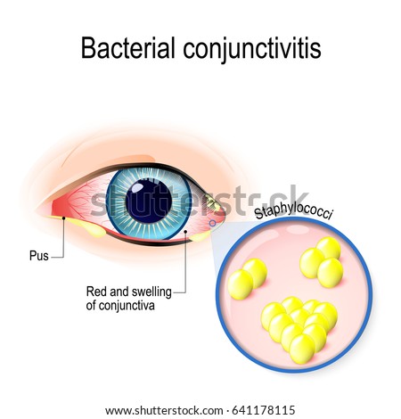
Bacterial pink eye

An infection may develop when bacteria enter the eye or the area around the eye. Some common infections that cause pink eye include:

  • Staph infection.
  • Haemophilus influenzae type b (Hib).
  • Cat-scratch disease.
  • Gonorrhea.

Symptoms of bacterial pink eye include:

  • Redness in the white of the eye.
  • Gray or yellow drainage from the eye. This drainage may cause the eyelashes to stick together.
  • Mild pain.
  • Swelling of the upper eyelid, which may make the lid appear to droop (pseudoptosis).

Bacterial pink eye may cause more drainage than viral pink eye. Bacterial infections usually last 7 to 10 days without antibiotic treatment and 2 to 4 days with antibiotic treatment. The person can usually return to daycare, school, or work 24 hours after an antibiotic has been started if symptoms have improved. Prescription antibiotic treatment usually kills the bacteria that cause pink eye.

Strabismus surgery in children: causes & treatments

With this condition, also known as crossed eyes or walleyes, your eyes aren’t always aligned. That means they don’t work together to look at an object. One may look in or out, or turn up or down. It can happen all the time or only when you’re stressed out or sick.

What Causes It?

Some children are born with it. Your child’s doctor will call this congenital strabismus. Many times, there’s no clear cause. There may be a problem with the part of his nervous system that controls eye muscles. Or he could have a tumor or eye disorder.

If it doesn’t appear until later in life, it will cause double vision. If an adult’s eyes cross without warning, he could have a serious condition like a stroke. If either one happens, see a doctor immediately.

Young children can suppress vision in a weaker eye, which lets them avoid double vision. However, that may lead to “lazy eye,” a condition your doctor will refer to as amblyopia. Depth perception and peripheral vision (vision off to the side) may be affected. It can cause eyestrain and headaches. If your eyes cross when you’re older, you may start to turn your head to see in certain directions and avoid double vision.

How Is It Treated?

Start treatment as soon as you can. If you don’t, the condition could continue into adulthood. Most adults with crossed eyes were born that way.

Talk to a pediatric ophthalmologist, an eye doctor who specializes in working with kids. He may start treatment with eyeglasses or a patch to force your child to use the off-kilter eye until he sees normally.

Sometimes, farsightedness is to blame. Glasses may solve the problem. The main goal is to get the problem eye working like it should before your child turns 8 years old. After that, permanent vision loss can set in.

Botox for Treating Squint or Strabismus in Children

Botulinum toxin injection can be used as a temporary cure for squint in children. This treatment is generally carried out under anaesthesia in case of children. This weakens the muscles that pull the eyes towards each other. However, this cannot be prescribed for all types of squint or strabismus. The effect of this may or may not be temporary, but for some types of squint, it has turned out to be very effective.

Is Surgery an Option?

Yes. It affects the muscles that move your child’s eye. It works best when done during childhood, but adults can have it, too

The surgeon opens the eyeball’s outer layer to reach a muscle. To strengthen the muscle, the surgeon removes a small section from one end and reattaches at the same location. This makes the muscle shorter, which turns the eye toward that side.

To weaken a muscle, the doctor moves it back or makes a partial cut across it. The eye turns away from that side.

Any double vision after surgery should go away within a few weeks as the brain adjusts to improved sight.

Bilopancreatic Diversion: Causes and details of procedure

A biliopancreatic diversion changes the normal process of digestion by making the stomach smaller. It allows food to bypass part of the small intestine so that you absorb fewer calories. Because of the risks, this surgery is for people who are more than severely obese and who haven’t been able to lose weight any other way. Super obesity means that you have a BMI(body mass index) of 50 or higher.

After surgery, you will feel full more quickly than when your stomach was its original size. This reduces the amount of food you will want to eat. Bypassing part of the intestine also means that you will absorb fewer calories. This leads to weight loss. But your best chance of keeping weight off after surgery is by adopting healthy habits, such as healthy eating and regular physical activity.

There are two biliopancreatic diversion surgeries: a biliopancreatic diversion and a biliopancreatic diversion with duodenal switch. Most surgeons will not perform duodenal switch surgery unless you are super obese (BMI of 50 or higher) and your weight is causing serious health problems.

  • In a biliopancreatic diversion , part of the stomach is removed. The remaining part of the stomach is connected to the lower portion of the small intestine. This is a high-risk surgery that can cause long-term health problems, because your body has a harder time absorbing food and nutrients. People who have this surgery must take vitamin and mineral supplements for the rest of their lives, which can be expensive.
  • In a biliopancreatic diversion with duodenal switch, a different part of the stomach is removed and the surgeon leaves the pylorus intact. The pylorus is the valve that controls food drainage from the stomach. This surgery is high-risk and can cause long-term health problems, because your body has a harder time absorbing food and nutrients. People who have this surgery must take vitamin and mineral supplements for the rest of their lives, which can be expensive. Another name for this surgery is duodenal switch.

These procedures can be done by making a large cut in the belly (an open procedure) or by making a small cut and using small tools and a camera to guide the surgery (laparoscopy).

What To Expect After Surgery

You will have some belly pain and may need pain medicine for the first week or so after surgery. The cut that the doctor makes (incision) may be tender and sore. Because the surgery makes your stomach smaller, you will get full more quickly when you eat. Food also may empty into the small intestine too quickly. This is called dumping syndrome. It can cause diarrhea and make you feel faint, shaky, and nauseated. It also can make it hard for your body to get enough nutrition. Having a duodenal switch reduces the risk of dumping syndrome.

Depending on how the surgery was done (open or laparoscopic), you’ll have to watch your activity during recovery. If you had open surgery, avoid heavy lifting or strenuous exercise while you are recovering so that your belly can heal. In this case, you will probably be able to return to work or your normal routine in 4 to 6 weeks.

Eating after surgery

Your doctor will give you specific instructions about what to eat after the surgery. For about the first month after surgery, your stomach can only handle small amounts of soft foods and liquids while you are healing. It is important to try to sip water throughout the day to avoid becoming dehydrated. You may notice that your bowel movements are not regular right after your surgery. This is common. Try to avoid constipation and straining with bowel movements.

Bit by bit, you will be able to add solid foods back into your diet. You must be careful to chew food well and to stop eating when you feel full. This can take some getting used to, because you will feel full after eating much less food than you are used to eating. If you do not chew your food well or do not stop eating soon enough, you may feel discomfort or nausea and may sometimes vomit. If you drink a lot of high calorie liquid such as soda or fruit juice, you may not lose weight. If you continually overeat, your stomach may stretch. If your stomach stretches, you will not benefit from your surgery.

This surgery removes the part of the intestine where many minerals and vitamins are most easily absorbed. Because of this, you may have a deficiency in iron, calcium, magnesium, or vitamins. It’s important to make sure you get enough nutrients in your daily meals to prevent vitamin and mineral deficiencies. You may need to work with a dietitian to plan meals. And you may need to take extra vitamin B12.

Why It Is Done

Weight loss surgery is suitable for people who are severely overweight and who have not been able to lose weight with diet, exercise, or medicine.

Most surgeons will not perform duodenal switch surgery unless you are super obese (body mass index (BMI) of 50 or higher) and your weight is causing serious health problem.

It is important to think of this surgery as a tool to help you lose weight. It is not an instant fix. You will still need to eat a healthy diet and get regular exercise. This will help you reach your weight goal and avoid regaining the weight you lose.

How Well It Works

Biliopancreatic diversion surgeries are effective. Most people lose 75% to 80% of their excess weight (the weight above what is considered healthy) and stay at their new weight. Ten years after weight loss surgery, many people have gained back 20% to 25% of the weight they lost. The long-term success is highest in people who are realistic about how much weight will be lost, keep appointments with the medical team, follow the recommended eating plan, and are physically active.

Risks

Risks common to all surgeries for weight loss include an infection in the incision, a leak from the stomach into the abdominal cavity or where the intestine is connected (resulting in an infection called peritonitis), and a blood clot in the legs (deep vein thrombosis, or DVT) or lung (pulmonary embolism). Some people develop gallstones or a nutritional deficiency condition such as anemia or osteoporosis.

Biliopancreatic diversion surgery has short-term and long-term risks, including:

  • Dumping syndrome. This causes nausea, weakness, sweating, faintness, and possibly diarrhea soon after eating. These symptoms get worse if you eat highly refined, high-calorie foods (like sweets). Sometimes you may become so weak that you have to lie down until the symptoms pass.
  • A higher risk of osteoporosis. This happens because your body can’t absorb nutrients as well as it used to.
  • Bad smelling stools and diarrhea. This can occur because of poor absorption of protein, fat, calcium, iron, and vitamins B12, A, D, E, and K.
  • Poor nutrition. Eating less and less absorption may mean that you are not getting enough nutrients, which can cause health problems. You will have to take vitamin supplements for the rest of your life.

What To Think About

Weight loss surgery does not remove fatty tissue. It is not cosmetic surgery.

Some studies show that people who have weight-loss surgery are less likely to die from heart problems, diabetes, or cancer compared to obese people who did not have the surgery.

Artificial Heart Valve Surgeries: Types & Procedures

When treatment for heart valve disease includes surgery, it can be traditional or minimally invasive balloon valvuloplasty.

What Happens During Traditional Heart Valve Surgery?

You’ll get medicine to help you sleep, and a surgeon will make a cut down the center of your sternum (also called your breastbone) so he can reach your heart. He’ll then repair or replace the heart valves that need it.

What Happens During Minimally Invasive Heart Valve Surgery?

It’s done through smaller cuts. This type of surgery lowers:

  • Blood loss
  • Trauma
  • How much time you’ll spend in the hospital

Your surgeon will review your test results first to see if you’re a candidate for this procedure.

Often, the surgeon and cardiologist will use something called a transesophageal echo before and after the surgery to see how the valve is working. It uses sound waves to give your doctor a real-time look at your heart and blood vessels.

What Is Heart Valve Repair Surgery?

The mitral valve is the one repaired most often. But the aortic, pulmonic, and tricuspid valves may be helped this way, too.

If your valve can be repaired, you’ll probably have one of the procedures below:

Commissurotomy: The surgeon takes away calcium deposits and other scar tissue from the valve’s leaflets (sometimes called flaps). This is usually done for people who have severely narrowed valves and shouldn’t have balloon valvotomy.

Decalcification: Calcium deposits are removed to allow the leaflets to be more flexible and close properly.

Reshape leaflets: If one of the leaflets is floppy, a part of it may be cut out, and then the flap will be sewn back together. This lets the valve close more tightly. This is also called quadrangular resection.

Chordal transfer: If the anterior leaflet of your mitral valve is floppy (your doctor may say it has prolapse), the tendons that connect your valves — called the chordae — are moved from your posterior leaflet to your anterior leaflet. Then, the posterior leaflet is fixed by the reshape leaflets procedure.

Annulus support: Your doctor may reshape or tighten the ring of tissue that supports your valve (called the valve annulus) if it’s too wide. He’ll sew a ring structure around it. The ring may be made of tissue or synthetic material.

Patched leaflets: Your surgeon may use tissue patches to repair any leaflets that have tears or holes.

The advantages of heart valve repair surgery include:

  • You’ll have less need for life-long blood thinner medication.
  • Your heart muscle will stay strong longer.

What If My Heart Valve Cannot Be Repaired?

When you have aortic or pulmonic valve disease, valve replacement surgery is usually done.

In rare cases, the aortic valve can be repaired.

If your heart valves cannot be repaired, your doctor will replace them.

During the surgery, the damaged valve is removed and a new valve is sewn to the tissue that supported the original. The new valve can be:

Mechanical: It’s made totally of mechanical parts that your body will accept. Something called a bi-leaflet valve is used most often. It’s two carbon leaflets in a ring covered by polyester knit fabric.

Their advantage is they’re designed to last many years.

There are also potential drawbacks. People who get these have to take blood thinner medication for the rest of their life to prevent clots from forming in it. These clots can raise your chance of having a stroke. Also, some people hear a ticking sound. It’s the valve leaflets opening and closing.

Biological: Tissue valves (your doctor may call them biologic or bioprosthetic valves) are made of human or animal tissue. It may come from pigs or cows. The valves may have some artificial parts to help give it support and help its placement.

With these, most people don’t need to take life-long blood thinners, unless they have other conditions (like atrial fibrillation) that make them needed. Some of these valves can last as long as 17 years.

Homograft: Also called allograft, it’s a valve removed from a donated human heart that’s preserved and frozen under sterile conditions. One may replace a diseased valve.

Are There Nonsurgical Options for Heart Valve Disease?

Balloon valvotomy can make the opening of a narrowed (stenotic) valve wider. It’s sometimes used for folks who have:

  • Mitral valve stenosis (narrowing of the mitral valve) with symptoms
  • Aortic stenosis (narrowing of the aortic valve), but aren’t able to have surgery
  • Pulmonic stenosis (narrowing of the pulmonic valve)

In this procedure, a catheter is put into a blood vessel in the groin and guided to the heart. The tip is steered inside the narrowed valve. Once there, a tiny balloon is inflated and deflated several times to widen the opening.

Once the cardiologist sees the valve has been made wide enough, the balloon is taken out.

During this, the cardiologist may do an echocardiogram (ultrasound of your heart) to get a better look at the valve.

Pages:12