Global Treatment Services Pvt. Ltd.

Global Treatment Services

Schwannomas: Symptoms & Treatments

Schwannomas are benign tumors that arise from the nerve sheath (covering) of cranial nerves along-side the cerebellum and brainstem.

The optimal treatment for the majority of symptomatic schwannomas is maximal surgical removal and/or focused radiation therapy (radiosurgery). Fortunately, for patients requiring schwannoma surgery, most large vestibular (acoustic) schwannomas can be removed through a retromastoid keyhole craniotomy while most trigeminal schwannomas can be removed through either a retromastoid approach or an endonasal endoscopic approach.

These tumors are typically benign and arise from the nerve sheath (covering) of cranial nerves along-side the cerebellum and brainstem. The most common schwannoma arises from the 8th cranial nerve (the vestibulo-cochlear nerve) or the 5th cranial nerve (the trigeminal nerve). In some instances, schwannomas are related to a genetic syndrome called Neurofibromatosis. Bilateral vestibular schwannomas are associated with NF-2.

Symptoms:

Vestibular (acoustic) schwannomas arise from one of the vestibular nerves in the internal auditory canal and initially cause hearing loss and tinnitus (ringing in the ear). As they enlarge into the cerebello-pontine angle, they can compress the brainstem and other cranial nerves, resulting in loss of balance and coordination, vertigo, facial numbness, facial weakness and difficulty swallowing.

Trigeminal schwannomas are less common than vestibular schwannomas. They generally arise in Meckel’s cave in the skull base and the pre-pontine space. These tumors typically cause facial pain (trigeminal neuralgia) or facial numbness. As they enlarge, they can grow farther into the cavernous sinus or into the brainstem, causing double vision, loss of coordination and other symptoms of brainstem compression.

Treatment:

Treatment for vestibular (acoustic) schwannomas is by surgical removal through akeyhole retrosigmoid craniotomy or other skull base approach or by radiosurgery. For tumors under 2.5 cm, either surgery or radiosurgery are reasonable treatment options. For tumors over 2.5 cm, surgical removal is generally recommended.
Treatment for trigeminal schwannomas is typically by surgical removal through a retrosigmoid craniotomy or other skull base approach, depending upon the location.

In some patients with a vestibular or trigeminal schwannoma in whom only a sub-total tumor removal is possible, radiosurgery or stereotactic radiotherapy may be effectively used to control further tumor growth. Chemotherapy is generally not used for treating schwannomas.


Gallstones: Symptoms, Causes & Treatments

What Are Gallstones?

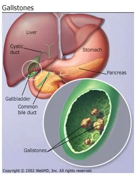
They aren’t really stones. They’re pieces of solid material that form in the gallbladder, a small organ located under the liver.

You might not even know you have them until they block a bile duct, causing pain that you need to get treated right away.

Types:
The two main kinds are:Cholesterol stones. These are usually yellow-green in color. They’re the most common kind, accounting for 80% of gallstones.

Pigment stones. These stones are smaller and darker. They’re made up of bilirubin, which comes from bile, a fluid your liver makes and your gallbladder stores.

Gallstones


What Causes Gallstones?

There may be several reasons, including:

  • Your genes
  • Your weight
  • Problems with your gallbladder
  • Diet

Bile can be part of the problem. Your body needs bile, but if it has too much cholesterol in it, that makes gallstones more likely.

It can also happen if your gallbladder can’t empty properly.

Pigment stones are more common in people with certain medical conditions, such as cirrhosis (a liver disease) or blood diseases such as sickle cell anemia.

What are  Risk factors?

You’re more likely to get gallstones if:

You’re obese. This is one of the biggest risk factors. Obesity can raise your cholesterol level and also make it harder for the gallbladder to empty completely.

You take birth control pills, hormone replacement therapy for menopause symptoms, or are pregnant. The extra estrogen is the problem. It can increase cholesterol and make it harder for the gallbladder to empty.

You have diabetes. People with this condition tend to have higher levels of triglycerides (a type of blood fat), which is a risk factor for gallstones.

You take medicine to lower your cholesterol. Some of these drugs boost the amount of cholesterol in bile, which may increase your chances of getting cholesterol stones.

You lost weight too quickly. Your liver makes extra cholesterol, which may lead to gallstones.

You’re fasting. Your gallbladder may not squeeze as much.

What Are the Symptoms?

You might not notice anything, or even know you have gallstones, unless your doctor tells you. But if you do get symptoms, they usually include:

  • Pain in your upper belly and upper back that can last for several hours
  • Nausea
  • Vomiting
  • Other digestive problems, including bloating, indigestion and heartburn, and gas

What’s the Treatment?

Many people with gallstones get surgery to take out the gallbladder. There are two different kinds of operations.

Laparoscopic cholecystectomy. This is the more common procedure. The surgeon passes instruments, a light, and a camera through several small cuts in the belly. He views the inside of the body on a video monitor. Afterward, you spend the night in the hospital.

Open cholecystectomy. The surgeon makes bigger cuts in the belly to remove the gallbladder. You stay in the hospital for a few days after the operation.

If gallstones are in your bile ducts, the doctor may use ERCP to find and remove them before or during gallbladder surgery.

Ischaemic Myelopathy: Symptoms, causes & Treatments

What is ischaemic myelopathy and what causes it?

Ischaemic myelopathy is a condition where the blood supply to part of the spinal cord (the bundle of nerves that run inside the spine) is suddenly interrupted by a ‘clot’ that blocks a small artery (blood vessel). This is similar to a stroke or heart attack in people, except that it is the spinal cord that is affected rather than the brain or heart.

‘Ischaemia’ is a loss of blood supply, and ‘myelopathy’ is a form of damage to the nerves of the spinal cord. The cause of the blockage of the spinal cord blood vessels is poorly understood. In the majority of cases the ‘clot’ which blocks the artery is similar in structure to the material that forms the discs (the cushions between the bones of the spine). This disc material is called ‘fibro-cartilage’. It is assumed that some of this disc material somehow moves from its normal location to become lodged in the blood vessels of the spinal cord. This is the reason why the condition is commonly referred to as‘fibrocartilaginous embolism’ (shortened to ‘FCE’).

Other less common reasons why the blood supply to the spinal cord can be interrupted include either an abscess or a tumour, either of which can press on important blood vessels and prevent blood flow to the nerves.


What are the common signs of ischaemic myelopathy?

Ischaemic myelopathy most commonly occurs in large breed dogs, although it is also encountered in smaller breeds and, very occasionally, in cats. The onset of neurological signs is always extremely rapid and often occurs during vigorous exercise. Neurological abnormalities may vary from mild weakness or incoordination, through to an inability to walk. Paralysis, incontinence and inability to feel pain are possible in severe cases. Ischaemic myelopathy is a non-painful condition, although at the initial onset some patients may cry out or yelp. Neurological signs do not tend to progress after the first 24 hours.


How is ischaemic myelopathy diagnosed?

Ischaemic myelopathy is diagnosed by ruling out other causes of sudden onset neurological signs, such as a ‘slipped disc’ (see information sheets on cervical disc disease and thoracolumbar disc disease), traumatic disc extrusion or a spinal fracture.

Advanced diagnostic imaging investigations are necessary in order to diagnose ischaemic myelopathy and to rule-out other conditions – an MRI scan is the imaging technique of choice in these circumstances. MRI scanning uses high powered magnets and a computer to generate images of the spine (this is the same technique and the same equipment which is used for body scanning in human patients). It provides detailed information on the location and extent of any blood vessel-related injury to the spinal cord. Myelography is an alternative imaging technique which can also be used for investigating spinal injuries. This involves injecting a dye (contrast agent) around the spinal cord and obtaining multiple X-rays to assess the flow of the dye. Injecting around the spinal cord is not without risk of causing further damage to already compromised nerve tissue, however. Unlike an MRI scan, myelography will not show damage to the spinal cord caused by ischaemic myelopathy, although it will enable some other conditions to be ruled out as the cause of the spinal cord injury. MRI scanning is less invasive than myelography, and with less risk of side-effects, and for most patients MRI provides the best option for investigation. Both MRI and myelography require the dog to have a general anaesthetic.


MRI scan showing an ischaemic myelopathy (FCE) in the neck (arrow)

MRI scan showing an ischaemic myelopathy (FCE) in the neck (arrow)


MRI scan showing an ischaemic myelopathy (FCE) in the back (arrow)

MRI scan showing an ischaemic myelopathy (FCE) in the back (arrow)


How can ischaemic myelopathy be treated?

There is no specific treatment for ischaemic myelopathy. Supportive care and nursing are essential in order to aid recovery. Bedding needs to be well padded in order to prevent bed sores. Regular physiotherapy and, in some cases, swimming at a hydrotherapy unit can help function to be regained and assist in building strength. Some patients may require a catheter to be placed in the bladder temporarily to aid urination.

What is the outcome for patients with ischaemic myelopathy?

The outcome in dogs with ischaemic myelopathy depends on two key factors – a) the location and b) the severity of the spinal cord injury. The findings on the neurological examination and MRI investigations can help to predict the chances of recovery. Dogs that are paralysed and also cannot feel pain in their limbs are unlikely to recover, unfortunately. However, the majority of less severely affected dogs will gradually improve over a period of weeks to months, to a point where they can freely exercise and have a good quality of life. A degree of weakness in one or more limbs may persist in some dogs. Recurrence of ischaemic myelopathy is very uncommon.


Hyperglycemia : symptoms, causes and treatments

Hyperglycemia facts

  • Hyperglycemia is an abnormally high blood glucose (blood sugar) level.
  • Hyperglycemia is a hallmark sign of diabetes (bothtype 1 diabetes and type 2 diabetes) and prediabetes.
  • Diabetes is the most common cause of hyperglycemia.
  • Other conditions that can cause hyperglycemia arepancreatitis, Cushing’s syndrome, unusual hormone-secreting tumors, pancreatic cancer, certain medications, and severe illnesses.
  • The main symptoms of hyperglycemia are increased thirst and a frequent need to urinate.
  • Severely elevated glucose levels can result in a medical emergency like diabetic ketoacidosis (DKA) or hyperglycemic hyperosmolar nonketotic syndrome (HHNS, also referred to as hyperglycemic hyperosmolar state).
  • Insulin is the treatment of choice for people with type 1 diabetes and for life-threatening increases in glucose levels.
  • People with type 2 diabetes may be managed with a combination of different oral and injectable medications.
  • Hyperglycemia due to medical conditions other than diabetes is generally treated by treating the underlying condition responsible for the elevated glucose


Diabetic Ketoacidosis Symptoms

People with diabetes don’t have the luxury of that auto-sensing. Not enough insulin and the glucose levels in the blood stream start to rise; too much insulin, and they plummet.

The consequences of hypoglycemia are easy to understand. No energy source, no function – and the first organ to go is the brain. It needs glucose to function and without it, the brain shuts down quickly. Confusion, lethargy, and coma occur quickly. Blood sugar is one of the first things checked on scene of a comatose patient, because it’s so easy to fix and very embarrassing for an EMT to miss.

What causes hyperglycemia?

A number of medical conditions can cause hyperglycemia, but the most common by far is diabetes mellitus. Diabetes affects over 8% of the total U.S. population. In diabetes, blood glucose levels rise either because there is an insufficient amount of insulin in the body or the body cannot use insulin well. Normally, the pancreas releases insulin after a meal so that the cells of the body can utilize glucose for fuel. This keeps blood glucose levels in the normal range.

Type 1 diabetes is responsible for about 5% of all cases of diabetes and results from damage to the insulin-secreting cells of the pancreas. Type 2 diabetes is far more common and is related to the body’s inability to effectively use insulin. In addition to type 1 and type 2, gestational diabetes is a form of diabetes that develops in pregnant women. Studies show that between 2% to 10% of all pregnant women get gestational diabetes.

Sometimes, hyperglycemia is not the result of diabetes. Other medical conditions that can cause hyperglycemia include:

How is hyperglycemia treated?

Mild or transient hyperglycemia may not need medical treatment, depending upon the cause. People with mildly elevated glucose or prediabetes can often lower their glucose levels by incorporating diet and lifestyle changes. To assure that you chose the right dietary and lifestyle changes , you should speak with your health care professional or use reliable resources such as the American Diabetes Association.

Insulin is the treatment of choice for people with type 1 diabetes and for life-threatening increases in glucose levels. People with type 2 diabetes may be managed with a combination of different oral and injectable medications. Some people with type 2 diabetes also take insulin.

Hyperglycemia due to medical conditions other than diabetes is generally treated by addressing the underlying condition responsible for the elevated glucose. In some cases, insulin may be needed to stabilize glucose levels during this treatment.